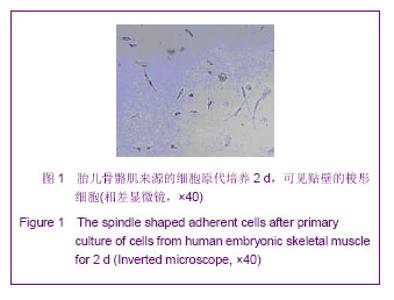
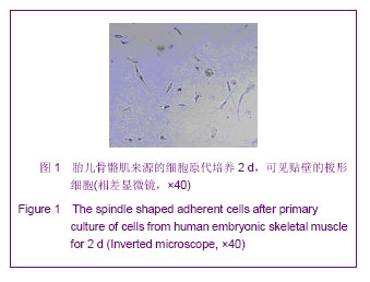
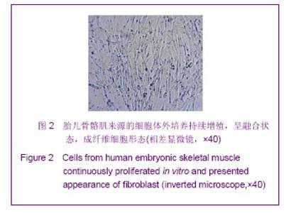
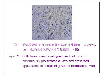
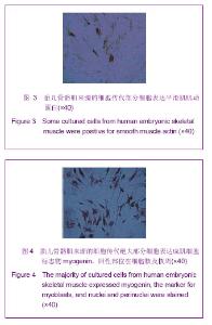
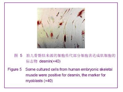
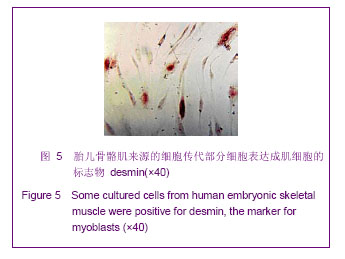
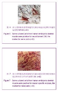
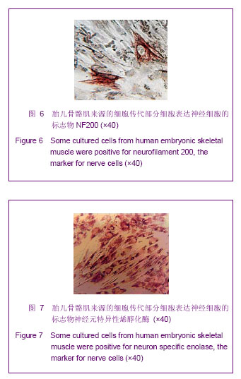
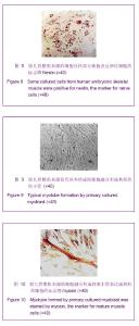
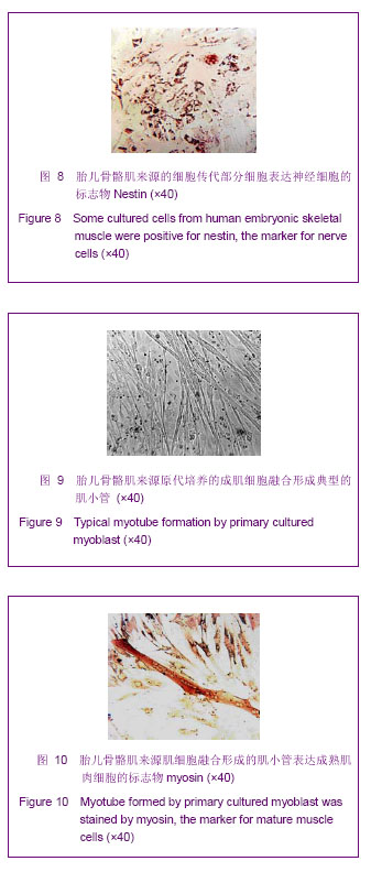
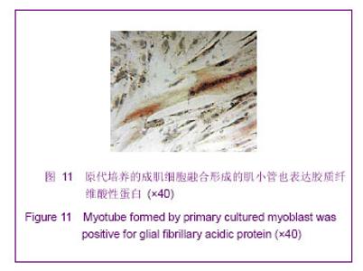
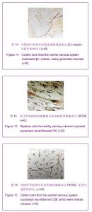
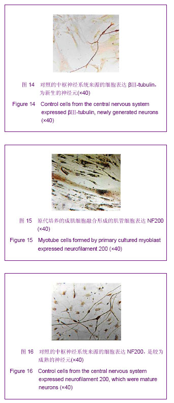
| [1] Ehrhardt J, Brimah K, Adkin C, et al.Human muscle precursor cells give rise to functional satellite cells in vivo. Neuromuscul Disord. 2007;17(8):631-638.[2] Salvatori G, Ferrari G, Mezzogiorno A, et al. Retroviral vector-mediated gene transfer into human primary myogenic cells leads to expression in muscle fibers in vivo. Hum Gene Ther.1993;4(6):713-723.[3] 杨立业,郑佳坤,郭礼和.人类神经前体细胞大鼠胎脑内移植研究[J].四川大学学报:医学版, 2005,36(5):613-617.[4] Lepper C,Conway SJ,Fan CM.Adult satellite cells and embryonic muscle progenitors have distinct genetic requirements.Nature.2009; 460(7255): 627-631.[5] 杨志明,岑石强,解慧琪,等.原代人胚成肌细胞体外培养增殖及分化特性[J].中国修复重建外科杂志,2001,15(5):261-264. [6] Morshead CM. Adult neural stem cells: attempting to solve the identity crisis. Dev Neurosci. 2004;26(2-4):93-100.[7] 王海英.5-氮胞苷诱导培养P19胚胎癌细胞表达心肌结构蛋白[J].中国组织工程研究,2012,16(11):2019-2022. [8] 王跃春,李业霞,段阿林,等.人脐带间充质干细胞的快速分离、纯化及冻存[J].中国病理生理杂志,2010,26(8):1658-1661.[9] 王跃春,张洹.人胎肝MSCs的分离培养及其类ESCs特性的研究[J].中国病理生理杂志,2009,25(2):409-412.[10] 张峻梅,岑石强,杨志明,等.流式细胞仪碘化丙啶染色法测定类胰岛素生长因子Ⅰ对人胚胎原代及传代成肌细胞生长周期的作用[J].中国组织工程研究与临床康复,2008,12(16): 3110- 3114. [11] 岑石强,李万里,黄富国,等.小肠黏膜下层复合上皮细胞及成肌细胞构建组织工程食管的初步研究[J].中国修复重建外科杂志, 2006,20(10):1040-1043. [12] 黄建荣,李建华,吴家昌,等.体外诱导人骨髓间充质干细胞分化为软骨细胞[J].中国临床康复,2006,23(33):128-133. [13] 刘兴茂,陈昭烈,刘红,等.在多孔胶原支架上构建三维工程的心肌组织[J].中国临床康复,2004,8(36):8387-8389. [14] 窦克非,杨跃进,阮英茆,等.成年犬骨骼肌成肌细胞培养方法改良及生长特性研究[J].中国循环杂志,2003,18(5): 382-384.[15] 杨志明,岑石强,解慧琪,等.原代人胚成肌细胞体外培养增殖及分化特性[J].中国修复重建外科杂志,2001,15(5):261-264. [16] Lapan AD, Gussoni E.Isolation and characterization of human fetal myoblasts. Methods Mol Biol. 2012;798:3-19.[17] Law PK,Bertorini TE,Goodwin TG,et al.Dystrophin production induced by myoblast transfer therapy in Duchenne muscular dystrophy.Lancet.1990; 336(8707):114-115.[18] Partridge TA, Morgan JE, Coulton GR, et al. Conversion of mdx myofibres from dystrophin-negative to -positive by injection of normal myoblasts. Nature.1989;337(6203): 176-179.[19] Huard J, Acsadi G, Jani A,et al.Gene transfer into skeletal muscles by isogenic myoblasts. Hum Gene Ther.1994;5: 949-958.[20] Goudenege S, Lebel C, Huot NB, et al.Myoblasts Derived From Normal hESCs and Dystrophic hiPSCs Efficiently Fuse With Existing Muscle Fibers Following Transplantation. Mol Ther. 2012;20(11):2153-2167. |