Chinese Journal of Tissue Engineering Research
Previous Articles Next Articles
Autologous bone marrow mesenchymal stem cells and related chemokines in fracture microenvironment
Zhang Xian-yu1, Wang Wei2, Chen Wei3, Chen Jian-mei4, Xu Hao4
- 1Department of Orthopedics, People’s Hospital of Shangrao, Shangrao 334000, Jiangxi Province, China; 2Department of Pathology, Shangrao Branch of Jiangxi Medical College, Shangrao 334000, Jiangxi Province, China; 3Department of Orthopedics, the Second Hospital of Fuzhou, Fuzhou 350007, Fujian Province, China; 4Department of Orthopedics, Fuzhou General Hospital of Nanjing Military Area Command of PLA, Fuzhou 350002, Fujian Province, China
-
Received:2012-11-12Revised:2012-12-16Online:2013-07-02Published:2013-07-02 -
Contact:Xu Hao, Chief physician, Professor, Master’s supervisor, Department of Orthopedics, Fuzhou General Hospital of Nanjing Military Area Command of PLA, Fuzhou 350002, Fujian Province, China xiuhao@medmail.com.cn -
About author:Zhang Xian-yu★, Master, Attending physician, Department of Orthopedics, People’s Hospital of Shangrao, Shangrao 334000, Jiangxi Province, China 50360042@qq.com -
Supported by:the Natural Science Foundation of Fujian Province, No. 2007J0110*; the Military Medicine and Health Science Foundation of China, No. 07Z037*; Medicine and Health Science Foundation of Nanjing Military Area Command of PLA During the Eleventh Five-year Plan, No. 06MA145*
CLC Number:
Cite this article
Zhang Xian-yu, Wang Wei, Chen Wei, Chen Jian-mei, Xu Hao . Autologous bone marrow mesenchymal stem cells and related chemokines in fracture microenvironment[J]. Chinese Journal of Tissue Engineering Research, doi: 10.3969/j.issn.2095-4344.2013.27.019.
share this article
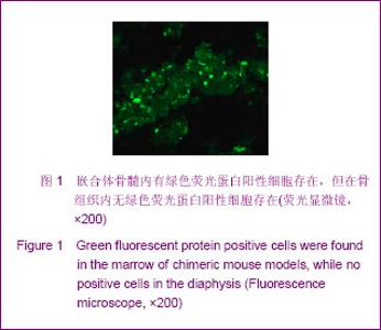
2.1 实验动物数量分析 成功建立荧光/普通C57BL/6小鼠嵌合体70只,3个月后存活63只。随机取42只建立荧光/普通C57BL/6小鼠嵌合体骨折模型;随机取7只检测正常骨组织内与骨髓间充质干细胞定向相关趋化因子表达水平。 2.2 骨髓间充质干细胞在骨折修复中的作用 正常骨组织内,未观察到明显的绿色荧蛋白阳性信号,提示骨髓间充质干细胞并未参与正常骨组织更新,见图1。"

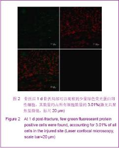
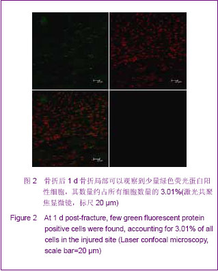
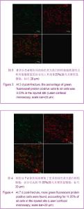
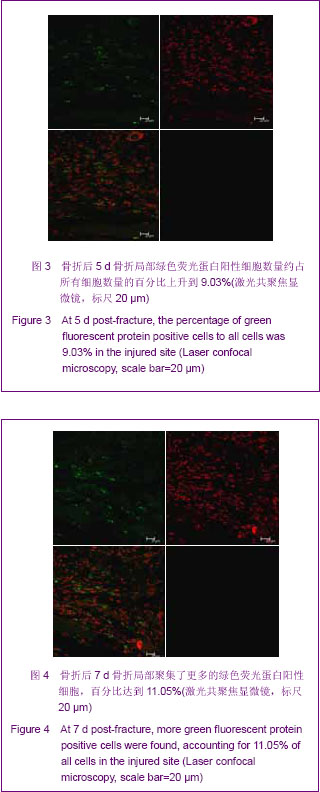
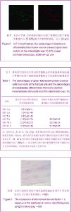
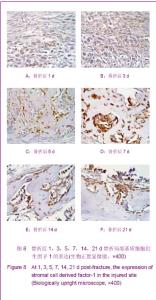
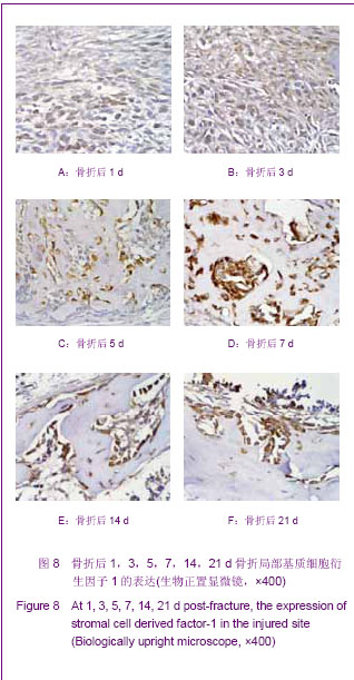
骨折后1,3,5,7,14,21 d,骨折部位可以观察到数量不等绿色荧蛋白阳性细胞聚集,数量呈逐渐上升趋势,见图2-4,提示骨折后骨髓间充质干细胞可以趋化到骨折局部。"
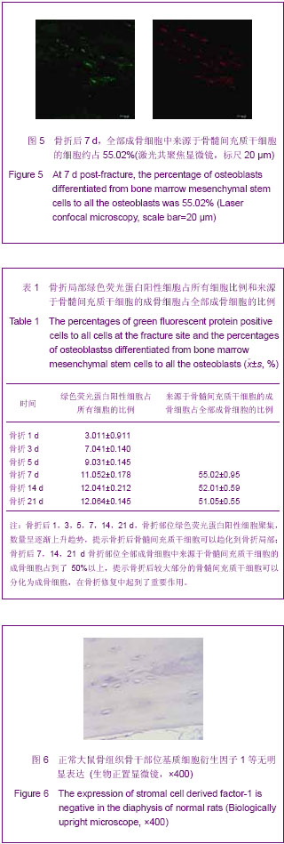
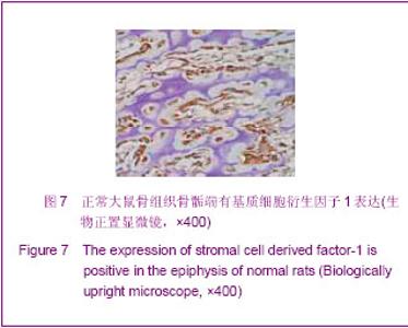
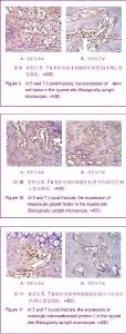
骨折后7,14,21 d,骨折部位全部成骨细胞中来源于骨髓间充质干细胞的成骨细胞占到了50%以上,见图5,提示骨折后较大部分的骨髓间充质干细胞可以分化为成骨细胞,在骨折修复中起到了重要作用。具体数据见表1。"
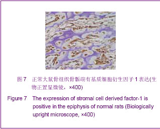
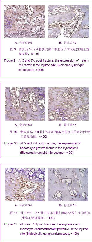
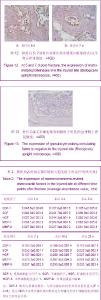
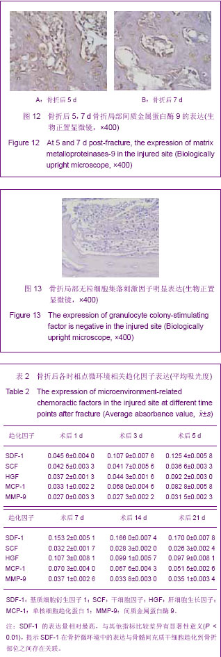
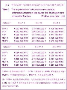
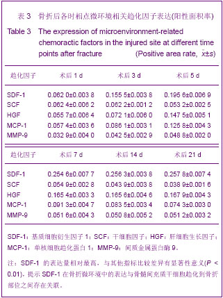
2.3 正常骨组织内与骨髓间充质干细胞定向相关趋化因子表达水平的检测结果 正常骨骨干部位未检出基质细胞衍生因子1、干细胞因子、肝细胞生长因子、粒细胞集落刺激因子、单核细胞趋化蛋白1、间质金属蛋白酶9因子表达,见图6。仅在干骺端软骨组织内的成骨样细胞胞浆内检出有基质细胞衍生因子1的表达,见图7。"
"
"
"
"
"
| [1] Westerhuis RJ, van Bezooijen RL, Kloen P.Use of bone morphogenetic proteins in traumatology. Injury. 2005;36(12): 1405-1412.[2] Parekkadan B, van Poll D, Suganuma K, et al. Mesenchymal stem cell-derived molecules reverse fulminant hepatic failure. PLoS One. 2007;2(9):e941.[3] Ohnishi S, Sumiyoshi H, Kitamura S, et al.Mesenchymal stem cells attenuate cardiac fibroblast proliferation and collagen synthesis through paracrine actions.FEBS Lett. 2007;581(21): 3961-3966.[4] Abdel Aziz MT, Atta HM, Mahfouz S,et al.Therapeutic potential of bone marrow-derived mesenchymal stem cells on experimental liver fibrosis.Clin Biochem. 2007;40(12):893-899.[5] Gupta N, Su X, Popov B,et al. Intrapulmonary delivery of bone marrow-derived mesenchymal stem cells improves survival and attenuates endotoxin-induced acute lung injury in mice. J Immunol. 2007;179(3):1855-1863.[6] Sadat S, Gehmert S, Song YH, et al. The cardioprotective effect of mesenchymal stem cells is mediated by IGF-I and VEGF. Biochem Biophys Res Commun. 2007;363(3): 674-679.[7] Ehrhardt D.GFP technology for live cell imaging.Curr Opin Plant Biol. 2003;6(6):622-628.[8] Blum JS, Temenoff JS, Park H, et al. Development and characterization of enhanced green fluorescent protein and luciferase expressing cell line for non-destructive evaluation of tissue engineering constructs.Biomaterials. 2004;25(27): 5809-5819.[9] Falanga V, Iwamoto S, Chartier M, et al. Autologous bone marrow-derived cultured mesenchymal stem cells delivered in a fibrin spray accelerate healing in murine and human cutaneous wounds.Tissue Eng. 2007;13(6):1299-1312.[10] Chung NG, Jeong DC, Park SJ, et al. Cotransplantation of marrow stromal cells may prevent lethal graft-versus-host disease in major histocompatibility complex mismatched murine hematopoietic stem cell transplantation. Int J Hematol. 2004;80(4):370-376.[11] Müller I, Kordowich S, Holzwarth C, et al. Application of multipotent mesenchymal stromal cells in pediatric patients following allogeneic stem cell transplantation.Blood Cells Mol Dis. 2008;40(1):25-32.[12] Wu GD, Bowdish ME, Jin YS, et al. Contribution of mesenchymal progenitor cells to tissue repair in rat cardiac allografts undergoing chronic rejection. J Heart Lung Transplant. 2005;24(12):2160-2169.[13] Bilic-Curcic I, Kalajzic Z, Wang L, et al. Origins of endothelial and osteogenic cells in the subcutaneous collagen gel implant. Bone. 2005;37(5):678-687.[14] Lazarini F, Tham TN, Casanova P, et al. Role of the alpha-chemokine stromal cell-derived factor (SDF-1) in the developing and mature central nervous system.Glia. 2003; 42(2):139-148.[15] Bhakta S, Hong P, Koc O.The surface adhesion molecule CXCR4 stimulates mesenchymal stem cell migration to stromal cell-derived factor-1 in vitro but does not decrease apoptosis under serum deprivation.Cardiovasc Revasc Med. 2006;7(1):19-24. [16] Kucia M, Jankowski K, Reca R, et al. CXCR4-SDF-1 signalling, locomotion, chemotaxis and adhesion. J Mol Histol. 2004;35(3):233-245.[17] Kögler G, Radke TF, Lefort A, et al. Cytokine production and hematopoiesis supporting activity of cord blood-derived unrestricted somatic stem cells. Exp Hematol. 2005;33(5): 573-583.[18] Sugimoto K, Adachi Y, Moriyama K,et al. Induction of the expression of SCF in mouse by lethal irradiation.Growth Factors. 2001;19(4):219-231. [19] Andrews RG, Knitter GH, Bartelmez SH,et al. Recombinant human stem cell factor, a c-kit ligand, stimulates hematopoiesis in primates. Blood. 1991;78(8):1975-1980.[20] Lapidot T, Dar A, Kollet O. How do stem cells find their way home. Blood. 2005;106(6):1901-1910.[21] Matsuda-Hashii Y, Takai K, Ohta H,et al. Hepatocyte growth factor plays roles in the induction and autocrine maintenance of bone marrow stromal cell IL-11, SDF-1 alpha, and stem cell factor. Exp Hematol. 2004;32(10):955-961.[22] Forte G, Minieri M, Cossa P, et al. Hepatocyte growth factor effects on mesenchymal stem cells: proliferation, migration, and differentiation.Stem Cells. 2006;24(1):23-33.[23] Bottaro DP, Rubin JS, Faletto DL,et al. Identification of the hepatocyte growth factor receptor as the c-met proto-oncogene product.Science. 1991;251(4995):802-804.[24] 刘红军,段海峰,鲁茁壮,等.肝细胞生长因子对骨髓间充质干细胞生物学特性的影响[J].中国实验血液学杂志,2005, 13(6) : 1044-1048.[25] Takano H, Ohtsuka M, Akazawa H, et al. Pleiotropic effects of cytokines on acute myocardial infarction: G-CSF as a novel therapy for acute myocardial infarction.Curr Pharm Des. 2003;9(14):1121-1127.[26] Seiler C, Pohl T, Wustmann K,et al. Promotion of collateral growth by granulocyte-macrophage colony-stimulating factor in patients with coronary artery disease: a randomized, double-blind, placebo-controlled study.Circulation. 2001; 104(17):2012-2017.[27] Fukuda K, Fujita J. Mesenchymal, but not hematopoietic, stem cells can be mobilized and differentiate into cardiomyocytes after myocardial infarction in mice. Kidney Int. 2005;68(5):1940-1943.[28] Kollet O, Spiegel A, Peled A, et al. Rapid and efficient homing of human CD34(+)CD38(-/low)CXCR4(+) stem and progenitor cells to the bone marrow and spleen of NOD/SCID and NOD/SCID/B2m(null) mice. Blood. 2001;97(10):3283- 3291.[29] 刘岐焕,程范军,高清平,等.重组人粒细胞集落刺激因子促进骨髓间充质干细胞增殖[J].中华器官移植杂志,2006,27(9):532-534.[30] 马胜忠,侯铁胜,张雪松,等.大鼠脊髓损伤后炎症细胞趋化因子MCP-1的表达及其意义[J].第二军医大学学报,2004,25(7): 766-768.[31] Che X, Ye W, Panga L, et al. Monocyte chemoattractant protein-1 expressed in neurons and astrocytes during focal ischemia in mice. Brain Res. 2001;902(2):171-177.[32] Wang L, Li Y, Chen J, et al. Ischemic cerebral tissue and MCP-1 enhance rat bone marrow stromal cell migration in interface culture. Exp Hematol. 2002;30(7):831-836.[33] 孟玲,刘国树,高艳红,等.骨髓间充质干细胞的体外趋化与单核细胞趋化蛋白1的作用[J].中国临床康复,2005,9(19):68-70. |
| [1] | Xu Feng, Kang Hui, Wei Tanjun, Xi Jintao. Biomechanical analysis of different fixation methods of pedicle screws for thoracolumbar fracture [J]. Chinese Journal of Tissue Engineering Research, 2021, 25(9): 1313-1317. |
| [2] | Zhang Chong, Liu Zhiang, Yao Shuaihui, Gao Junsheng, Jiang Yan, Zhang Lu. Safety and effectiveness of topical application of tranexamic acid to reduce drainage of elderly femoral neck fractures after total hip arthroplasty [J]. Chinese Journal of Tissue Engineering Research, 2021, 25(9): 1381-1386. |
| [3] | Chen Xinmin, Li Wenbiao, Xiong Kaikai, Xiong Xiaoyan, Zheng Liqin, Li Musheng, Zheng Yongze, Lin Ziling. Type A3.3 femoral intertrochanteric fracture with augmented proximal femoral nail anti-rotation in the elderly: finite element analysis of the optimal amount of bone cement [J]. Chinese Journal of Tissue Engineering Research, 2021, 25(9): 1404-1409. |
| [4] | Du Xiupeng, Yang Zhaohui. Effect of degree of initial deformity of impacted femoral neck fractures under 65 years of age on femoral neck shortening [J]. Chinese Journal of Tissue Engineering Research, 2021, 25(9): 1410-1416. |
| [5] | Zhang Chao, Lü Xin. Heterotopic ossification after acetabular fracture fixation: risk factors, prevention and treatment progress [J]. Chinese Journal of Tissue Engineering Research, 2021, 25(9): 1434-1439. |
| [6] | Zhou Jihui, Li Xinzhi, Zhou You, Huang Wei, Chen Wenyao. Multiple problems in the selection of implants for patellar fracture [J]. Chinese Journal of Tissue Engineering Research, 2021, 25(9): 1440-1445. |
| [7] | Wang Debin, Bi Zhenggang. Related problems in anatomy mechanics, injury characteristics, fixed repair and three-dimensional technology application for olecranon fracture-dislocations [J]. Chinese Journal of Tissue Engineering Research, 2021, 25(9): 1446-1451. |
| [8] | Hu Kai, Qiao Xiaohong, Zhang Yonghong, Wang Dong, Qin Sihe. Treatment of displaced intra-articular calcaneal fractures with cannulated screws and plates: a meta-analysis of 15 randomized controlled trials [J]. Chinese Journal of Tissue Engineering Research, 2021, 25(9): 1465-1470. |
| [9] | Hou Jingying, Yu Menglei, Guo Tianzhu, Long Huibao, Wu Hao. Hypoxia preconditioning promotes bone marrow mesenchymal stem cells survival and vascularization through the activation of HIF-1α/MALAT1/VEGFA pathway [J]. Chinese Journal of Tissue Engineering Research, 2021, 25(7): 985-990. |
| [10] | Liang Xueqi, Guo Lijiao, Chen Hejie, Wu Jie, Sun Yaqi, Xing Zhikun, Zou Hailiang, Chen Xueling, Wu Xiangwei. Alveolar echinococcosis protoscolices inhibits the differentiation of bone marrow mesenchymal stem cells into fibroblasts [J]. Chinese Journal of Tissue Engineering Research, 2021, 25(7): 996-1001. |
| [11] | Geng Yao, Yin Zhiliang, Li Xingping, Xiao Dongqin, Hou Weiguang. Role of hsa-miRNA-223-3p in regulating osteogenic differentiation of human bone marrow mesenchymal stem cells [J]. Chinese Journal of Tissue Engineering Research, 2021, 25(7): 1008-1013. |
| [12] | Lun Zhigang, Jin Jing, Wang Tianyan, Li Aimin. Effect of peroxiredoxin 6 on proliferation and differentiation of bone marrow mesenchymal stem cells into neural lineage in vitro [J]. Chinese Journal of Tissue Engineering Research, 2021, 25(7): 1014-1018. |
| [13] | Zhu Xuefen, Huang Cheng, Ding Jian, Dai Yongping, Liu Yuanbing, Le Lixiang, Wang Liangliang, Yang Jiandong. Mechanism of bone marrow mesenchymal stem cells differentiation into functional neurons induced by glial cell line derived neurotrophic factor [J]. Chinese Journal of Tissue Engineering Research, 2021, 25(7): 1019-1025. |
| [14] | Pei Lili, Sun Guicai, Wang Di. Salvianolic acid B inhibits oxidative damage of bone marrow mesenchymal stem cells and promotes differentiation into cardiomyocytes [J]. Chinese Journal of Tissue Engineering Research, 2021, 25(7): 1032-1036. |
| [15] | Wang Shiqi, Zhang Jinsheng. Effects of Chinese medicine on proliferation, differentiation and aging of bone marrow mesenchymal stem cells regulating ischemia-hypoxia microenvironment [J]. Chinese Journal of Tissue Engineering Research, 2021, 25(7): 1129-1134. |
| Viewed | ||||||
|
Full text |
|
|||||
|
Abstract |
|
|||||