Chinese Journal of Tissue Engineering Research ›› 2013, Vol. 17 ›› Issue (23): 4224-4231.doi: 10.3969/j.issn.2095-4344.2013.23.007
Previous Articles Next Articles
Adipose-derived stem cells differentiate into vascular endothelial cells
Liu Lin, Zhang Ya, Zhou Yun, Zhai Jing-mei, Cao Xu
- Institute of Pediatric Medicine, Soochow University, Suzhou 215003, Jiangsu Province, China
-
Online:2013-06-04Published:2013-06-04 -
Contact:: Zhang Ya, Chief examiner, Master’s supervisor, Institute of Pediatric Medicine, Soochow University, Suzhou 215003, Jiangsu Province, China zhangya62@yahoo.com.cn -
About author:Liu Lin, Research intern, Institute of Pediatric Medicine, Soochow University, Suzhou 215003, Jiangsu Province, China flykf@163.com -
Supported by:Social Development Science and Technology Program of Suzhou City, No. SYS201244*;
Clinical Science and Technology Projects of Jiangsu Province, No. BL2012051*
CLC Number:
Cite this article
Liu Lin, Zhang Ya, Zhou Yun, Zhai Jing-mei, Cao Xu. Adipose-derived stem cells differentiate into vascular endothelial cells[J]. Chinese Journal of Tissue Engineering Research, 2013, 17(23): 4224-4231.
share this article

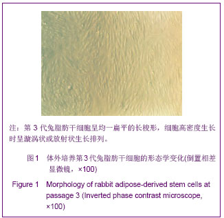
2.1 兔脂肪干细胞生物学特性 分离出的原代兔脂肪干细胞接种6 h开始有少量细胞散在贴壁;24 h后大部分细胞以群落或散在的方式贴壁生长,细胞呈小圆形、短梭形或多角形;培养48 h首次全量换液,除去未贴壁细胞后,大部分贴壁细胞呈宽大扁平的长梭形或多角形。 细胞在最初3-5 d,生长较缓慢,过后进入指数增殖期;9-10 d长满瓶底,消化后按1∶2传代培养。传代细胞增长速度较原代细胞明显增快,两三天传1代,第3代兔脂肪干细胞呈均一扁平的长梭形,细胞高密度生长时呈漩涡状或放射状生长排列,见图1。"
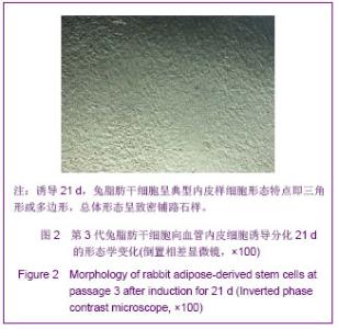
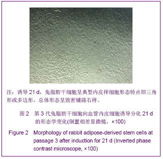
兔脂肪干细胞生长曲线呈S形,接种后第一至二天为潜伏期,3 d后细胞增殖加速并进入指数增殖期,8 d达到高峰,以后细胞生长速度减慢,进入生长平台期,其生长规律符合干细胞生长特征。 第3代兔脂肪干细胞免疫荧光法检测Vimentin为阳性。体外培养至15代细胞的增殖速度无明显减慢,细胞形态无明显变化。 2.2 兔脂肪干细胞向血管内皮细胞诱导分化的形态学观察结果 诱导3 d,大部分诱导组细胞由长梭形逐渐变短,变粗,成为多角形;诱导21 d后,细胞呈典型内皮样细胞形态特点即三角形或多边形,总体形态呈致密铺路石样,见图2。"
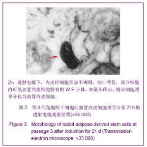
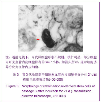
透射电镜下,内皮样细胞形态不规则,细胞核扁平,核仁明显,胞浆内有较多的线粒体、高尔基体和粗面内质网,部分细胞内可见血管内皮细胞特有的细胞器,即W-P小体(Weibel-Palade body),见图3。对照组光镜下观察细胞形态无改变。"
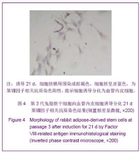
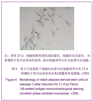
2.3 诱导前后细胞的表面标志变化 流式细胞仪检测诱导组诱导21 d的细胞结果显示CD31阳性表达,CD44阴性表达;检测对照组第3代兔脂肪干细胞结果显示CD44阳性表达,CD31阴性表达。 2.4 第Ⅷ因子相关抗原免疫组织化学染色结果 诱导组核膜周围染成棕褐色,细胞核呈淡蓝色,为阳性,见图4。 未加一抗的阴性对照与对照组细胞胞浆和核膜未有染色,仅细胞核呈淡蓝色。"
| [1]Krenning G, van Luyn MJ, Harmsen MC,et al.Endothelial progenitor cell-based neovascularization: implications for therapy. Trends Mol Med.2009;15(4):180-189.[2]Oikawa A,Katare R,Herman A, et al.Importance of mouse hematopoietic stem cells in neovascularisation.Cardiovasc Res. 2012;93(1): S92-S127.[3]Nourse MB,Halpin DE,Scatena M,et al. VEGF induces differentiation of functional endothelium from human embryonic stem cells: implications for tissue engineering. Am Heart Assoc. 2010; 30: 80-89.[4]Duffy GP, Ahsan T, O'Brien T,et al. Bone marrow–derived mesenchymal stem cells promote angiogenic processes in a time-and dose-dependent manner in vitro. Tissue Eng Part A. 2009;15(9): 2459-2470.[5]Zuk PA,Zhu M,Mizuno H,et al.Multilineage cells from human adipose tissue: implications for cell-based therapies.Tissue Eng.2001;7(2): 211 -228.[6]Zuk PA,Zhu M, Ashjian P,et al.Human adipose tissue is a source of multipotent stem cells.Mol Biol Cell.2002;13(12): 4279-4295.[7]Hong SJ,Traktev DO,March KL.Therapeutic potential of adipose- derived stem cells in vasular growth and tissue repair.Curr Opin Organ Transplant.2010;15(1):86-91.[8]Levi B,James AW,Nelson ER,et al.Human adipose-derived stromal cells stimulate autogenous skeletal repair via paracrine Hedgehog signaling with calvarial osteoblasts.Stem Cells Dev.2011;20(2):243-257.[9]Zhu M,Zhou Z,Chen Y,et al.Supplementation of fat grafts with adipose-derived regenerative cells improves long-term graft retention.Ann Plast Surg.2010;64 (2):222-228. [10]Gimble JM,Bunnel BA,Chiu ES,et al.Concise review:adipose- derived stromal vascular fraction cells and stem cells:let's not get lost in translation.Stem Cell.2011;29 (5):749-754.[11]Behr B, Tang C, Germann G,et al.Locally applied VEGFA increases the osteogenic healing capacity of human adipose derived stem cells by promoting osteogenic and endothelial differentiation. Stem Cells. 2011;29(2):286-296.[12]Aluigi MG,Coradeghini R,Guida C,et al.Pre-adipocytes commitment to neurogenesis 1:preliminary localisation of cholinergic molecules.Cell Biol Int.2009;33(5):594-601.[13]Choi YS, Dusting GJ, Stubbs S,et al.Differentiation of human adipose‐derived stem cells into beating cardiomyocytes. J Cell Mol Med.2010;14(4): 878-889.[14]Jeong JH.Adipose stem cells and skin repair.Curr Stem Cell Ther.2010;5(2):137-140. [15]Banas A. Purification of adipose tissue mesenchymal stem cells and differentiation toward hepatic-like cells. Methods Mol Biol.2012;826: 61-72.[16]Lin G,Wang G,Liu G,et al.Treatment of type 1 diabetes with adipose tissue-derived stem cells expressing pancreatic duodenal homeobox 1.Stem Cells Dev. 2009;18(10): 1399-1406.[17]The Ministry of Science and Technology of the People’s Republic of China. Guidance suggestion of caring laboratory animals.2006-09-30.中华人民共和国科学技术部.关于善待实验动物的指导性意见.2006-09-30.[18]Zhang Y,Zhou Y,Jia LS.Zhongguo Zuzhi Gongcheng Yanjiu yu Linchuang Kangfu.2011;15 (47):8777 -8781.张亚,周云,贾立山.多孔丝素复合脂肪间充质干细胞支架修复兔尿道缺损能促进血管的形成[J].中国组织工程研究与临床康复, 2011,15 (47):8777 -8781.[19]Zhang Y,Zhou Y,Jia LS,et al.Zhonghua Xiaoer Waike Zazhi. 2011;32(5):380-383.张亚,周云,贾立山, 等.脂肪间充质干细胞的体外诱导和成血管作用[J].中华小儿外科杂志,2011,32(5):380-383.[20]Jeschke MG, Hermanutz V, Wolf SE, et al.Polyurethane vascular prostheses decreas neointimal formation compared with expanded polytetrafluoroethylene.J Vasc Surg.1999;29 (1):168-176.[21]Tang GH, Pinney SP, Broumand SR,et al. Excellent outcomes with use of synthetic vascular grafts for treatment of mycotic aortic pseudoaneurysms after heart transplantation. Ann Thorac Surg.2011;92(6):2112-2116.[22]Peter A,Schneider,Harry F,et al.Thromboembolic potential of synthetic vascular grafts in baboons.J Vasc Surg.1989;10(1) : 75-82.[23]Losi P,Lomhardi S,Briganti E, et al. Luminal surface microgeometry affects platelet adhesion in small-diameter synthetic grafts. Biomaterials .2004;25 (18):4447-4455.[24]Bznz Y, Rieben R. Endothelial cell protection in xeno transplantation: looking after a key player in rejection. Xenotransplantation. 2006;13:19-30.[25]Miranville A, Heeschen C, Sengenès C, et al. Improvement of postnatal neovascularization by human adipose tissue-derived stem cells. Circulation. 2004; 110(3):349-355.[26]Cao Y,Meng Y,Zhao CH,et al.Zhongguo Yixue Kexue Yuanbao. 2005;27(6):678-682.曹莹,孟艳,赵春华,等.脂肪来源成体干细胞分化为内皮细胞的潜能[J].中国医学科学院学报,2005,27(6):678-682.[27]Murohara T.Autologous adipose tissue as a new source of progenitor cells for therapeutic angiogenesis. J Cardiol. 2009; 53(2):155-163.[28]Zhu XY,Zhang XZ,Xu L,et al.Transplantation of adipose-derived stem cells overexpressing hHGF into cardiac tissue. Biochem Biophys Res Commun.2009;379(4):1084-1090.[29]Jia LS, Zhou Y,Zhang Y, et al.Zhongguo Zuzhi Gongcheng Yanjiu yu Linchuang Kangfu.2009;13(19):3698-3702.贾立山,周云,张亚,等.体外诱导大鼠骨髓间充质干细胞向血管内皮细胞的分化[J].中国组织工程研究与临床康复,2009,13(19): 3698-3702. [30]Gurevitch O,Slavin S,Resnick I,et al.Mesenehymal progenitor cells in red and yellow bone marrow.Folia Biol(Praha). 2009; 55(1):27-34.[31]Parker AM,Shang H,Khurgel M,et al.Low serum and serum-free culture of multipotential human adipose stem cells.Cytotherapy.2007;9(7):637-646.[32]Matushansky I,Hemando E,Socci ND,et al.Derivation of sarcomas from mesechymal stem cells via inactivation of the Wnt pathway.Clini Invest.2007;117(11):3248-3257.[33]Dai J,Rabie AB.VEGF:an essential mediator of both anglogenesis and endochondral ossification.J Dent Res. 2007; 86(10):937-950.[34]Schäffler A, Büchler C. Concise review: Adipose tissue-derived stromal cells-basic and clinical implications for novel cell-based therapies.Stem Cells.2007;25(4):818-827.[35]Chiou M,Xu Y,Longaker MT.Mitogenic and chondrogenic effects of fibroblast growth factor-2 in adipose-derived mesenchymal cells. Biochem Biophys Res Commun. 2006; 343(2):644-652.[36]De Ugarte DA,Alfonso Z,Zuk PA,et al.Differential expression of stem cell mobilization -associated molecules on multi-lineage cells from adipose tissue and bone marrow. Immunol Lett. 2003;89:267-270.[37]Götherström C, Ringdén O, Westgren M,et al. Immunomodulatory effects of human foetal liver-derived mesenchymal stem cells. Bone Marrow Transplant. 2003; 32(3): 265-272.[38]Pusztaszeri MP, Seelentag W, Bosman FT. Immunohistochemical expression of endothelial markers CD31,CD34, von willebrand factor,and Fli-1 in Normal Human Tissues. J Histochem Cytochem. 2006;54(4):385-395.[39]Gimbrone MA, Cotran RS, Folkman J, et al. Human vascular endothelial cells in culture growth and DNA synthesis. J Cell Biol.1974;60(3):673-684.[40]Weibel ER, Palade GE. New cytoplasmic components in arterial endothelia. J Cell Biol.1964;23: 101-112.[41]Shi DB,Fu WG,He HB,et al.Zhongguo Zuzhi Gongcheng Yanjiu yu Linchuang Kangfu.2008;12(20):3831-3836.施德兵,符伟国,何红兵,等.实验犬浅静脉内皮细胞原位获取及体外培养[J].中国组织工程研究与临床康复,2008,12(20):3831- 3836. |
| [1] | Pu Rui, Chen Ziyang, Yuan Lingyan. Characteristics and effects of exosomes from different cell sources in cardioprotection [J]. Chinese Journal of Tissue Engineering Research, 2021, 25(在线): 1-. |
| [2] | Zhang Xiumei, Zhai Yunkai, Zhao Jie, Zhao Meng. Research hotspots of organoid models in recent 10 years: a search in domestic and foreign databases [J]. Chinese Journal of Tissue Engineering Research, 2021, 25(8): 1249-1255. |
| [3] | Wang Zhengdong, Huang Na, Chen Jingxian, Zheng Zuobing, Hu Xinyu, Li Mei, Su Xiao, Su Xuesen, Yan Nan. Inhibitory effects of sodium butyrate on microglial activation and expression of inflammatory factors induced by fluorosis [J]. Chinese Journal of Tissue Engineering Research, 2021, 25(7): 1075-1080. |
| [4] | Wang Xianyao, Guan Yalin, Liu Zhongshan. Strategies for improving the therapeutic efficacy of mesenchymal stem cells in the treatment of nonhealing wounds [J]. Chinese Journal of Tissue Engineering Research, 2021, 25(7): 1081-1087. |
| [5] | Liao Chengcheng, An Jiaxing, Tan Zhangxue, Wang Qian, Liu Jianguo. Therapeutic target and application prospects of oral squamous cell carcinoma stem cells [J]. Chinese Journal of Tissue Engineering Research, 2021, 25(7): 1096-1103. |
| [6] | Xie Wenjia, Xia Tianjiao, Zhou Qingyun, Liu Yujia, Gu Xiaoping. Role of microglia-mediated neuronal injury in neurodegenerative diseases [J]. Chinese Journal of Tissue Engineering Research, 2021, 25(7): 1109-1115. |
| [7] | Li Shanshan, Guo Xiaoxiao, You Ran, Yang Xiufen, Zhao Lu, Chen Xi, Wang Yanling. Photoreceptor cell replacement therapy for retinal degeneration diseases [J]. Chinese Journal of Tissue Engineering Research, 2021, 25(7): 1116-1121. |
| [8] | Jiao Hui, Zhang Yining, Song Yuqing, Lin Yu, Wang Xiuli. Advances in research and application of breast cancer organoids [J]. Chinese Journal of Tissue Engineering Research, 2021, 25(7): 1122-1128. |
| [9] | Wang Shiqi, Zhang Jinsheng. Effects of Chinese medicine on proliferation, differentiation and aging of bone marrow mesenchymal stem cells regulating ischemia-hypoxia microenvironment [J]. Chinese Journal of Tissue Engineering Research, 2021, 25(7): 1129-1134. |
| [10] | Zeng Yanhua, Hao Yanlei. In vitro culture and purification of Schwann cells: a systematic review [J]. Chinese Journal of Tissue Engineering Research, 2021, 25(7): 1135-1141. |
| [11] | Kong Desheng, He Jingjing, Feng Baofeng, Guo Ruiyun, Asiamah Ernest Amponsah, Lü Fei, Zhang Shuhan, Zhang Xiaolin, Ma Jun, Cui Huixian. Efficacy of mesenchymal stem cells in the spinal cord injury of large animal models: a meta-analysis [J]. Chinese Journal of Tissue Engineering Research, 2021, 25(7): 1142-1148. |
| [12] | Hou Jingying, Yu Menglei, Guo Tianzhu, Long Huibao, Wu Hao. Hypoxia preconditioning promotes bone marrow mesenchymal stem cells survival and vascularization through the activation of HIF-1α/MALAT1/VEGFA pathway [J]. Chinese Journal of Tissue Engineering Research, 2021, 25(7): 985-990. |
| [13] | Shi Yangyang, Qin Yingfei, Wu Fuling, He Xiao, Zhang Xuejing. Pretreatment of placental mesenchymal stem cells to prevent bronchiolitis in mice [J]. Chinese Journal of Tissue Engineering Research, 2021, 25(7): 991-995. |
| [14] | Liang Xueqi, Guo Lijiao, Chen Hejie, Wu Jie, Sun Yaqi, Xing Zhikun, Zou Hailiang, Chen Xueling, Wu Xiangwei. Alveolar echinococcosis protoscolices inhibits the differentiation of bone marrow mesenchymal stem cells into fibroblasts [J]. Chinese Journal of Tissue Engineering Research, 2021, 25(7): 996-1001. |
| [15] | Fan Quanbao, Luo Huina, Wang Bingyun, Chen Shengfeng, Cui Lianxu, Jiang Wenkang, Zhao Mingming, Wang Jingjing, Luo Dongzhang, Chen Zhisheng, Bai Yinshan, Liu Canying, Zhang Hui. Biological characteristics of canine adipose-derived mesenchymal stem cells cultured in hypoxia [J]. Chinese Journal of Tissue Engineering Research, 2021, 25(7): 1002-1007. |
| Viewed | ||||||
|
Full text |
|
|||||
|
Abstract |
|
|||||