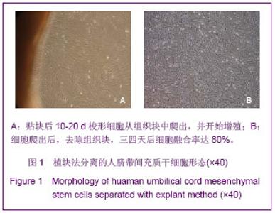
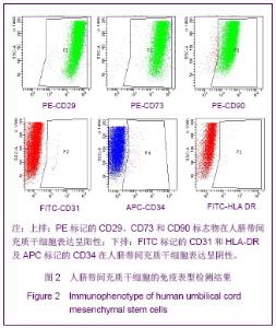
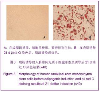
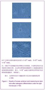
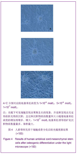
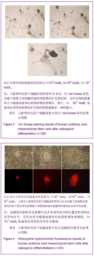
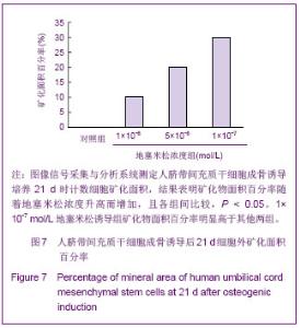
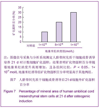
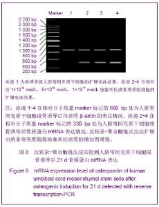
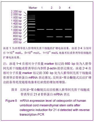
| [1]Xu RN, Shi M, Wang FS. Zhongguo Mianyixue Zazhi. 2010; 26(1):82-84.徐若男,施明,王福生.人脐带间充质干细胞的特征及其临床应用前景[J].中国免疫学杂志,2010,26(1):82-84.[2]Guo ZK, Wang LS, Wu ZZ. Zhongguo Yiyao Shengwu Jishu. 2006;V1(1):58-61.郭子宽,王立生,吴祖泽.间充质干细胞在组织再生和免疫调节治疗中的应用[J].中国医药生物技术,2006,V1(1):58-61.[3]Karahuseyinoglu S,Cinar O,Kilic E,et al. Biology of stem cells in human umbilical cord stroma: in situ and in vitro surveys. Stem Cells. 2007;25(2):319-331.[4]Baksh D,Yao R,Tuan RS,et al. Comparison of proliferative and multilineage differentiation potential of human mesenchymal stem cells derived from umbilical cord and bone marrow. Stem Cells. 2007;25(6):1384-1392.[5]Dominici M, Le Blanc K, Mueller I, et al. Minimal criteria for defining multipotent mesenchymal stromal cells. The International Society for Cellular Therapy position statement. Cytotherapy. 2006;8 (4):315-317.[6]Janderova L,McNeil M,Murrell AN,et al. Human mesenchymal stem cells as an in vitro model for human adipogenesis. Obes Res. 2003;11(1):65-74.[7]Hung SC,Chang CF,Ma HL,et al.Gene expression profiles of early adipogenesis in human mesenehymal stem cells. Gene. 2004; 340(1):141-150.[8]Hong JX, Zhang QZ, Han JL, et al. Zhongguo Zuzhi Gongcheng Yanjiu yu Linchuang Kangfu. 2010;14(1):42-47.洪敬欣,张茜真,韩俊领,等.人脐带间充质干细胞在3种不同培养体系中的生长状况及腺病毒感染效率[J].中国组织工程研究与临床康复,2010,14(1):42-47.[9]Li D, Shi C, Hong JX, et al. Zhongguo Yiyao Daobao. 2011; 8(24):19-22.李铎,石钏,洪敬欣,等.组织块法分离人脐带间充质干细胞的研究[J].中国医药导报,2011,8(24):19-22.[10]Xu Y, Li CH, Meng HX, et al. Zhongguo Zuzhi Gongcheng Yanjiu yu Linchuang Kangfu. 2009;13(32):6289-6294.徐燕,李长虹,孟恒星,等.人脐带间充质干细胞分离培养条件的优化及其生物学特性[J].中国组织工程研究与临床康复,2009, 13(32):6289-6294.[11]Jin KL. Beijing: Science Press. 2011.金坤林.干细胞临床应用-基础、伦理和原则[M]. 北京:科学出版社, 2011. [12]Fan Q, Zhao JM, Su W, et al. Zhongguo Zuzhi Gongcheng Yanjiu yu Linchuang Kangfu. 2011;15(14):2477-2481.范锲,赵劲民,苏伟,等.地塞米松对大鼠骨髓间充质干细胞凋亡的影响[J].中国组织工程研究与临床康复,2011,15(14):2477- 2481.[13]Zuk PA, Zhu M, Ashjian P, et al. Human adipose tissue is a source of multipotent stem cells. Mol Biol Cell. 2002;13(12): 4279-4295.[14]Pang JH,Huang HY,Zhang Q,et al. Chuangshang Waike Zazhi. 2009;11(3):248-252.庞金辉,黄煌渊,张权,等.不同浓度地塞米松对人骨髓间充质干细胞生物学特性的影响[J].创伤外科杂志,2009,11(3):248-252.[15]Wang W,Chen LJ,Zhang C, et al. Zhongguo Xiufu Chongjian Waike Zazhi. 2011;25(12):1486-1492.王伟,陈磊杰,张晨,等.不同浓度地塞米松诱导脂肪干细胞成骨分化的实验研究[J].中国修复重建外科杂志,2011,25(12): 1486-1492.[16]Wu Y, Chen B, Wang L. Sichuan Jiepouxue Zazhi. 2008; 16(1):17-19.吴岩,陈波,王兰.骨髓间充质干细胞在骨组织工程中的研究进展[J].四川解剖学杂志,2008,16(1):17-19.[17]Xu HS, Liu JZ. Zhongguo Jiaoxing Waike Zazhi. 2009;17(20): 1553-1556.许红生,刘金钊.间充质干细胞在软骨组织工程中的应用[J].中国矫形外科杂志,2009,17(20):1553-1556.[18]Pittenger MF, Mackay AM, Beck SC, et al. Multilineage potential of adult human mesenchymal stem cells. Science. 1999; 284 (5411): 143-147.[19]Wagner W, Wein F, Seckinger A, et al. Comparative characteristics of mesenchymal stem cells from human bone marrow, adipose tissue, and umbilical cord blood. Exp Hematol. 2005;33 (11):1402-1416. [20]Karahuseyinoglu S, Cinar O, Kilic E, et al. Biology of stem ceils in human umbilical cord stroma; in situ and in vitro surveys. Stem Cells. 2007;25 (2):319-331.[21]Fang YY, Ma J. Zhongguo Zuzhi Gongcheng Yanjiu yu Linchuang Kangfu. 2010;14(12):2265-2267.方彦艳,马健.设想:脐带是理想的骨组织工程材料[J].中国组织工程研究与临床康复,2010,14(12):2265-2267.[22]Coelho MJ, Fernandes MH. Human bone cell cultures in biocompatibity testing. Part Ⅱ:effect of ascorbic acid.β-glycerophosphate and dexamethasone on ostoeblast differentiation. Biomaterials. 2000;21(11):1095-1102.[23]Liao QH, Huang Z, Cai DH. Zhongguo Zuzhi Gongcheng Yanjiu yu Linchuang Kangfu. 2009;13(1):88-91.廖庆辉,黄震,蔡德鸿. 抗坏血酸、β-甘油磷酸钠和地塞米松联合诱导大鼠骨髓间充质干细胞向成骨细胞的分化[J].中国组织工程研究与临床康复,2009,13(1):88-91.[24]Mountziaris PM, Tzouanas SN, Mikos AG. Dose effect of tumor necrosis actor-alpha on in vitro osteogenic differentiation of mesenchymal stem cells on biodegradable olymeric microfiber scaffolds. Biomaterials. 2010;31(7): 1666-1675. [25]Zhang JP. Xiandai Zhongxiyi Jiehe Zazhi. 2012;21(24):2642- 2646.张建萍.不同浓度地塞米松在体外定向诱导分化兔骨髓间充质干细胞为成骨细胞的实验研究[J].现代中西医结合杂志,2012, 21(24):2642-2646.[26]Shi WH, Pang JH, Cao CF, et al. Shoudu Yiyao. 2009; 16(6): 41-43.文俊,庞金辉,曹成福,等.地塞米松对人骨髓间充质干细胞增殖、成骨分化及凋亡的影响[J].首都医药,2009,16(6):41-43.[27]Zhang WX, Liu MF, Ye QH, et al. Guangdong Yiyao. 2012; 33(2):170-172.张文信,刘美芬,叶青合,等.地塞米松对兔骨髓间充质干细胞凋亡及细胞周期的影响[J].广东医学,2012,33(2):170-172.[28]Jaiswal N, Haynesworth SE, Caplan AI, et al. Osteogenic diferentiation of purified, culture-expanded human mesenchymal stem cells in vitro. J Cell Biochem. 1997; 64(2):295-312.[29]Cheng SL, Yang JW, Rifas L, et al. Differentiation of human bone marrow osteogenic stromal cells in vitro: induction of the osteoblast phenotype by dexamethasone. Endocrinology. 1994;134(1): 277-286.[30]Lin L, Li X, Yang MW, et al. Shandong Yiyao. 2009;49(26): 28-29.林乐,李旭,杨茂伟,等.地塞米松对骨髓间充质干细胞Runx2表达的影响[J].山东医药,2009,49(26):28-29 |