Chinese Journal of Tissue Engineering Research ›› 2013, Vol. 17 ›› Issue (16): 2961-2967.doi: 10.3969/j.issn.2095-4344.2013.16.016
Previous Articles Next Articles
Collagen scaffold facilitates the three-dimensional culture of myocardial cells from adult rats in vitro
Wang Hui-ling1, Li Qiong1, Guo Zhi-kun1, Sun Yong-kun2, Kuboki Y2
- 1 Key Laboratory for Medical Tissue Regeneration of Henan Province, Xinxiang Medical University, Xinxiang 453003, Henan Province, China
2 Hokkaido University, Hokkaido, Japan
-
Received:2012-07-24Revised:2012-09-02Online:2013-04-16Published:2013-04-16 -
Contact:Guo Zhi-kun, Doctor, Professor, Key Laboratory for Medical Tissue Regeneration of Henan Province, Xinxiang Medical University, Xinxiang 453003, Henan Province, China -
About author:Wang Hui-ling★, Studying for master’s degree, Key Laboratory for Medical Tissue Regeneration of Henan Province, Xinxiang Medical University, Xinxiang 453003, Henan Province, China mcolourful@126.com -
Supported by:an Province, No. 2005HANCET215*; the Tackle Key Program of Henan Province, No. 072102310012*
CLC Number:
Cite this article
Wang Hui-ling, Li Qiong, Guo Zhi-kun, Sun Yong-kun, Kuboki Y. Collagen scaffold facilitates the three-dimensional culture of myocardial cells from adult rats in vitro[J]. Chinese Journal of Tissue Engineering Research, 2013, 17(16): 2961-2967.
share this article
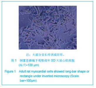
2.1 成年大鼠心室肌细胞的形态特点 成年大鼠心室肌细胞分离后,大部分呈长杆状或矩形,见图1,高倍镜下横纹清晰,界限分明,少数呈圆形。其中夹杂一些大小不等的细胞碎片。大部分细胞呈静止状态,极少数细胞保持自律性收缩。每个心脏细胞产量可达1×10⁷细胞数。 2.2 成年大鼠心肌细胞活性分析 活性细胞的细胞膜完整,所以锥虫蓝不能进入细胞内而拒染。死亡或受损伤的细胞由于细胞膜的完整性被破坏,被锥虫蓝染成蓝色,结果显示80%的心肌细胞锥虫蓝染色阴性。"
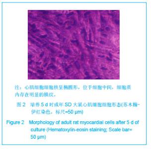
2.3 二维培养心肌细胞的形态特点 见图2。 接种的心肌细胞加血清培养24-48 h后,细胞发生形态结构变化[22-23],由杆状变成扁平状,细胞边界逐渐变得圆顿,部分细胞培养24-48 h后发生贴壁。苏木精-伊红染色可见心肌细胞核呈椭圆形,位于细胞中间,细胞质内存在明显的横纹,见图2。"

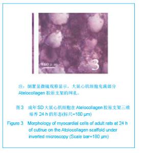
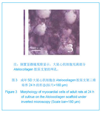
2.4 三维培养心肌细胞的形态特点 分离纯化的心肌细胞种到Atelocollagen胶原支架后约24 h大部分开始贴支架壁生长,但是仍然不具有自律性,见图3,细胞之间夹杂少量崩解的细胞碎片。"
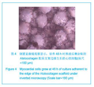
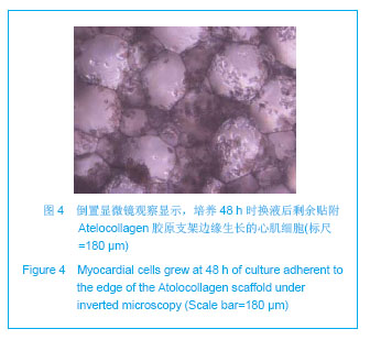
48 h换液后支架网孔中崩解的细胞被换掉,剩下大部分细胞贴附在支架壁边缘生长,见图4。 "
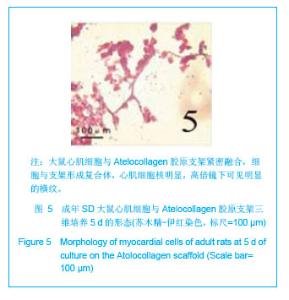
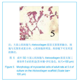
苏木精-伊红染色显示,培养48 h的心肌细胞与支架紧密融合,细胞与支架形成复合体。培养第5天,心肌细胞核明显,高倍镜下可见明显的横纹,见图5。"

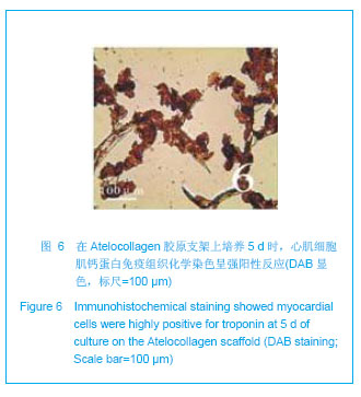
肌钙蛋白免疫组织化学染色呈棕黄色,肌钙蛋白阳性表达的细胞为心肌细胞,见图6。"
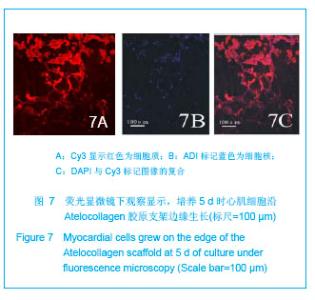
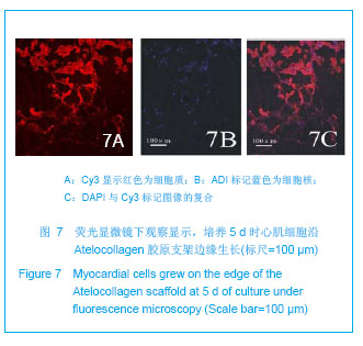
免疫荧光染色显示:cy3显示红色为细胞质,见图7A,DAPI标记蓝色为细胞核,见图7B,将DAPI和cy3标记的图像复合后,见图7C,细胞核周围显示红色荧光者为心肌细胞。免疫组织化学和免疫荧光显色均显示在支架内生长的大部分是心肌细胞,并且紧贴支架,生长良好。"
| [1]Alcon A,Cagavi BE,Qyang Y.Regenerating functional heart tissue for myocardial repair.Cell Mol Life Sci.2012;69(16): 2635-2656.[2]Curtis MW,Russell B.Cardiac tissue engineering. Cardiovasc Nurs.2009; 24(2): 87-92.[3]Nunes SS,Song H,Chiang CK,et al.Stem cell-based cardiac tissue engineering. Cardiovasc Transl Res.2011;4(5): 592-602.[4]Beitnes JO,Lunde K,Brinchmann JE,et al.Stem cells for cardiac repair in acute myocardial infarction.Expert Rev Cardiovasc Ther. 2011;9(8): 1015-1025.[5]Teng W,Guo ZK,Li Q,et al.Jiepou Xuebao. 2010;41(6): 885-890.滕伟,郭志坤,李琼,等. 在Atelocollagen胶原支架上体外三维培养乳大鼠心肌[J].解剖学报,2010,41(6): 885-890.[6]Guo Z,Iku S,Zheng X,et al.Three-Dimensional Geometry of Honeycomb Collagen Promotes Higher Beating Rate of Myocardial Cells in Culture. Artif Organs. 2012;36(9):816-819.[7]Huang ZB,Xiao JF,Shen JX.Jiguang Shengwu Xuebao. 2010; 19(3): 307-313.黄泽炳,肖剑锋,沈建新.胞外镁离子对SD成年大鼠心肌细胞胞内钙信号的影响[J].激光生物学报,2010,19(3): 307-313.[8]Wei LL,Mo SR.Zhongguo Zuzhi Gongcheng Yanjiu. 2012; 16(11): 1969-1972.韦丽兰,莫书荣.成年大鼠心肌细胞的急性分离方法[J].中国组织工程研究,2012,16(11): 1969-1972.[9]Li D,Tang B,Yang DC,et al.Zhongguo Shiyan Dongwu Xuebao. 2011;19(2): 150-152.李德,唐兵,杨大春,等.Ⅱ型胶原酶加压灌流提高成年大鼠心肌细胞分离效率[J].中国实验动物学报,2011,19(2): 150-152.[10]Liao H,Mi T,Tu ZY,et al. Zhongguo Zuzhi Gongcheng Yanjiu yu Linchuang Kangfu. 2009;13(33): 6536-6539.廖华,糜涛,涂志业,等.成年大鼠心肌细胞分离方法的改良[J].中国组织工程研究与临床康复,2009,13(33): 6536-6539.[11]Li H,Xiao YB.Disan Junyi Daxue Xuebao. 2004;26(7): 644-646.李洪,肖颖彬 .成年大鼠心室肌细胞的分离、培养与鉴定[J].第三军医大学学报,2004,26(7): 644-646.[12]Zhu XX,Niu XL,Zhu XL,et al.Jiefangjun Yixue Zazhi.2008; 33(9): 1089-1091.朱肖星,牛小麟,朱萧玲,等.不同活性的胶原酶对大鼠心肌细胞分离的影响[J].解放军医学杂志,2008,33(9): 1089-1091.[13]Chang H,Zhang L,Yu ZB.Zhongguo Yingyong Shenglixue Zazhi. 3011;27(1): 57-61,136.常惠,张琳,余志斌.培养成年大鼠心肌细胞存活的形态标志[J].中国应用生理学杂志,3011,27(1): 57-61,136.[14]Gong HB,Wang J,Wang L.Zhongguo Weixunhuan. 2009; 13(1):55-59.宫海滨,王洁,王雷.不同培养基对成年大鼠心肌细胞形态、存活率、收缩功能的影响[J].中国微循环,2009,13(1): 55-59.[15]Pavlovic D,McLatchie LM,Shattock MJ.The rate of loss of T-tubules in cultured adult ventricular myocytes is species dependent. Exp Physiol.2010; 95(4): 518-527.[16]Smyrnias I,Mair W,Harzheim D,et al. Comparison of the T-tubule system in adult rat ventricular and atrial myocytes, and its role in excitation-contraction coupling and inotropic stimulation. Cell Calcium. 2010; 47(3): 210-223.[17]Zhai YJ,Dong XH,Zhou LN,et al.Disi Junyi Daxue Xuebao. 2005;25(8): 705-707.翟迎九, 董雪红,周丽诺,等.成年SD大鼠心肌细胞的培养[J].第四军医大学学报,2005,25(8): 705-707.[18]Wang T,Fu Y,Fang XS,et al.Lingnan Jizhen Yixue Zazhi. 2007; 12(5): 323-325.王彤,符岳,方向韶,等.普通大鼠心肌细胞的分离培养和收缩舒张观察[J].岭南急诊医学杂志,2007,12(5): 323-325.[19]Wang Y,Sun H,Fan YM,et al.Xuzhou Yixueyuan Xuebao. 2005; 25(5): 393-396.王影,孙红,范乐明,等.成年大鼠心肌细胞的分离和培养技术[J].徐州医学院学报,2005,25(5): 393-396.[20]Li YG,Shi YK,Chen HW,et al.Huaxi Yixue. 2005;20(2): 308-309.李勇刚, 石应康,陈焕文,等.成年大鼠心肌细胞的分离和培养[J].华西医学,2005,20(2): 308-309.[21]Xu XF,Li WB,Chen BT,et al.Shoudu Yike Daxue Xuebao. 2000;21(2): 104-107.许秀芳,李温斌,陈宝田,等.成年大鼠心肌细胞培养方法的建立和形态学观察[J].首都医科大学学报,2000,21(2): 104-107.[22]Kato S,Takemura G,Maruyama R,et al. Apoptosis, rather than oncosis, is the predominant mode of spontaneous death of isolated adult rat cardiac myocytes in culture.Jpn Circ J. 2001;65(8): 743-748.[23]Hein S,Kostin S,Schaper J.Adult rat cardiac myocytes in culture: 'Second-floor' cells and coculture experiments. Exp Clin Cardiol.2006;11(3): 175-182. [24]Bugaisky LB,Zak R.Differentiation of adult rat cardiac myocytes in cell culture. Circ Res.1989;64(3): 493-500.[25]Guo ZK.Beijing:Renmin Junyi Chbanshe.2005:6.郭志坤.正常心脏组织学图谱[M].北京:人民军医出版社,2005: 6.[26]Ohno S,Hirano S,Kanemaru S,et al.Implantation of an atelocollagen sponge with autologous bone marrow-derived mesenchymal stromal cells for treatment of vocal fold scarring in a canine model. Ann Otol Rhinol Laryngol. 2011;120(6): 401-408.[27]Lee KI,Moon SH,Kim H,et al. Tissue engineering of the intervertebral disc with cultured nucleus pulposus cells using atelocollagen scaffold and growth factors. Spine (Phila Pa 1976). 2012;37(6): 452-458.[28]Ko EC,Fujihara Y,Ogasawara T,et al.BMP-2 Embedded Atelocollagen Scaffold for Tissue-Engineered Cartilage Cultured in the Medium Containing Insulin and Triiodothyronine-A New Protocol for Three-Dimensional In Vitro Culture of Human Chondrocytes. Tissue Eng Part C Methods.2012;18(5):374-386.[29]Zheng XJ,Guo ZK,Chang KS,et al.Jiepou Xuebao. 2008;39(4): 582-585.郑先杰,郭志坤,常联生,等.Atelocoilagen胶原支架在三维培养大鼠心肌中的生物相容性[J].解剖学报,2008,39(4): 582-585.[30]Akhyari P,Fedak PW,Weisel RD,et al.Mechanical stretch regimenenhances the formation of bioengineered autologous cardiac muscle grafts. Circulation. 2002;106(Supp l1): Ⅰ137-Ⅰ142.[31]Egorova MV,Afanas'ev SA,Popov SV.A simple method for isolation of cardiomyocytes from adult rat heart. Bull Exp Biol Med. 2005;140(3): 370-373.[32]Xu X, Colecraft HM.Primary culture of adult rat heart myocytes. Vis Exp. 2009;(28): pii: 1308.[33]Eckel J,van EG,Reinauer H. Adult cardiac myocytes in primary culture: cell characteristics and insulin-receptor interaction. Am J Physiol.1985;249(2 Pt 2): H212-221.[34]Jacobson SL,Piper HM.Cell cultures of adult cardiomyocytes as models of the myocardium.Mol Cell Cardiol.1986;18(7): 661-678.[35]Dubus I,Samuel JL,Marotte F,et al.Beta-adrenergic agonists stimulate the synthesis of noncontractile but not contractile proteins in cultured myocytes isolated from adult rat heart. Circ Res. 1990;66(3): 867-874.[36]Schwarzfeld TA,Jacobson SL.Isolation and development in cell culture of myocardial cells of the adult rat. Mol Cell Cardiol. 1981;13(6): 563-575.[37]Yamamoto S,Yasui K,Palade PT,et al.Spontaneous death of isolated adult rat cardiocytes in culture in association with internucleosomal cleavage of genomic DNA. Apoptosis.1997; 2(2): 178-188.[38]Louch WE,Bito V,Heinzel FR,et al.Reduced synchrony of Ca2+ release with loss of T-tubules-a comparison to Ca2+ release in human failing cardiomyocytes.Cardiovasc Res. 2004;62(1): 63-73.[39]Leach RN, Desai JC,Orchard CH. Effect of cytoskeleton disruptors on L-type Ca channel distribution in rat ventricular myocytes. Cell Calcium. 2005;38(5): 515-526.[40]Jacobson SL,Piper HM.Cell cultures of adult cardiomyocytes as models of the myocardium. Mol Cell Cardiol.1986;18(7): 661-678. |
| [1] | Li Jing, Xie Jianshan, Cui Huilin, Cao Ximei, Yang Yanping, Li Hairong. Expression and localization of diacylglycerol kinase zeta and protein kinase C beta II in mouse back skin with different coat colors [J]. Chinese Journal of Tissue Engineering Research, 2021, 25(8): 1196-1200. |
| [2] | Li Li, Ma Li. Immobilization of lactase on magnetic chitosan microspheres and its effect on enzymatic properties [J]. Chinese Journal of Tissue Engineering Research, 2021, 25(4): 576-581. |
| [3] | Yang Caihui, Liu Qicheng, Dong Ming, Wang Lina, Zuo Meina, Lu Ying, Niu Weidong. Serine/threonine protein kinases can promote bone destruction in mouse models of chronic periapical periodontitis [J]. Chinese Journal of Tissue Engineering Research, 2021, 25(23): 3654-3659. |
| [4] | Yan Peng, Ma Yufei, Cui Jingfu, Hao Shaofei, Liu Jinhui, Guan Chunlei, Wang Xiaoran, Yang Xiaoyu. Mechanism of anodic block electrical stimulation of sacral nerve root to reconstruct bladder function [J]. Chinese Journal of Tissue Engineering Research, 2021, 25(23): 3684-3689. |
| [5] | Zhou Anqi, Tang Yufei, Wu Bingfeng, Xiang Lin. Designing of periosteum tissue engineering: combination of generality and individuality [J]. Chinese Journal of Tissue Engineering Research, 2021, 25(22): 3551-3557. |
| [6] | Lang Limin, He Sheng, Jiang Zengyu, Hu Yiyi, Zhang Zhixing, Liang Minqian. Application progress of conductive composite materials in the field of tissue engineering treatment of myocardial infarction [J]. Chinese Journal of Tissue Engineering Research, 2021, 25(22): 3584-3590. |
| [7] | Li Shao, Liang Yongkang, Gao Yi, Peng Qing. Establishment and functional in vitro characteristics of three-dimensional collagen HepaRG microsphere [J]. Chinese Journal of Tissue Engineering Research, 2021, 25(16): 2541-2547. |
| [8] | Xie Jian, Su Jiansheng. Advantages and characteristics of electrospun aligned nanofibers as scaffolds for tissue engineering [J]. Chinese Journal of Tissue Engineering Research, 2021, 25(16): 2575-2581. |
| [9] | Ji Qi, Yu Zhengwen, Zhang Jian. Problems and trends of technique and clinical application of metallic biomaterials prepared by three-dimensional printing technology [J]. Chinese Journal of Tissue Engineering Research, 2021, 25(16): 2597-2604. |
| [10] | Han Ningning, Zuo Jinfu, Sun Miao, Tang Shengjian, Liu Fangjun. Application and progress of umbilical cord mesenchymal stem cells in bone tissue engineering [J]. Chinese Journal of Tissue Engineering Research, 2021, 25(13): 2079-2086. |
| [11] | Qian Nannan, Zhang Qian, Yang Rui, Ao Jun, Zhang Tao. Mesenchymal stem cells in the treatment of spinal cord injury: cell therapy and combination of new drugs and biomaterials [J]. Chinese Journal of Tissue Engineering Research, 2021, 25(13): 2114-2120. |
| [12] | Jia Wei, Zhang Mandong, Chen Weiyi, Wang Chenyan, Guo Yuan. Effects of femoral prosthetic materials on artificial knee arthroplasty performance [J]. Chinese Journal of Tissue Engineering Research, 2021, 25(10): 1477-1481. |
| [13] | Wang Qian, Li Lu, Shu Jingyuan, Dong Zhiheng, Jin Youshi, Wang Qingshan. Micro-morphology and phase of zirconia-based nano-hydroxyapatite functional gradient biomaterials [J]. Chinese Journal of Tissue Engineering Research, 2021, 25(10): 1517-1521. |
| [14] | Zhu Mingqi, Zhou Jingxu, Lin Lizhu, Chen Zeren, Liao Zhixiao . In vitro co-culture of decellularized matrix of porcine colon with colon cancer HCT116 cells [J]. Chinese Journal of Tissue Engineering Research, 2021, 25(10): 1533-1538. |
| [15] | Li Li, Ma Li, Li He. Preparation and characterization of magnetic chitosan microspheres [J]. Chinese Journal of Tissue Engineering Research, 2020, 24(4): 577-582. |
| Viewed | ||||||
|
Full text |
|
|||||
|
Abstract |
|
|||||