Chinese Journal of Tissue Engineering Research ›› 2013, Vol. 17 ›› Issue (10): 1827-1834.doi: 10.3969/j.issn.2095-4344.2013.10.019
Previous Articles Next Articles
Pioglitazone promotes the proliferation of rat bone marrow endothelial progenitor cells
Zhang Hui-feng1,2, Wang Li2, Ma Yue-hua2, Hu Zi-ying2, Zhao Zhi-gang2
- 1 Department of Endocrinology, First Affiliated Hospital of Zhengzhou University, Zhengzhou 450052, Henan Province, China
2 Department of Endocrinology, People’s Hospital of Henan Province, Zhengzhou 450003, Henan Province, China
-
Received:2012-12-09Revised:2013-01-04Online:2013-03-05Published:2013-03-05 -
Contact:Zhao Zhi-gang, Chief physician, Department of Endocrinology, People’s Hospital of Henan Province, Zhengzhou 450003, Henan Province, China -
About author:Zhang Hui-feng☆, Studying for doctorate, Department of Endocrinology, First Affiliated Hospital of Zhengzhou University, Zhengzhou 450052, Henan Province, China; Department of Endocrinology, People’s Hospital of Henan Province, Zhengzhou 450003, Henan Province, China zhf666777@163.com
CLC Number:
Cite this article
Zhang Hui-feng, Wang Li, Ma Yue-hua, Hu Zi-ying, Zhao Zhi-gang. Pioglitazone promotes the proliferation of rat bone marrow endothelial progenitor cells[J]. Chinese Journal of Tissue Engineering Research, 2013, 17(10): 1827-1834.
share this article
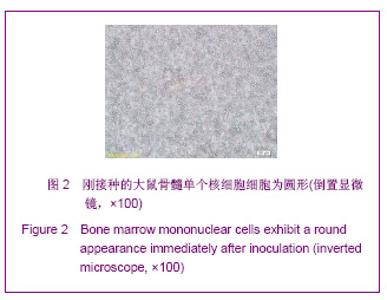
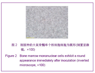
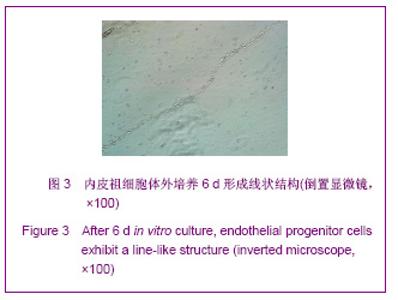
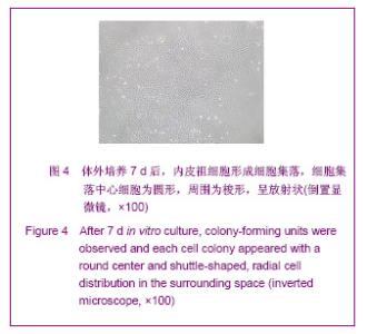
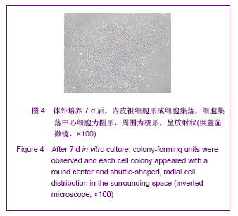
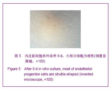
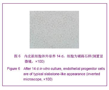
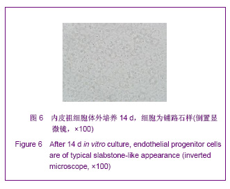
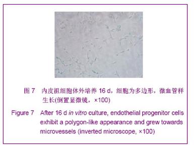
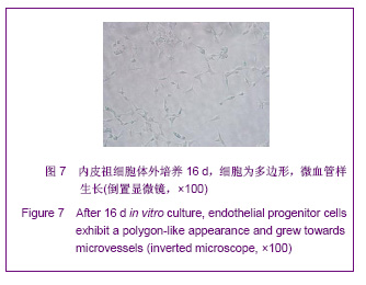
2.1 分离培养的大鼠骨髓内皮祖细胞的形态 新分离的细胞为圆形,大小不一,平均每只大鼠可获得1×10⁸个细胞。二次贴壁的细胞于培养3 d内贴壁,刚开始为圆形,逐渐变大、伸出伪足,变为梭形、纺锤形、多角形,以梭形细胞为主。培养2 d后可见部分细胞贴壁变形。培养前 4 d细胞增殖不明显,第5-10天迅速增殖,并可见细胞集落及线状结构形成,第10天可达80%融合。细胞集落中心为圆形细胞,周围为梭形,呈放射状。培养10 d以后细胞增殖缓慢。第14-16天,细胞为典型铺路石样,见图2-7。细胞融合至80%时,以1∶2消化传代。"
"

"
"

"
"
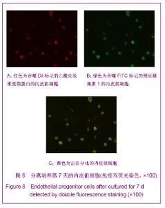
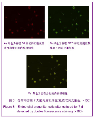
2.2 分离培养的大鼠骨髓内皮祖细胞的鉴定结果 免疫双荧光染色见正在分化的内皮祖细胞既可吞噬Dil标记的乙酰化低密度脂蛋白,又可吞噬FITC标记的荆豆凝集素1,在激光共聚焦显微镜下表现为黄色荧光,见图8。"
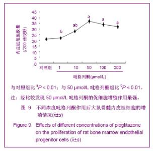
2.3 不同浓度的吡格列酮对内皮祖细胞增殖的影响 实验采用MTT法观察内皮祖细胞的增殖情况。结果发现10-200 μmol/L的吡格列酮均可明显促进内皮祖细胞的增殖(P < 0.01),并且50 μmol/L的吡格列酮对内皮祖细胞增殖的促进作用明显强于1,10 μmol/L,但与100,200 μmol/L吡格列酮的促增殖作用差异无显著性意义(P > 0.05),见图9。因此选择50 μmol/L吡格列酮用于后续实验。"

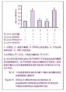
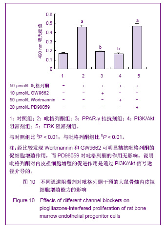
2.4 吡格列酮对内皮祖细胞增殖影响的机制 MTT结果显示,与对照组比较,吡格列酮组细胞增殖明显增多 (P < 0.01);与吡格列酮组比较,PPAR-γ拮抗剂组和PI3K/Akt阻滞剂组细胞增殖明显降低,而ERK阻滞剂组与吡格列酮组细胞增殖差异无显著性意义(P > 0.05)。说明吡格列酮对内皮祖细胞增殖的促进作用可能是通过PI3K/Akt信号途径介导的,见图10。"
| [1] Ferguson JE 3rd, Kelley RW, Patterson C. Mechanisms of endothelial differentiation in embryonic vasculogenesis. Arterioscler Thromb Vasc Biol. 2005;25(11):2246-2254.[2] Asahara T, Murohara T, Sullivan A, et al. Isolation of putative progenitor endothelial cells for angiogenesis. Science. 1997; 275(5302):964-967.[3] Hill JM, Zalos G, Halcox JP, et al. Circulating endothelial progenitor cells, vascular function, and cardiovascular risk. N Engl J Med. 2003;348(7):593-600.[4] Fadini GP, Miorin M, Facco M, et al. Circulating endothelial progenitor cells are reduced in peripheral vascular complications of type 2 diabetes mellitus. J Am Coll Cardiol. 2005;45(9):1449-1457.[5] Fadini GP, Sartore S, Agostini C, et al. Significance of endothelial progenitor cells in subjects with diabetes. Diabetes Care. 2007;30(5):1305-1313.[6] Fadini GP, Sartore S, Albiero M, et al. Number and function of endothelial progenitor cells as a marker of severity for diabetic vasculopathy. Arterioscler Thromb Vasc Biol. 2006;26(9): 2140-2146.[7] Ikeda U, Shimpo M, Murakami Y, et al. Peroxisome proliferator-activated receptor-gamma ligands inhibit nitric oxide synthesis in vascular smooth muscle cells. Hypertension. 2000;35(6):1232-1236.[8] Guo YF, Zhao Y, Song DL, et al. Yixue Jianyan yu Linchuang. 2007;18(2):15-18. 郭永芳,赵莹,宋达琳,等.冠心病合并代谢综合征危险因素患者胰岛素抵抗与氧化应激的关系[J].医学检验与临床,2007,18(2): 15-18.[9] Dimmeler S, Aicher A, Vasa M, et al. HMG-CoA reductase inhibitors (statins) increase endothelial progenitor cells via the PI 3-kinase/Akt pathway. J Clin Invest. 2001;108(3): 391-397.[10] Cesari F, Gori AM, Romagnuolo I, et al. Endothelial progenitor cells and vascular health: effects of lifestyle's modifications. Monaldi Arch Chest Dis. 2012;78(2):66-72.[11] Walter DH, Rittig K, Bahlmann FH, et al. Statin therapy accelerates reendothelialization: a novel effect involving mobilization and incorporation of bone marrow-derived endothelial progenitor cells. Circulation. 2002;105(25): 3017-3024.[12] Aicher A, Heeschen C, Mildner-Rihm C, et al. Essential role of endothelial nitric oxide synthase for mobilization of stem and progenitor cells. Nat Med. 2003;9(11):1370-1376.[13] Strehlow K, Werner N, Berweiler J, et al. Estrogen increases bone marrow-derived endothelial progenitor cell production and diminishes neointima formation. Circulation. 2003;107 (24): 3059-3065.[14] Werner N, Junk S, Laufs U, et al. Intravenous transfusion of endothelial progenitor cells reduces neointima formation after vascular injury. Circ Res. 2003;93(2):e17-24. [15] Wollert KC, Meyer GP, Lotz J, et al. Intracoronary autologous bone-marrow cell transfer after myocardial infarction: the BOOST randomised controlled clinical trial. Lancet. 2004;364 (9429):141-148.[16] Beltrami AP, Barlucchi L, Torella D, et al. Adult cardiac stem cells are multipotent and support myocardial regeneration. Cell. 2003;114(6):763-776.[17] Tobler K, Freudenthaler A, Baumgartner-Parzer SM, et al. Reduction of both number and proliferative activity of human endothelial progenitor cells in obesity. Int J Obes (Lond). 2010;34(4):687-700. [18] Braissant O, Foufelle F, Scotto C, et al. Differential expression of peroxisome proliferator-activated receptors (PPARs): tissue distribution of PPAR-alpha, -beta, and -gamma in the adult rat. Endocrinology. 1996;137(1):354-366.[19] Lowell BB. PPARgamma: an essential regulator of adipogenesis and modulator of fat cell function. Cell. 1999; 99(3):239-242.[20] Wang CH, Ciliberti N, Li SH, et al. Rosiglitazone facilitates angiogenic progenitor cell differentiation toward endothelial lineage: a new paradigm in glitazone pleiotropy. Circulation. 2004;109(11):1392-1400. [21] Onuta G, Rienstra H, de Boer JF, et al. Rosiglitazone attenuates transplant arteriosclerosis after allogeneic aorta transplantation in rats. Transplantation. 2007;84(4):517-526.[22] Bolten CW, Payne MA, McDonald WG, et al. Thiazolidinediones inhibit the progression of established hypertension in the Dahl salt-sensitive rat. Diab Vasc Dis Res. 2007;4(2):117-123.[23] Gensch C, Clever YP, Werner C, et al. The PPAR-gamma agonist pioglitazone increases neoangiogenesis and prevents apoptosis of endothelial progenitor cells. Atherosclerosis. 2007; 192(1):67-74.[24] Katso R, Okkenhaug K, Ahmadi K, et al. Cellular function of phosphoinositide 3-kinases: implications for development, homeostasis, and cancer. Annu Rev Cell Dev Biol. 2001;17: 615-675.[25] Asahara T, Masuda H, Takahashi T, et al. Bone marrow origin of endothelial progenitor cells responsible for postnatal vasculogenesis in physiological and pathological neovascularization. Circ Res. 1999;85(3):221-228.[26] Han JK, Kim HL, Jeon KH, et al. Peroxisome proliferator-activated receptor-{delta} activates endothelial progenitor cells to induce angio-myogenesis through matrix metallo-proteinase-9-mediated insulin-like growth factor-1 paracrine networks. Eur Heart J. 2011.[27] Wang CH, Verma S, Hsieh IC, et al. Enalapril increases ischemia-induced endothelial progenitor cell mobilization through manipulation of the CD26 system. J Mol Cell Cardiol. 2006;41(1):34-43.[28] You D, Cochain C, Loinard C, et al. Combination of the angiotensin-converting enzyme inhibitor perindopril and the diuretic indapamide activate postnatal vasculogenesis in spontaneously hypertensive rats. J Pharmacol Exp Ther. 2008;325(3):766-773.[29] Verma S, Kuliszewski MA, Li SH, et al. C-reactive protein attenuates endothelial progenitor cell survival, differentiation, and function: further evidence of a mechanistic link between C-reactive protein and cardiovascular disease. Circulation. 2004;109(17):2058-2067.[30] Thum T, Fraccarollo D, Galuppo P, et al. Bone marrow molecular alterations after myocardial infarction: Impact on endothelial progenitor cells. Cardiovasc Res. 2006;70(1): 50-60.[31] Müller P, Kazakov A, Jagoda P, et al. ACE inhibition promotes upregulation of endothelial progenitor cells and neoangiogenesis in cardiac pressure overload. Cardiovasc Res. 2009;83(1):106-114.[32] Imanishi T, Hano T, Nishio I. Angiotensin II accelerates endothelial progenitor cell senescence through induction of oxidative stress. J Hypertens. 2005;23(1):97-104.[33] Yu Y, Fukuda N, Yao EH, et al. Effects of an ARB on endothelial progenitor cell function and cardiovascular oxidation in hypertension. Am J Hypertens. 2008;21(1):72-77.[34] Yao EH, Fukuda N, Matsumoto T, et al. Losartan improves the impaired function of endothelial progenitor cells in hypertension via an antioxidant effect. Hypertens Res. 2007; 30(11):1119-1128.[35] Pelliccia F, Pasceri V, Cianfrocca C, et al. Angiotensin II receptor antagonism with telmisartan increases number of endothelial progenitor cells in normotensive patients with coronary artery disease: a randomized, double-blind, placebo-controlled study. Atherosclerosis. 2010;210(2): 510-515.[36] Honda A, Matsuura K, Fukushima N, et al. Telmisartan induces proliferation of human endothelial progenitor cells via PPARgamma-dependent PI3K/Akt pathway. Atherosclerosis. 2009;205(2):376-384.[37] Benson SC, Pershadsingh HA, Ho CI, et al. Identification of telmisartan as a unique angiotensin II receptor antagonist with selective PPARgamma-modulating activity. Hypertension. 2004; 43(5):993-1002.[38] Steinmetz M, Brouwers C, Nickenig G, et al. Synergistic effects of telmisartan and simvastatin on endothelial progenitor cells. J Cell Mol Med. 2010;14(6B):1645-1656.[39] Bahlmann FH, de Groot K, Mueller O, et al. Stimulation of endothelial progenitor cells: a new putative therapeutic effect of angiotensin II receptor antagonists. Hypertension. 2005; 45(4):526-529. |
| [1] | Pu Rui, Chen Ziyang, Yuan Lingyan. Characteristics and effects of exosomes from different cell sources in cardioprotection [J]. Chinese Journal of Tissue Engineering Research, 2021, 25(在线): 1-. |
| [2] | Zhang Xiumei, Zhai Yunkai, Zhao Jie, Zhao Meng. Research hotspots of organoid models in recent 10 years: a search in domestic and foreign databases [J]. Chinese Journal of Tissue Engineering Research, 2021, 25(8): 1249-1255. |
| [3] | Wang Zhengdong, Huang Na, Chen Jingxian, Zheng Zuobing, Hu Xinyu, Li Mei, Su Xiao, Su Xuesen, Yan Nan. Inhibitory effects of sodium butyrate on microglial activation and expression of inflammatory factors induced by fluorosis [J]. Chinese Journal of Tissue Engineering Research, 2021, 25(7): 1075-1080. |
| [4] | Wang Xianyao, Guan Yalin, Liu Zhongshan. Strategies for improving the therapeutic efficacy of mesenchymal stem cells in the treatment of nonhealing wounds [J]. Chinese Journal of Tissue Engineering Research, 2021, 25(7): 1081-1087. |
| [5] | Liao Chengcheng, An Jiaxing, Tan Zhangxue, Wang Qian, Liu Jianguo. Therapeutic target and application prospects of oral squamous cell carcinoma stem cells [J]. Chinese Journal of Tissue Engineering Research, 2021, 25(7): 1096-1103. |
| [6] | Xie Wenjia, Xia Tianjiao, Zhou Qingyun, Liu Yujia, Gu Xiaoping. Role of microglia-mediated neuronal injury in neurodegenerative diseases [J]. Chinese Journal of Tissue Engineering Research, 2021, 25(7): 1109-1115. |
| [7] | Li Shanshan, Guo Xiaoxiao, You Ran, Yang Xiufen, Zhao Lu, Chen Xi, Wang Yanling. Photoreceptor cell replacement therapy for retinal degeneration diseases [J]. Chinese Journal of Tissue Engineering Research, 2021, 25(7): 1116-1121. |
| [8] | Jiao Hui, Zhang Yining, Song Yuqing, Lin Yu, Wang Xiuli. Advances in research and application of breast cancer organoids [J]. Chinese Journal of Tissue Engineering Research, 2021, 25(7): 1122-1128. |
| [9] | Wang Shiqi, Zhang Jinsheng. Effects of Chinese medicine on proliferation, differentiation and aging of bone marrow mesenchymal stem cells regulating ischemia-hypoxia microenvironment [J]. Chinese Journal of Tissue Engineering Research, 2021, 25(7): 1129-1134. |
| [10] | Zeng Yanhua, Hao Yanlei. In vitro culture and purification of Schwann cells: a systematic review [J]. Chinese Journal of Tissue Engineering Research, 2021, 25(7): 1135-1141. |
| [11] | Kong Desheng, He Jingjing, Feng Baofeng, Guo Ruiyun, Asiamah Ernest Amponsah, Lü Fei, Zhang Shuhan, Zhang Xiaolin, Ma Jun, Cui Huixian. Efficacy of mesenchymal stem cells in the spinal cord injury of large animal models: a meta-analysis [J]. Chinese Journal of Tissue Engineering Research, 2021, 25(7): 1142-1148. |
| [12] | Hou Jingying, Yu Menglei, Guo Tianzhu, Long Huibao, Wu Hao. Hypoxia preconditioning promotes bone marrow mesenchymal stem cells survival and vascularization through the activation of HIF-1α/MALAT1/VEGFA pathway [J]. Chinese Journal of Tissue Engineering Research, 2021, 25(7): 985-990. |
| [13] | Shi Yangyang, Qin Yingfei, Wu Fuling, He Xiao, Zhang Xuejing. Pretreatment of placental mesenchymal stem cells to prevent bronchiolitis in mice [J]. Chinese Journal of Tissue Engineering Research, 2021, 25(7): 991-995. |
| [14] | Liang Xueqi, Guo Lijiao, Chen Hejie, Wu Jie, Sun Yaqi, Xing Zhikun, Zou Hailiang, Chen Xueling, Wu Xiangwei. Alveolar echinococcosis protoscolices inhibits the differentiation of bone marrow mesenchymal stem cells into fibroblasts [J]. Chinese Journal of Tissue Engineering Research, 2021, 25(7): 996-1001. |
| [15] | Fan Quanbao, Luo Huina, Wang Bingyun, Chen Shengfeng, Cui Lianxu, Jiang Wenkang, Zhao Mingming, Wang Jingjing, Luo Dongzhang, Chen Zhisheng, Bai Yinshan, Liu Canying, Zhang Hui. Biological characteristics of canine adipose-derived mesenchymal stem cells cultured in hypoxia [J]. Chinese Journal of Tissue Engineering Research, 2021, 25(7): 1002-1007. |
| Viewed | ||||||
|
Full text |
|
|||||
|
Abstract |
|
|||||