Chinese Journal of Tissue Engineering Research ›› 2020, Vol. 24 ›› Issue (13): 2080-2086.doi: 10.3969/j.issn.2095-4344.1867
Previous Articles Next Articles
Scleraxis lentivirus-transfected human amniotic mesenchymal stem cells promote tendon-bone healing in rabbits
Zou Gang, Zhang Jun, You Qi, Tang Jingfeng, Jin Ying, Yang Jibin, Zhu Xizhong, Liu Yi
- First Department of Orthopedics, Affiliated Hospital of Zunyi Medical University, Zunyi 563000, Guizhou Province, China
-
Received:2019-06-03Revised:2019-06-11Accepted:2019-07-12Online:2020-05-08Published:2020-03-10 -
Contact:Liu Yi, Professor, Master’s supervisor, First Department of Orthopedics, Affiliated Hospital of Zunyi Medical University, Zunyi 563000, Guizhou Province, China -
About author:Zou Gang, Master, Associate chief physician, First Department of Orthopedics, Affiliated Hospital of Zunyi Medical University, Zunyi 563000, Guizhou Province, China -
Supported by:the Joint Fund of Guizhou Provincial Science and Technology Department, No. LH[2017]7105
CLC Number:
Cite this article
Zou Gang, Zhang Jun, You Qi, Tang Jingfeng, Jin Ying, Yang Jibin, Zhu Xizhong, Liu Yi. Scleraxis lentivirus-transfected human amniotic mesenchymal stem cells promote tendon-bone healing in rabbits[J]. Chinese Journal of Tissue Engineering Research, 2020, 24(13): 2080-2086.
share this article
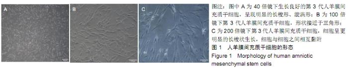
2.1 人羊膜间充质干细胞的形态 倒置显微镜下观察显示:生长良好的第3代人羊膜间充质干细胞在40倍镜下呈现明显的长梭形、漩涡形,见图1A;100倍镜下细胞接触更紧密,形状接近于三角形,见图1B;200倍镜下细胞呈更明显的长梭状生长,细胞与细胞之间相互黏附,见图1C。 "
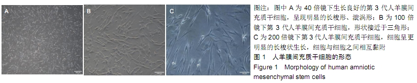
2.2 慢病毒感染人羊膜间充质干细胞 课题组前期实验发现6.5 μL Scleraxis慢病毒原液感染人羊膜间充质干细胞效果最好,96 h后荧光表达量最强,见图2。 "

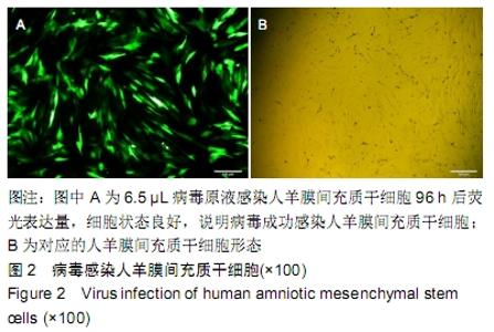
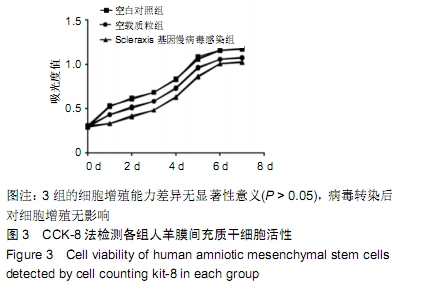
2.3 CCK-8法检测各组人羊膜间充质干细胞增殖活性 各组细胞生长曲线均呈S型。空白对照组、空载质粒组、Scleraxis基因慢病毒感染组之间的细胞增殖能力无明显差异(P > 0.05),说明Scleraxis慢病毒感染人羊膜间充质干细胞后对其增殖无影响,见图3。 "
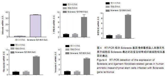
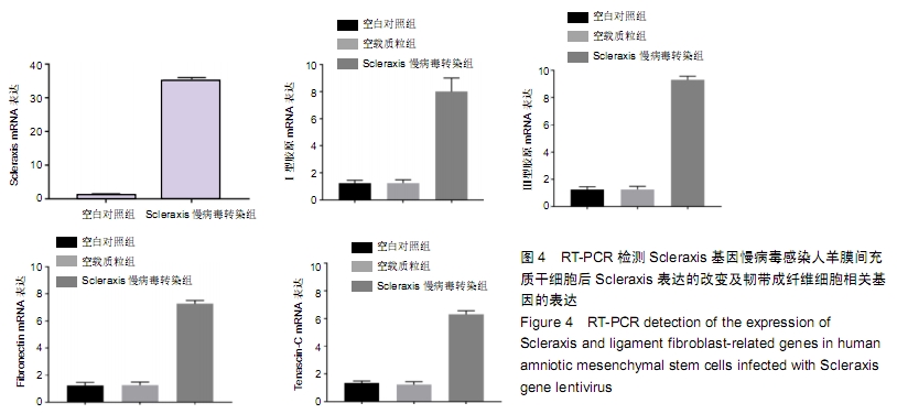
2.4 实时荧光定量PCR检测慢病毒感染人羊膜间充质干细胞后Scleraxis的表达及韧带相关基因的表达 携带有Scleraxis基因的慢病毒能够成功感染人羊膜间充质干细胞,感染后其mRNA表达量成倍的增加,Scleraxis基因慢病毒感染组韧带相关基因的mRNA表达量高于空白对照组和空载质粒组,差异有显著性意义(P < 0.05),空白对照组和空载质粒组的韧带相关基因mRNA表达量差异无显著性意义(P > 0.05),见图4。 "
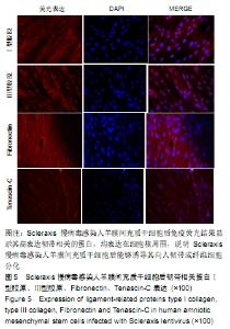
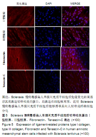
2.5 免疫荧光检测人羊膜间充质干细胞经Scleraxis慢病毒转染后其韧带相关蛋白的表达 Scleraxis慢病毒感染人羊膜间充质干细胞组韧带相关蛋白Ⅰ、Ⅲ型胶原、Fibronectin、Tenascin-C均有表达,主要在胞浆及细胞周围表达,空白对照组和空载质粒组均未观察到Ⅰ、Ⅲ型胶原、Fibronectin、Tenascin-C的表达,见图5。 "
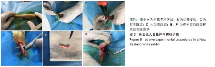
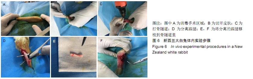
2.6 体内动物实验过程 动物体内实验步骤,见图6。 "
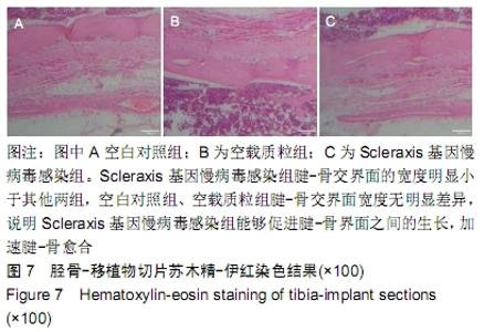
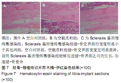
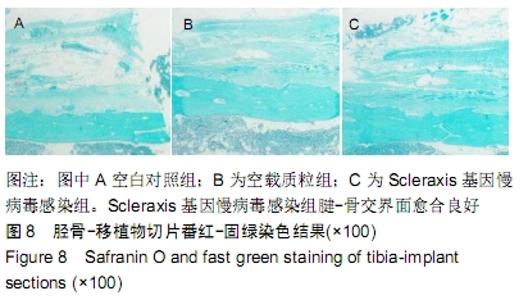
2.7 胫骨-移植物切片染色 术后3个月取胫骨-移植物标本,Scleraxis基因慢病毒感染组苏木精-伊红染色显示腱-骨交界面胶原排列整齐,交界面逐渐变窄;番红固绿染色 显示腱-骨交界面愈合良好,见图7,8。 "
"

| [1] RALLES S, AGEL J, OBERMEIER M, et al. Incidence of Secondary Intra-articular Injuries With Time to Anterior Cruciate Ligament Reconstruction. Am J Sports Med. 2015;43(6): 1373-1379. [2] ATESOK K, FU FH, WOLF MR, et al. Augmentation of tendon-to- bone healing. J Bone Joint Surg Am. 2014;96(6):513-521. [3] BALDINO L, CARDEA S, MAFFULLI N, et al. Regeneration techniques for bone-to-tendon and muscle-to-tendon interfaces reconstruction. Br Med Bull. 2016;117(1):25-37. [4] BUNKER DL, ILIE V, ILIE V, et al. Tendon to bone healing and its implications for surgery. Muscles Ligaments Tendons J. 2014;4(3): 343-350. [5] COTTRELL JA, TURNER JC, ARINZEH TL, et al. The Biology of Bone and Ligament Healing. Foot Ankle Clin. 2016;21(4):739-761. [6] CARLBERG AL, TUAN RS, HALL DJ. Regulation of scleraxis function by interaction with the bHLH protein E47. Mol Cell Biol Res Commun. 2000;3(2):82-86. [7] HU JS, OLSON EN, KINGSTON RE. HEB, a helix-loop-helix protein related to E2A and ITF2 that can modulate the DNA-binding ability of myogenic regulatory factors. Mol Cell Biol. 1992;12(3): 1031-1042. [8] LIU Y, WATANABE H, NIFUJI A, et al. Overexpression of a single helix-loop-helix-type transcription factor, scleraxis, enhances aggrecan gene expression in osteoblastic osteosarcoma ROS17/2.8 cells. J Biol Chem. 1997;272(47):29880-29885. [9] ALBERTON P, POPOV C, PRÄGERT M, et al. Conversion of human bone marrow-derived mesenchymal stem cells into tendon progenitor cells by ectopic expression of scleraxis. Stem Cells Dev. 2012;21(6):846-858. [10] MADRY H, KOHN D, CUCCHIARINI M. eDirect FGF-2 gene transfer via recombinant adeno-associated virus vectors stimulates cell proliferation, collagen production, and the repair of experimental lesions in the human ACL. Am J Sports Med. 2013; 41(1):194-202. [11] WANG CJ, WENG LH, HSU SL, et al. pCMV-BMP-2-transfected cell-mediated gene therapy in anterior cruciate ligament reconstruction in rabbits. Arthroscopy. 2010;26(7):968-976. [12] CHEN B, LI B, QI YJ, et al. Enhancement of tendon-to-bone healing after anterior cruciate ligament reconstruction using bone marrow-derived mesenchymal stem cells genetically modified with bFGF/BMP2. Sci Rep. 2016;6:25940. [13] KAWAKAMI Y, TAKAYAMA K, MATSUMOTO T, et al. Anterior Cruciate Ligament-Derived Stem Cells Transduced With BMP2 Accelerate Graft-Bone Integration After ACL Reconstruction. Am J Sports Med. 2017;45(3):584-597. [14] PENG W, ZHANG J, ZHANG H, et al. Effects of lentiviral transfection containing bFGF gene on the biological characteristics of rabbit BMSCs. J Cell Biochem. 2018;119(10): 8389-8397. [15] 周文然,李新,王文波,等.人羊膜间充质干细胞生物学特征及向多巴胺能神经元样细胞的分化[J].中国组织工程研究,2014,18(23): 3682-3690. [16] MAIDHOF R, RAFIUDDIN A, CHOWDHURY F, et al. Timing of mesenchymal stem cell delivery impacts the fate and therapeutic potential in intervertebral disc repair. J Orthop Res. 2017;35(1): 32-40. [17] NOGAMI M, KIMURA T, SEKI S, et al. A Human Amnion-Derived Extracellular Matrix-Coated Cell-Free Scaffold for Cartilage Repair: In Vitro and In Vivo Studies. Tissue Eng Part A. 2016; 22(7-8):680-688. [18] 洪佳琼,高雅,宋洁,等.人羊膜间充质干细胞与骨髓间充质干细胞的生物学特性及免疫抑制作用的比较[J].中国实验血液学杂志, 2016, 24(3):858-864. [19] LI Y, LIU Z, JIN Y, et al. Differentiation of Human Amniotic Mesenchymal Stem Cells into Human Anterior Cruciate Ligament Fibroblast Cells by In Vitro Coculture. Biomed Res Int. 2017;2017: 7360354. [20] 邹刚,李豫皖,金瑛,等.TGF-β1联合VEGF对人羊膜间充质干细胞向韧带成纤维细胞体外分化作用的研究[J].中国修复重建外科杂志, 2017,31(5): 582-591. [21] 李豫皖,朱喜忠,金瑛,等.体外定向诱导人羊膜间充质干细胞向骨、软骨及脂肪细胞的分化[J].中国组织工程研究,2017,21(1):122-127. [22] 朱喜忠,刘子铭,吴术红,等.Scleraxis慢病毒基因感染人羊膜间充质干细胞向肌腱细胞的定向分化[J].中国组织工程研究,2017,21(33): 5382-5387. [23] HONG Y. ACL injury: incidences, healing, rehabilitation, and prevention: part of the Routledge Olympic special issue collection. Res Sports Med. 2012;20(3-4):155-156. [24] ZAFFAGNINI S, SIGNORELLI C, BONANZINGA T, et al. Technical variables of ACL surgical reconstruction: effect on post-operative static laxity and clinical implication. Knee Surg Sports Traumatol Arthrosc. 2016;24(11):3496-3506. [25] AHN JH, LEE YS, KO TS, et al. Accuracy and Reproducibility of the Femoral Tunnel With Different Viewing Techniques in the ACL Reconstruction. Orthopedics. 2016;39(6):e1085-e1091. [26] SCHWEITZER R, CHYUNG JH, MURTAUGH LC, et al. Analysis of the tendon cell fate using Scleraxis, a specific marker for tendons and ligaments. Development. 2001;128(19):3855-3866. [27] CSERJESI P, BROWN D, LIGON KL, et al. Scleraxis: a basic helix-loop-helix protein that prefigures skeletal formation during mouse embryogenesis. Development.1995;121(4):1099-1110. [28] PRYCE BA, BRENT AE, MURCHISON ND, et al. Generation of transgenic tendon reporters, ScxGFP and ScxAP, using regulatory elements of the scleraxis gene. Dev Dyn. 2007;236(6):1677-1682. [29] MENDIAS CL, GUMUCIO JP, BAKHURIN KI, et al. Physiological loading of tendons induces scleraxis expression in epitenon fibroblasts. J Orthop Res. 2012;30(4):606-612. [30] MURCHISON ND, PRICE BA, Conner DA, et al. Regulation of tendon differentiation by scleraxis distinguishes force-transmitting tendons from muscle-anchoring tendons. Development. 2007; 134(14): 2697-2708. [31] ALBERTON P, POPOV C, PRÄGERT M, et al. Conversion of human bone marrow-derived mesenchymal stem cells into tendon progenitor cells by ectopic expression of scleraxis. Stem Cells Dev. 2012;21(6): 846-858. [32] GULOTTA LV, RODEO SA. Emerging ideas: Evaluation of stem cells genetically modified with scleraxis to improve rotator cuff healing. Clin Orthop Relat Res. 2011;469(10):2977-2980. [33] GULOTTA LV, KOVACEVIC D, Packer JD, et al. Bone marrow-derived mesenchymal stem cells transduced with scleraxis improve rotator cuff healing in a rat model. Am J Sports Med. 2011;39(6): 1282-1289. [34] CAI J, WANG J, YE K, et al. Dual-layer aligned-random nanofibrous scaffolds for improving gradient microstructure of tendon-to-bone healing in a rabbit extra-articular model. Int J Nanomedicine. 2018;13:3481-3492. [35] ZHANG P, ZHI Y, FANG H, et al. Effects of polyvinylpyrrolidone- iodine on tendon-bone healing in a rabbit extra-articular model. Exp Ther Med. 2017;13(6):2751-2756. [36] LI H, GE Y, WU Y, et al. Hydroxyapatite coating enhances polyethylene terephthalate artificial ligament graft osseointegration in the bone tunnel. Int Orthop. 2011;35(10): 1561-1567. [37] ZHI Y, LIU W, ZHANG P, et al. Electrospun silk fibroin mat enhances tendon-bone healing in a rabbit extra-articular model. Biotechnol Lett. 2016;38(10):1827-1835. |
| [1] | Zhang Tongtong, Wang Zhonghua, Wen Jie, Song Yuxin, Liu Lin. Application of three-dimensional printing model in surgical resection and reconstruction of cervical tumor [J]. Chinese Journal of Tissue Engineering Research, 2021, 25(9): 1335-1339. |
| [2] | Zeng Yanhua, Hao Yanlei. In vitro culture and purification of Schwann cells: a systematic review [J]. Chinese Journal of Tissue Engineering Research, 2021, 25(7): 1135-1141. |
| [3] | Xu Dongzi, Zhang Ting, Ouyang Zhaolian. The global competitive situation of cardiac tissue engineering based on patent analysis [J]. Chinese Journal of Tissue Engineering Research, 2021, 25(5): 807-812. |
| [4] | Chen Junyi, Wang Ning, Peng Chengfei, Zhu Lunjing, Duan Jiangtao, Wang Ye, Bei Chaoyong. Decalcified bone matrix and lentivirus-mediated silencing of P75 neurotrophin receptor transfected bone marrow mesenchymal stem cells to construct tissue-engineered bone [J]. Chinese Journal of Tissue Engineering Research, 2021, 25(4): 510-515. |
| [5] | Wu Zijian, Hu Zhaoduan, Xie Youqiong, Wang Feng, Li Jia, Li Bocun, Cai Guowei, Peng Rui. Three-dimensional printing technology and bone tissue engineering research: literature metrology and visual analysis of research hotspots [J]. Chinese Journal of Tissue Engineering Research, 2021, 25(4): 564-569. |
| [6] | Ma Ziyue, Ju Xiaochen, Zhang Lei, Sun Rongxin. Tendon-bone healing in anterior cruciate ligament reconstruction with and without remnant preservation [J]. Chinese Journal of Tissue Engineering Research, 2021, 25(4): 582-587. |
| [7] | Chang Wenliao, Zhao Jie, Sun Xiaoliang, Wang Kun, Wu Guofeng, Zhou Jian, Li Shuxiang, Sun Han. Material selection, theoretical design and biomimetic function of artificial periosteum [J]. Chinese Journal of Tissue Engineering Research, 2021, 25(4): 600-606. |
| [8] | Liu Fei, Cui Yutao, Liu He. Advantages and problems of local antibiotic delivery system in the treatment of osteomyelitis [J]. Chinese Journal of Tissue Engineering Research, 2021, 25(4): 614-620. |
| [9] | Li Xiaozhuang, Duan Hao, Wang Weizhou, Tang Zhihong, Wang Yanghao, He Fei. Application of bone tissue engineering materials in the treatment of bone defect diseases in vivo [J]. Chinese Journal of Tissue Engineering Research, 2021, 25(4): 626-631. |
| [10] | Zhang Zhenkun, Li Zhe, Li Ya, Wang Yingying, Wang Yaping, Zhou Xinkui, Ma Shanshan, Guan Fangxia. Application of alginate based hydrogels/dressings in wound healing: sustained, dynamic and sequential release [J]. Chinese Journal of Tissue Engineering Research, 2021, 25(4): 638-643. |
| [11] | Chen Jiana, Qiu Yanling, Nie Minhai, Liu Xuqian. Tissue engineering scaffolds in repairing oral and maxillofacial soft tissue defects [J]. Chinese Journal of Tissue Engineering Research, 2021, 25(4): 644-650. |
| [12] | Xing Hao, Zhang Yonghong, Wang Dong. Advantages and disadvantages of repairing large-segment bone defect [J]. Chinese Journal of Tissue Engineering Research, 2021, 25(3): 426-430. |
| [13] | Chen Siqi, Xian Debin, Xu Rongsheng, Qin Zhongjie, Zhang Lei, Xia Delin. Effects of bone marrow mesenchymal stem cells and human umbilical vein endothelial cells combined with hydroxyapatite-tricalcium phosphate scaffolds on early angiogenesis in skull defect repair in rats [J]. Chinese Journal of Tissue Engineering Research, 2021, 25(22): 3458-3465. |
| [14] | Wang Hao, Chen Mingxue, Li Junkang, Luo Xujiang, Peng Liqing, Li Huo, Huang Bo, Tian Guangzhao, Liu Shuyun, Sui Xiang, Huang Jingxiang, Guo Quanyi, Lu Xiaobo. Decellularized porcine skin matrix for tissue-engineered meniscus scaffold [J]. Chinese Journal of Tissue Engineering Research, 2021, 25(22): 3473-3478. |
| [15] | Mo Jianling, He Shaoru, Feng Bowen, Jian Minqiao, Zhang Xiaohui, Liu Caisheng, Liang Yijing, Liu Yumei, Chen Liang, Zhou Haiyu, Liu Yanhui. Forming prevascularized cell sheets and the expression of angiogenesis-related factors [J]. Chinese Journal of Tissue Engineering Research, 2021, 25(22): 3479-3486. |
| Viewed | ||||||
|
Full text |
|
|||||
|
Abstract |
|
|||||