Chinese Journal of Tissue Engineering Research ›› 2018, Vol. 22 ›› Issue (34): 5518-5525.doi: 10.3969/j.issn.2095-4344.0993
Previous Articles Next Articles
Ultra-small superparamagnetic iron oxide particles enhance MRI diagnosis of lymph node metastasis of the head and neck
Li Wenjin, Niu Jinliang, Zhu Li, Wang Tao, Wang Yu
- Second Hospital of Shanxi Medical University, Taiyuan 030001, Shanxi Province, China
-
Received:2018-08-07Online:2018-12-08Published:2018-12-08 -
Contact:Niu Jinliang, MD, Professor, Second Hospital of Shanxi Medical University, Taiyuan 030001, Shanxi Province, China -
About author:Li Wenjin, MD, Associate chief physician, Second Hospital of Shanxi Medical University, Taiyuan 030001, Shanxi Province, China -
Supported by:the Scientific Research Project of Shanxi Provincial Health Department, No. 20100141; the Shanxi Basic Research Foundation for Youth Science and Technology Research, No. 2011021035-4; the Graduate Innovation Project of Shanxi Province, No. 20093066; Doctoral Start Fund of Shanxi Medical University Second Hospital, No. 2013-6; the Teaching Fund of Shanxi Medical University Second Hospital, No. 201603-5; Science and Technology Innovation Project for High Educations of Shanxi Provincial Department of Education, No. 20141105; Shanxi Provincial Returned Overseas Students Research Funding Project of Shanxi Provincial Office for Study Abroad, No. 2016-122; the Shanxi Provincial Project Funding for Overseas Returnees, Shanxi Provincial Department of Human Resources and Social Sciences
CLC Number:
Cite this article
Li Wenjin, Niu Jinliang, Zhu Li, Wang Tao, Wang Yu. Ultra-small superparamagnetic iron oxide particles enhance MRI diagnosis of lymph node metastasis of the head and neck[J]. Chinese Journal of Tissue Engineering Research, 2018, 22(34): 5518-5525.
share this article
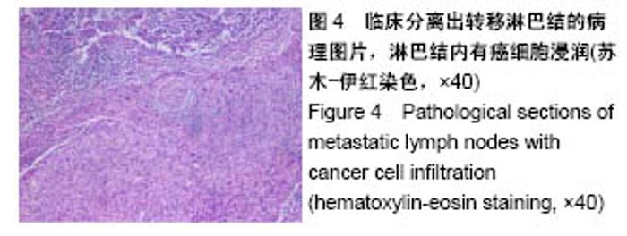
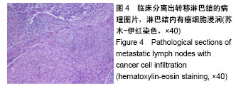
2.1 实验动物数量分析 纳入新西兰兔20只,均进入结果分析,无死亡和感染。 2.2 淋巴结病理组织学检查结果 20只兔共分离出57个淋巴结,其中25个病理检测证实淋巴结转移,见图4,32个淋巴结未发生转移。57个淋巴结中腮腺淋巴结20个、颌下淋巴结27个。腮腺淋巴结发生转移的有19个,颌下淋巴结发生转移的有6个。"
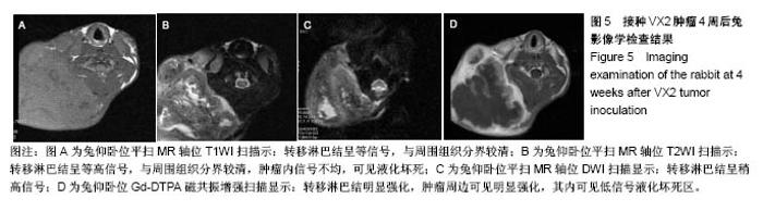
转移的淋巴结在病理学检查发现有4个在淋巴结皮质存在癌细胞浸润,其中1个发现癌细胞浸润突破淋巴结皮质;有3个在淋巴结髓质可见癌细胞浸润;有10个在淋巴结皮质及髓质均可见癌细胞浸润;8个发现浸润的癌组织中可见坏死灶,表现为细胞核固缩或溶解,细胞内结构消失。 2.3 MRI检查结果 常规MRI检查:20只兔中分离出57个淋巴结。淋巴结MRI平扫T1WI多呈中低信号,T2WI呈中高信号,或中心信号不均匀,圆形或球形,液化坏死区T2WI表现为明显高信号,被膜侵犯淋巴结边界欠清晰(图5A,B)。按淋巴结形态分为转移淋巴结及未转移淋巴结标准,符合转移淋巴结标准的23个,与病理学淋巴结检查结果对照,13枚淋巴结为真阳性,真阳性率52%(13/25),假阳性10枚,假阳性率40%(10/25);MRI诊断未转移淋巴结34枚,病理学阴性淋巴结32枚,真阴性率69%(22/32),假阴性率38%(12/32)。 DWI检测方法的ADC值:DWI显示与未转移淋巴结相比,转移淋巴结信号强度增加,液化坏死区DWI为低信号(图5C)。ADC图测量发现未转移淋巴结的ADC值为(1.39±0.12)×10-3 mm2/s,转移淋巴结的ADC值为(0.76±0.08)×10-3 mm2/s,两组的ADC值差异有显著性意义(P < 0.05) 。 Gd-DTPA增强扫描SI比率值:结合病理学检查结果,Gd-DTPA增强扫描转移淋巴结强化明显,呈薄环状、不规则或锯齿状,或较均匀强化,中央淋巴结坏死区未见强化,未转移淋巴结强化不明显(图5D)。定量测量发现未转移淋巴结T1WI Gd-DTPA增强扫描SI比率值为1.499±0.012,转移淋巴结Gd-DTPA增强扫描SI比率值为2.934±0.020,二者差异具有显著性意义(P < 0.05)。"

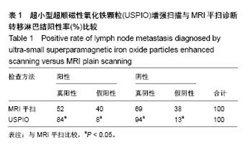
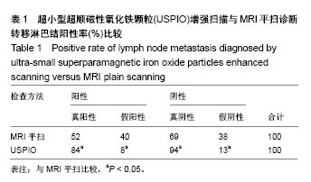
USPIO增强扫描结果:定性分析:未转移淋巴结USPIO增强扫描T2WI上淋巴结呈均匀的低信号,形态规则,伴宽窄不一的环形强化区,或均匀的低信号区内可见中性点状高信号区。转移淋巴结USPIO增强扫描T2WI上淋巴结无明显信号减低区,或结内不规则信号减低区。按USPIO增强扫描诊断良恶性淋巴结的标准:符合转移淋巴结标准的23枚,其中21枚淋巴结均病理学淋巴结转移,真阳性率为84%(21/25),假阳性为2枚,假阳性率8%(2/25);MRI诊断未转移淋巴结34枚,病理学结果对照,真阴性率为94%(30/32),假阴性率为13%(4/32)。 定量分析:未转移淋巴结:注射USPIO 24 h后,T2﹡WI信号下降,增强扫描前SNR值为38.61±11.48,增强扫描后SNR值为15.30±3.69,△SNR= -57.20±16.03;转移淋巴结:USPIO增强后T2﹡WI信号下降,SNR下降不明显,增强扫描前SNR值为37.43±9.46,增强扫描后SNR值为32.30±2.67,△SNR=-16.20±5.03。增强扫描前后△SNR差值具有显著性意义。 2.4 USPIO增强扫描与MRI平扫诊断淋巴结阳性率比较 按照以上转移及未转移淋巴结诊断标准,USPIO增强扫描与MRI平扫诊断淋巴结阳性率比较见表1,两种不同检查方法对淋巴结的转移诊断阳性率差异有显著性意义(P < 0.05)。"
| [1] Hoffmann TK, Schuler PJ, Laban S, et al. Response evaluation in head and neck oncology: definition and prediction. ORL J Otorhinolaryngol Relat Spec. 2017;79(1):14-23.[2] Maruyama T, Nishihara K, Saio M, et al. Kikuchi-Fujimoto disease in the regional lymph nodes with node metastasis in a patient with tongue cancer: a case report and literature review. Oncol Lett. 2017; 14(1):257-263.[3] Van den Brekel M, Castelijns J, Snow G. The size of lymph nodes in the neck on sonograms as a radiologic criterion for metastasis: how reliable is it? A JNR Am J Neuroradiol. 1998;19:695-700.[4] Kim SH, Oh SN, Choi HS, et al. USPIO enhanced lymph node MRI using 3D multi-echo GRE in a rabbit model. Contrast Media Mol Imaging. 2016;11(6):544-549.[5] Lahaye MJ, Engelen SM, Kessels AG, et al. USPIO-enhanced MR imaging for nodal staging in patients with primary rectal cancer:predictive criteria. Radiology. 2008;246(3):804-811.[6] Crott R, Lawson G, Nollevaux MC, et al. Comprehensive cost analysis of sentinel node biopsy in solid head and neck tumors using a time-driven activity-based costing approach. Eur Arch Otorhinolaryngol. 2016;273(9):2621-2628.[7] Werner JA. Untersuchungen zum Lymphgef äβsystem der oberen Luft-und Speisewege. Aachen: Shaker. 1995.[8] Kim SH, Oh SN, Choi HS, et al. Ultra-small superparamagnetic iron oxide mediated magnetic hyperthermia in treatment of neck lymph node metastasis in rabbit pyriform sinus VX2 carcinoma. Tumour Biol. 2015;36(10):8035-8040.[9] Payabvash S, Meric K.,Cayci Z. Differentiation of benign from malignant cervical lymph nodes in patients with head and neck cancer using PET/CT imaging.Clin Imaging. 2016;40(1):101-105.[10] Dunne AA, Plehn S, Schulz S, et al. Lymph node tepography of the head and neck in New Zealand white rabbits. Lab Anim. 2003;37: 37-43.[11] 李文晋,牛金亮,朱莉,等.头颈部不同部位肿瘤动物模型的建立及生长转移特性比较[J].中国组织工程研究,2016,20(5):748-753. [12] Van den Brekel MW, Stel HV, Castelijns JA, et al. Cervical lymph node metastases:assessing of radiological criteria. Radiology. 1990;177(2):379-384.[13] Steinkamp HJ, Hosten N, Richter C, et al. Enlarged cervical lymph nodes at helical CT. Radiology. 1994;191(3):795-798.[14] Iannessi A. Ouvrier MJ. Thariat J, et al. Imaging in head and neck cancers. Bull Cancer. 2014;101(5):469-480.[15] Sun J, Li B, Li CJ, et al. Computed tomography versus magnetic resonance imaging for diagnosing cervical lymph node metastasis of head and neck cancer: a systematic review and meta-analysis. Onco Targets Ther. 2015;8:1291-1313.[16] Eida S, Sumi M, Yonetsu K, et al. Combination of helical CT and Doppler sonography in the follow-up of patients with clinical N0 stage neck disease and oral cancer. AJNR. 2003;24(3):312-318.[17] 李文晋,牛金亮,金慧兰,等.头颈部肿瘤颈部淋巴结隐匿性转移的病理实验研究[J].中国药物与临床,2012,12(12):1533-1535. [18] Poant? L, Pop S, Cosgarea M, et al. The role of contrast enhanced ultrasound in the assessment of superficial lymph nodes. Rom J Intern Med, 2012;50(3):189-193.[19] Manca G, Vanzi E, Rubello D, et al. (18)F-FDG PET/CT quantification in head and neck squamous cell cancer: principles, technical issues and clinical applications. Eur J Nucl Med Mol Imaging.2016;43(7):1360-1375. [20] Lu L, Li Y, Li W. The role of intravoxel incoherent motion MRI in predicting early treatment response to chemoradiation for metastatic lymph nodes in nasopharyngeal carcinoma. Adv Ther. 2016;33(7):1158-1168.[21] 蓝美红,牛金亮,李文晋,等.相对表观弥散系数与钆喷替酸葡甲胺增强MRI诊断兔头颈转移淋巴结的对比研究[J].中国中西医结合影像学杂志, 2012,10(2):105-108.[22] Triantafyllou M, Studer UE, Birkhäuser FD, et al. Ultrasmall superparamagnetic particles of iron oxide allow for the detection of metastases in normal sized pelvic lymph nodes of patients with bladder and/or prostate cancer. Eur J Cancer. 2013;49(3):616-624.[23] Froehlich JM, Triantafyllou M, Fleischmann A, et al. Does quantification of USPIO uptake-related signal loss allow differentiation of benign and malignant normal-sized pelvic lymph nodes? Contrast Media Mol Imaging. 2012;7(3):346-355.[24] Schramm N. Macrophage imaging for differentiation between infectious spondylitis/spondylodiscitis and sterile vertebral inflammation: new options for the use of USPIO-enhanced MRI beyond lymph node characterization. Radiology. 2010;50(3): 205-207.[25] Wu L, Cao Y, Liao C, et al. Diagnostic performance of USPIO-enhanced MRI for lymph- node metastases in different body regions: a meta-analysis. Eur J Radiol. 2011;80(2):582-589.[26] Choi SH, Kim KH, Moon WK, et al. Comparison of lymph node metastases assessment with the use of USPIO-enhanced MR imaging at 1.5 T versus 3.0 T in a rabbit model. J Magn Reson Imaging. 2010;31(1):134-141.[27] Bandini M, Gandaglia G, Fossati N, et al. An Explanatory Case on the Limitations of Lymph Node Staging in Recurrent Prostate Cance. Urol Case Rep. 2017;12:34-36.[28] Kawai Y, Sumi M, Nakamura T. Turbo short tau inversion recovery imaging for metastatic node screening in patients with head and neck cancer. Am J Neuroradiol. 2006;27(6):1283-1287.[29] Smits LP, Coolen BF, Panno MD, et al. Noninvasive differentiation between hepatic steatosis and steatohepatitis with mr imaging enhanced with USPIOs in patients with nonalcoholic fatty liver disease: a proof-of-concept study. Radiology. 2016;278(3):782-791.[30] Seyfer P, Pagenstecher A, Mandic R, et al. Cancer and inflammation: differentiation by USPIO-enhanced MR imaging. J Magn Reson Imaging. 2014;39(3):665-672.[31] Liang L, Luo X, Lian Z, et al. Lymph node metastasis in head and neck squamous carcinoma: efficacy of intravoxel incoherent motion magnetic resonance imaging for the differential diagnosis. Eur J Radiol. 2017;90:159-165. |
| [1] | Zhang Tongtong, Wang Zhonghua, Wen Jie, Song Yuxin, Liu Lin. Application of three-dimensional printing model in surgical resection and reconstruction of cervical tumor [J]. Chinese Journal of Tissue Engineering Research, 2021, 25(9): 1335-1339. |
| [2] | Zeng Yanhua, Hao Yanlei. In vitro culture and purification of Schwann cells: a systematic review [J]. Chinese Journal of Tissue Engineering Research, 2021, 25(7): 1135-1141. |
| [3] | Xu Dongzi, Zhang Ting, Ouyang Zhaolian. The global competitive situation of cardiac tissue engineering based on patent analysis [J]. Chinese Journal of Tissue Engineering Research, 2021, 25(5): 807-812. |
| [4] | Wu Zijian, Hu Zhaoduan, Xie Youqiong, Wang Feng, Li Jia, Li Bocun, Cai Guowei, Peng Rui. Three-dimensional printing technology and bone tissue engineering research: literature metrology and visual analysis of research hotspots [J]. Chinese Journal of Tissue Engineering Research, 2021, 25(4): 564-569. |
| [5] | Chang Wenliao, Zhao Jie, Sun Xiaoliang, Wang Kun, Wu Guofeng, Zhou Jian, Li Shuxiang, Sun Han. Material selection, theoretical design and biomimetic function of artificial periosteum [J]. Chinese Journal of Tissue Engineering Research, 2021, 25(4): 600-606. |
| [6] | Liu Fei, Cui Yutao, Liu He. Advantages and problems of local antibiotic delivery system in the treatment of osteomyelitis [J]. Chinese Journal of Tissue Engineering Research, 2021, 25(4): 614-620. |
| [7] | Li Xiaozhuang, Duan Hao, Wang Weizhou, Tang Zhihong, Wang Yanghao, He Fei. Application of bone tissue engineering materials in the treatment of bone defect diseases in vivo [J]. Chinese Journal of Tissue Engineering Research, 2021, 25(4): 626-631. |
| [8] | Zhang Zhenkun, Li Zhe, Li Ya, Wang Yingying, Wang Yaping, Zhou Xinkui, Ma Shanshan, Guan Fangxia. Application of alginate based hydrogels/dressings in wound healing: sustained, dynamic and sequential release [J]. Chinese Journal of Tissue Engineering Research, 2021, 25(4): 638-643. |
| [9] | Chen Jiana, Qiu Yanling, Nie Minhai, Liu Xuqian. Tissue engineering scaffolds in repairing oral and maxillofacial soft tissue defects [J]. Chinese Journal of Tissue Engineering Research, 2021, 25(4): 644-650. |
| [10] | Xing Hao, Zhang Yonghong, Wang Dong. Advantages and disadvantages of repairing large-segment bone defect [J]. Chinese Journal of Tissue Engineering Research, 2021, 25(3): 426-430. |
| [11] | Chen Siqi, Xian Debin, Xu Rongsheng, Qin Zhongjie, Zhang Lei, Xia Delin. Effects of bone marrow mesenchymal stem cells and human umbilical vein endothelial cells combined with hydroxyapatite-tricalcium phosphate scaffolds on early angiogenesis in skull defect repair in rats [J]. Chinese Journal of Tissue Engineering Research, 2021, 25(22): 3458-3465. |
| [12] | Wang Hao, Chen Mingxue, Li Junkang, Luo Xujiang, Peng Liqing, Li Huo, Huang Bo, Tian Guangzhao, Liu Shuyun, Sui Xiang, Huang Jingxiang, Guo Quanyi, Lu Xiaobo. Decellularized porcine skin matrix for tissue-engineered meniscus scaffold [J]. Chinese Journal of Tissue Engineering Research, 2021, 25(22): 3473-3478. |
| [13] | Mo Jianling, He Shaoru, Feng Bowen, Jian Minqiao, Zhang Xiaohui, Liu Caisheng, Liang Yijing, Liu Yumei, Chen Liang, Zhou Haiyu, Liu Yanhui. Forming prevascularized cell sheets and the expression of angiogenesis-related factors [J]. Chinese Journal of Tissue Engineering Research, 2021, 25(22): 3479-3486. |
| [14] | Liu Chang, Li Datong, Liu Yuan, Kong Lingbo, Guo Rui, Yang Lixue, Hao Dingjun, He Baorong. Poor efficacy after vertebral augmentation surgery of acute symptomatic thoracolumbar osteoporotic compression fracture: relationship with bone cement, bone mineral density, and adjacent fractures [J]. Chinese Journal of Tissue Engineering Research, 2021, 25(22): 3510-3516. |
| [15] | Liu Liyong, Zhou Lei. Research and development status and development trend of hydrogel in tissue engineering based on patent information [J]. Chinese Journal of Tissue Engineering Research, 2021, 25(22): 3527-3533. |
| Viewed | ||||||
|
Full text |
|
|||||
|
Abstract |
|
|||||