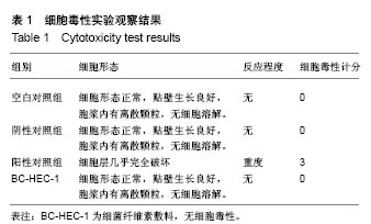
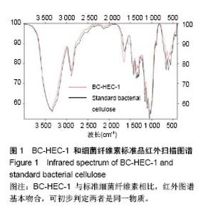
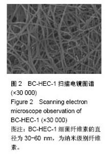
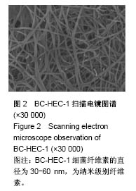
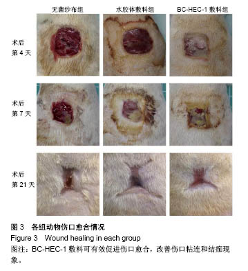
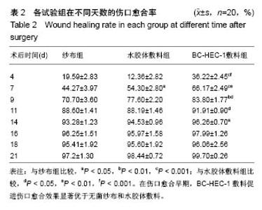
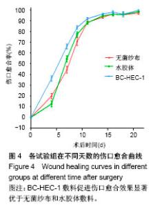
| [1] Gurtner GC, Werner S, Barrandon Y, et al. Wound repair and regeneration. Nature. 2008;453(7193):314-321.[2] 刘德伍.现代敷料研究现状[J].中国临床康复,2002,6(22): 3436-3437.[3] 冯正,李江萍,李红.新型敷料与传统干纱布换药在外科切口感染中的应用效果对比[J].当代护士,2016,23(12):100-102.[4] 廖敏,梁文卿,袁先容.新型敷料在伤口护理中的应用[J].中国保健营养,2016,26(27):206.[5] Chaudhari AA, Vig K, Baganizi DR, et al. Future prospects for scaffolding methods and biomaterials in skin tissue engineering: a review. Int J Mol Sci. 2016;17(12):1974.[6] 刘公洪,廖毅.湿性敷料治疗在烧伤创面的应用进展[J].西南军医, 2012,14(1):113-114.[7] Field CK, Kerstein MD. Overview of wound healing in a moist environment. Am J Surg. 1994;167(1A):2S.[8] Sood A, Granick MS, Tomaselli NL. Wound Dressings and Comparative Effectiveness Data. Adv Wound Care. 2014;3(8): 511.[9] 李晓明,刘苹,张波.创面敷料的研究现状[J].重庆医学,2017,46(20): 2851-2853.[10] 张劲峰,郝建波,张劲鹏,等.生物敷料的研究进展[J].中国修复重建外科杂志,2015,29(2):254-259.[11] 李晶,薛斌.新型医用敷料的分类及特点[J].中国组织工程研究, 2013,17(12):2225-2232.[12] Bäckdahl H, Helenius G, Bodin A, et al. Mechanical properties of bacterial cellulose and interactions with smooth muscle cells. Biomaterials. 2006;27(9):2141.[13] Helenius G, Bäckdahl H, Bodin A, et al. In vivo, biocompatibility of bacterial cellulose. J Biomed Mater Res Part A. 2006;76(2):431.[14] Czaja WK, Young DJ, Kawecki M, et al. The future prospects of microbial cellulose in biomedical applications. Biomacromolecules. 2007;8(1):1.[15] 李莹.基于细菌纤维素的皮肤伤口敷料的制备和评价[D].武汉:中科技大学,2015.[16] 南方,赖琛,奚廷斐.细菌纤维素与纳米银复合制备创伤敷料[J].中国组织工程研究,2015,19(43):7023-7028.[17] Khalid A, Khan R, Ul-Islam M, et al. Bacterial cellulose-zinc oxide nanocomposites as a novel dressing system for burn wounds. Carbohydr Polym. 2017;164:214.[18] Pandey M, Mohamad N, Low W L, et al. Microwaved bacterial cellulose-based hydrogel microparticles for the healing of partial thickness burn wounds. Drug Deliv Transl Res. 2017; 7(1):1-11.[19] 蒋琪霞,张晓霞,刘亚红,等.伤口护理中不同敷料的疗效观察[J].解放军护理杂志,2000,17(6):10-11.[20] 首家保,陈彬,芦慎,等.不同敷料修复创面的实验研究及临床应用评价[J].中国组织工程研究,2012,16(8):1479-1482.[21] 马霞,陈世文,王瑞明,等.纳米材料细菌纤维素对大鼠皮肤创伤的促愈作用[J].中国组织工程研究,2006,10(37):45-47.[22] 汪涌,何清濂.生长因子与皮肤伤口愈合[J].中华整形外科杂志, 1999,(6):462-464.[23] 蔡泽浩,刘伟,钱云良,等.TGF-β细胞内信号转导与伤口过度愈合[J].上海交通大学学报(医学版),2004,24(7):587-589.[24] 谷庆阳.TGF-β及其信号分子SMAD3在伤口愈合过程中的作用[J].国外医学:生理、病理科学与临床分册,2002,22(1):67-69. [25] Darby IA, Laverdet B, Bonté F, et al. Fibroblasts and myofibroblasts in wound healing. Clin Cosmet Investig Dermatol. 2014;7(default):301-311.[26] Pierce G F, Mustoe T A, Altrock B W, et al. Role of platelet-derived growth factor in wound healing. J Cell Biochem. 1991;45(4):319.[27] 何亮家.结肠伤口愈合模型中TGF-β1、EGF和PDGF-BB的时序表达[J].国际骨科学杂志,1999,(2):124-125.[28] 王雪,牛星焘,陈东明.PDGF在大鼠断层供皮区创面愈合过程中表达变化的研究[J].中国组织化学与细胞化学杂志,1997,(3):76-79. |