Chinese Journal of Tissue Engineering Research ›› 2018, Vol. 22 ›› Issue (26): 4215-4221.doi: 10.3969/j.issn.2095-4344.0787
Previous Articles Next Articles
Biopolymer materials for cartilage tissue engineering
Feng Nai-bo, Chang Fei, Han Yu, Wang Xin
- Second Hospital of Jilin University, Changchun 130000, Jilin Province, China
-
Received:2017-12-19 -
Contact:Chang Fei, Associate professor, Second Hospital of Jilin University, Changchun 130000, Jilin Province, China -
About author:Feng Nai-bo, Master candidate, Second Hospital of Jilin University, Changchun 130000, Jilin Province, China -
Supported by:the National Natural Science Foundation of China, No. 81671804; the Natural Science Foundation of Jilin Provincial Science and Technology Department, No. 20160101109JC
CLC Number:
Cite this article
Feng Nai-bo, Chang Fei, Han Yu, Wang Xin. Biopolymer materials for cartilage tissue engineering[J]. Chinese Journal of Tissue Engineering Research, 2018, 22(26): 4215-4221.
share this article
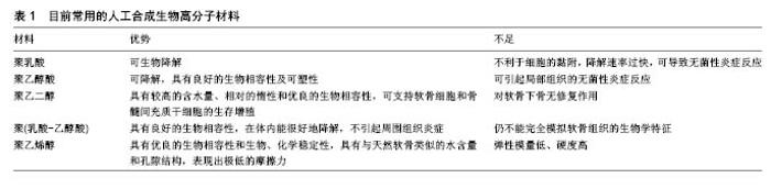
在体内,细胞在无外源支架的情况下通过细胞-细胞的相互作用、化学刺激等作用构建成组织,在构建软骨组织时还存在机械力作用和细胞自分化等作用。在体外实验,许多组织工程策略已经围绕支架的结构设计来指导细胞向特定组织的增殖分化。例如,负载骨形态发生蛋白2的羟基磷灰石支架在临床上成功地促进了骨髓间充质干细胞在支架上的定植和成骨分化[5]。骨修复支架材料的选择已经形成体系(如胶原蛋白、羟基磷灰石和磷酸三钙复合材料),但是对于软骨修复的支架材料的选择仍在不断尝试创新之中。现常用的高分子化合物材料分为天然和人工合成两种类型。 2.1 天然生物高分子材料 天然高分子化合物材料被认为是第一种用于临床的生物可降解材料[6]。天然高分子化合物材料与细胞具有更好的生物相容性,使得它们在生物系统应用中具有更优越的性能。目前被应有的包括天然高分子蛋白质(丝素蛋白、胶原、纤维蛋白原等)、多糖(纤维素、淀粉、糖胺聚糖、壳聚糖等)和聚核苷酸(DNA、RNA)等。 2.1.1 壳聚糖 壳聚糖是甲壳素脱N-乙酰基的产物。由于壳聚糖易溶于有机酸,方便参与化学反应,为壳聚糖的开发利用提供了有利条件[7]。较高孔隙率是生物高分子材料制备支架必备特征之一,它为组织生长所必需的细胞增殖、分化提供一个平台,并起到物理引导作用。壳聚糖作为一种多孔生物高分子材料,它为细胞生长提供了足够的表面积,并且其内部高度互通的孔隙结构和适宜的孔径为细胞的营养物质运输交换提供便利条件[8]。 Breyner等[9]在三维壳聚糖支架中加入软骨细胞培养基后,通过检测基因表达和Ⅱ型胶原蛋白量,证明了三维壳聚糖支架能够促进骨髓间充质干细胞在支架上黏附增殖并向软骨细胞分化。然而,美中不足的是,壳聚糖作为软骨组织工程的重要生物高分子材料,其质地硬并且较脆,也就是说壳聚糖的抗剪切力能力相对较弱。因此,在许多后期材料设计中,将壳聚糖与其他材料结合对其性能进行了优化。Bettini等[10]证实高浓度棉子糖使壳聚糖的表面更为光滑,亲水性也更强,并且具有优越的弹性性能。Ravanetti团队[11]也用棉子糖修饰壳聚糖,设立对照实验,证实修饰后的壳聚糖在兔软骨缺损模型中表现出更优良的软骨修复作用。其他常见修饰方式,如通过微交联的方法获得的壳聚糖复合材料[12-14],同样在体外和体内试验中表现出良好的理化性能,是软骨组织工程重要的生物高分子材料之一。 2.1.2 丝素蛋白 另一种常见天然生物高分子材料是丝素蛋白。丝素蛋白用于软骨组织工程有许多天然优势如:良好的生物相容性、较低的免疫原性和可控的降解速率等[15-16]。同时,丝素蛋白还用另一大优势即容易在其结构上连接短肽对其进行修饰,可以促进细胞的黏附定植[17-18]。先前报道称,直接组装的丝素蛋白支架可以支持干细胞的附着生长,并且在加入软骨细胞的培养条件下能够促进干细胞向软骨细胞分化[19-20],这为丝素蛋白用于软骨组织工程提供了理论基础。同样的,尽管丝素蛋白作为生物高分子材料展现出独特的天然优势,但是仍有不足之处,例如极易断裂、力学性能差、不易成型及吸水性不良等。因此丝素蛋白的性能优化成为后期研究重点。Silva团队结合壳聚糖与丝素蛋白的生物特点,将两者组成优势互补的复合材料,在体外进行了力学性能、细胞实验等基础研究,表现出优越的生物性 能[21],在随后Deng等[22]在兔膝关节软骨缺损模型中验证了壳聚糖-丝素蛋白复合材料用于软骨组织工程的可行性。丝素蛋白的优化方案还有与明胶[23]、透明质酸等结合[24],实验表明这种多材料复合即弥补了丝素蛋白的缺陷,同时将多种材料的优势发挥出来,形成一种各项性能优异的支架复合材料。静电纺丝微球也是生物高分子材料在软骨修复组织工程的应用设计形式之一。静电纺丝是一种多功能支架的制备技术,这种技术可以丝素蛋白为原料制备出与天然细胞外基质空间相似的仿生纳米纤维支架,通过调整制备过程中的工艺参数,可以获得空间结构、直径及生物机械性能等可控的微球。这种三维微球支架可以支持细胞的生长、增殖、分化,在软骨修复应用中,可以装载骨髓间充质干细胞等细胞促进软骨形成[25-27]。 2.1.3 胶原 胶原也是一种在软骨组织工程应用广泛的生物材料[28-29]。胶原凝胶是一种高度水合的三维网状结构,其内封存着大量的水分子,它与软骨基质性状相似,并且具有较低的免疫原性,因此在软骨修复方面受到重视[30],同时后期实验证明,胶原凝胶材料可以刺激移植细胞产生新的胶原,更利于组织修复。目前胶原支架已经应用于临床,但是所有的支架材料都是Ⅰ型胶原蛋白,它是所用组织中都含有的胶原蛋白类型,在软骨组织中主要的基质成分是Ⅱ型胶原蛋白。同时体外实验表明与单纯Ⅰ型胶原蛋白支架相比,Ⅰ型、Ⅱ型胶原蛋白混合材料或者单纯Ⅱ型胶原蛋白材料设计的支架更利于骨髓间充质干细胞向软骨细胞的分化[31-33]。Breinan等[34]在犬软骨缺损模型中,对比微骨折术、微骨折+Ⅱ型胶原蛋白支架、自体软骨细胞抑制+Ⅱ型胶原蛋白支架3组治疗方案,15周的数据采集显示:3组软骨缺损修复面积分别达到56%、86%、62%,其中3组均70%为纤维软骨修复,其余部分为纤维组织覆盖。在不同的动物模型(羊、兔)体内实验也显示,Ⅱ型胶原蛋白支架对软骨缺损具有良好的修复效果[35-36]。因此越来越多的研究者更加重视Ⅱ型胶原蛋白的研发利用。但是由于胶原凝胶较差的机械性能,其在软骨组织工程中的应用受到限制。为了优化胶原蛋白性能,越来越多的研究者用过交联等方法进行修饰胶原材料,同时采用负载细胞或者生长因子的方式实现结构、功能的完善[37]。 2.2 人工合成生物高分子材料 人工合成的生物高分子材料也在全层软骨缺损修复方面表现出优良的机械性能(表1)。人工合成生物高分子材料同样具有优越的生物相容性,并且具有可调性的孔隙率、孔隙连接度,使得人工合成生物高分子材料展现出更大的优势和应用前景。 2.2.1 聚乳酸 聚乳酸是一种常见的生物可降解材料, 它可以在自然条件下完全降解生成水和二氧化碳。是一种少有的经过美国食品药品监督管理局(FDA)批准用于临床的生物高分子材料。但是在临床使用中常出现一些不良反应,如聚乳酸不利于细胞的黏附并导致细胞识别信号缺乏。此外聚乳酸的降解速率过快,与软骨组织的新生速率不匹配,可能导致软骨修复不完全的问题。并且聚乳酸的降解产物呈酸性,会引起周围组织的无菌性炎症反应[38]。鉴于这一材料的局限性,复合生物材料的设计满足了生物医学的要求。在适当的比例混合聚乳酸和丝素蛋白设计的复合生物材料,能满足在软骨组织工程支架的要求[39]。Haaparanta团队对比了聚乳酸/胶原、聚乳酸/壳聚糖、聚乳酸/胶原/壳聚糖三种复合材料在软骨缺损的修复效果。结果显示,胶原和壳聚糖均增加了支架的弹性模量并且所有支架具有较高的孔隙率和孔隙连通度,但是胶原支架有较强的摄水能力,含有壳聚糖的支架由于摄水能力较差,出现了支架收缩,并且胶原/聚乳酸支架具有更好的机械强度,更适合用于软骨组织工程[40]。 2.2.2 聚乙醇酸 聚乙醇酸也是一种具有良好生物相容性的可降解材料,同时它具有良好的可塑性,这使得聚乙醇酸在医学领域有着广泛应用。Lohan等[41]使用聚乙醇酸进行了体内和体外实验,验证了其在软骨组织工程中软骨修复的可行性。同时有研究者将聚乙醇酸负载骨髓间充质干细胞移植到兔子软骨缺损区域,达到了预期修复效果[42]。然而聚乙醇酸和聚乳酸一样,其降解产物呈酸性可引起局部组织的无菌性炎症。因此聚乙醇酸的应用仍需要在后期实验中优化设计,并进行长期的修复效果研究。 2.2.3 聚乙二醇 聚乙二醇水凝胶相比于其他人工合成的高分子材料如聚乳酸、聚乙醇酸等具有更高的含水量,同时具有相对的惰性和优良的生物相容性,可以支持软骨细胞和骨髓间充质干细胞的生存增殖[43-44]。研究显示聚乙二醇支架在修复软骨缺损时,新生软骨为较多的透明软骨,表明了此支架主要增强了透明软骨的形成,但是对软骨下骨无修复作用。相比于其他支架较常见的新生软骨类型为纤维软骨,聚乙二醇支架显示出在软骨组织工程应用的独特优势[45]。 2.2.4 聚(乳酸-乙醇酸) 聚(乳酸-乙醇酸)是一种在组织工程应用的经典高分子材料[46-47],也是经FDA批准用于临床的生物高分子材料之一。聚(乳酸-乙醇酸)兼有聚乳酸和聚乙醇酸两种材料的优势,被广泛应用在生物医学领域,如手术缝线、组织修复材料、药物控制释放等领域。它具有良好的生物相容性,在体内能很好地降解,不引起周围组织炎症。在软骨组织工程领域,为了更好地模拟软骨组织的生物学特征,许多研究者进行了聚(乳酸-乙醇酸)的改进,使聚(乳酸-乙醇酸)支架更方便细胞的黏附和增殖。如通过聚合物化学降解技术,用NaOH对聚(乳酸-乙醇酸)支架进行处理,体外实验显示与未处理的支架相比,处理后的聚(乳酸-乙醇酸)支架明显了增强了软骨细胞的功能[48]。在动物实验中,NaOH处理过的聚(乳酸-乙醇酸)支架也表现出更优异的软骨修复效果[49]。也有团队将聚乙二醇与聚(乳酸-乙醇酸)进行组合使用,体内和体外实验结果显示出这是一项在软骨修复方面有应用前景的设计[50-51]。Li等[52]在此研究基础上设计出聚(乳酸-乙醇酸)-聚乙二醇-聚(乳酸-乙醇酸)多层支架,与其他对照组相比,新生软骨表面更光滑,并且含有更多的糖胺聚糖和Ⅱ型胶原,也就证明了这类新生软骨更接近于天然软骨成分。这些研究结果为下一步聚(乳酸-乙醇酸)在软骨组织工程的应用提供更佳优化方案。 2.2.5 聚乙烯醇 聚乙烯醇是一种亲水性网络结构的化合物,可通过物理或者化学交联的方式封存大量水分子,由于其优良的生物相容性和生物、化学稳定性,被认为是一种有前途用于临床的高分子材料[53]。同时研究表明,聚乙烯醇有着与天然软骨类似的水含量和孔隙结构,并且表现出极低的摩擦力[54]。但是聚乙烯醇材料同时也有弹性模量低、硬度高等特点,Li等[55]研究发现在关节软骨-聚乙烯醇的接触处,聚乙烯醇支架表层出现严重损坏,证实这种磨损机制为表面疲劳和接触磨损。因此单一聚乙烯醇作为软骨组织工程材料仍需要进行一系列摩擦力学研究。 2.3 生物高分子复合多层支架 在前面的介绍中也可以了解到,很多时候单一支架材料并不能满足软骨修复的要求,例如天然高分子材料虽然具有良好的生物相容性、孔隙率、孔隙连接度和含水量等优势,但是大多数仍有机械性能差、抗剪切能力弱等不足之处。相反的,人工合成生物高分子材料具有足够的生物强度,但是同时却表现出弹性性能较弱、降解产物对组织有损伤等特点。因此多种材料的联合应用越来越受到重视,并且在实验中展现出更优越的性能。为了模拟天然软骨单位结构,Chen等[56]设计了一种双层支架,支架上层材料选用胶原海绵,机械强度相对较低,用来作为软骨层支架;下层材料为聚(乳酸-乙醇酸),具有较高的机械强度,可以支持软骨下骨的修复;同时用支架装载骨髓间充质干细胞植入犬膝关节软骨缺损模型中,在植入支架4个月后检测新生软骨发现,在软骨的两层检查出相应的软骨样和骨样组织。Kon团队研发了一种3层软骨支架,在支架的上层有Ⅰ型胶原蛋白组成,中间层由60%Ⅰ型胶原蛋白和40%镁-羟基磷灰石组成,下层为30%Ⅰ型胶原蛋白和70%镁-羟基磷灰石复合材料[57]。作者将其在绵羊股骨软骨缺损模型中,并且分别加入或者不加入自体软骨细胞,然后再与不使用支架的对照组进行软骨修复效果分析。结果显示支架修复组软骨修复效果明显好于对照组,但是加入或不加入自体软骨细胞的两种支架组治疗效果并无明显差异。因此这项实验中显示3层支架对软骨与软骨下骨的有序修复起到重要作用。同一实验组对马软骨缺损模型的再生修复或得了类似的结果,并证明新生软骨多为透明样软骨组织[58]。先前研究中,Bai-wen Qi团队设计了壳聚糖-聚乙烯醇复合材料支架,结合壳聚糖的生物机械性能优点,在一定程度上优化聚乙烯醇的弹性模量低、硬度高这一缺陷,在体内实验中表现出良好的修复效果。以聚乙烯醇作为主要成分的复合材料已被广泛用于软骨组织工程,包括透明质酸-聚乙烯醇[59]、胶原-聚乙烯醇[60]、聚乙烯醇-聚(乳酸-乙醇酸)等[61],均表现出良好的生物机械性能特点,为聚乙烯醇在软骨组织工程应用提供新的思路。Maqsood Ahmed团队设计不同比例的聚乳酸/聚(乳酸-乙醇酸) (85∶15和50∶50),然后与胶原混合,设计出不同的支架进行研究,表征了不同支架的湿润性和硬度等物理性能,以及细胞增殖、细胞外基质生成和基因表达等生物性能。表明复合支架是一项软骨组织工程支架设计开发方法[62]。 多层支架的安全性能和修复效果在临床上也有研究[63]。有研究选择了30例Ⅲ-Ⅳ期膝关节病变患者,支架选用了Elizaveta Kon团队的实验设计,缺陷部位包括股骨内外侧髁、髌骨、胫骨平台等,平均缺损区域直径约为2.9 cm2。最终28例患者随访期为2年,进行国际膝关节委员会IKDC评分[64]、Tegner膝关节活动评分[65];其中24例患者进行了核磁共振检查,观察软骨修复组织。随访1,2年的IKDC评分和Tegner膝关节活动评分比较显示出有统计学意义的关节功能改善;然而1年和2年的随访结果中体育活动水平仍低于伤前水平;70%患者软骨缺损区域达到了一体化修复效果,但是只有少数显示是完整的软骨下骨修复。这项临床研究缺少対照组,并且随访时间相对较短,软骨缺损位置、大小、深度也不统一,并且病例数量有限,同时不同患者的年龄、活动量也存在差异,手术操作也存在相关附加伤害,因此并不能精确评估修复效果。但是基于动物实验和有限的临床实验报道,软骨组织工程采用多层支架修复是一项短期疗效可靠、安全的方法。 2.4 生物高分子材料支架的开发应用 随着研究的不断创新,研究者越来越注重对高分子支架自带优越性能的开发利用:将细胞、细胞因子等装载于支架之中[66-67]。此设计结合了细胞或者细胞因子与支架的特点,利用其支架空隙与细胞组成的三维结构,模拟天然细胞外基质结构,促进细胞向软骨组织的分化,从而达到软骨修复目的。骨髓间充质干细胞是一种具有多向分化潜能的细胞,具有潜在的治疗软骨缺损的理论优势,在组织修复工程中应用也是最为广泛。先前大量实验证明,采用负载骨髓间充质干细胞的支架修复软骨缺损后,新生的软骨和软骨下骨质量更好,软骨的表面也更为光滑,并且新生软骨与周围固有组织整合性更完美[22,68-69]。组织生长因子已被证实可以促进软骨修复和细胞外基质修复,在软骨修复中起到重要作用,因此有研究将组织生长因子装载于支架中协助软骨修复,增加新生软骨单位与周围组织整合度[49,70]。报道显示,在组织生长因子β1的作用下,骨髓间充质干细胞向软骨细胞分化,同时负载骨髓间充质干细胞和组织生长因子β1的壳聚糖/聚乙烯醇水凝胶系统,能够同时发挥骨髓间充质干细胞本身的抗炎修复效果和软骨分化作用,新生软骨强度达到理想状态,与周围固有组织契合良好[71]。在支架建立的三维空间将骨髓间充质干细胞、软骨细胞或者其它细胞因子等负载其中,协同修复软骨缺损,达到更好的修复效果并且使新生软骨更接近于天然软骨性能。 "

| [1] Huey DJ,Hu JC,Athanasiou KA.Unlike bone, cartilage regeneration remains elusive.Science (New York, NY). 2012;338:917-921.[2] Langer R,Tirrell DA.Designing materials for biology and medicine. Nature.2004;428:487-492.[3] Atesok K,Doral MN,Karlsson J,et al.Multilayer scaffolds in orthopaedic tissue engineering.Knee Surg Sports Traumatol Arthrosc. 2016;24(7): 2365-2373.[4] Lopa S,Madry H.Bioinspired scaffolds for osteochondral regeneration.Tissue Eng Part A. 2014;20(15-16):2052-2076. [5] He D,Genecov DG,Herbert M,et al.Effect of recombinant human bone morphogenetic protein-2 on bone regeneration in large defects of the growing canine skull after dura mater replacement with a dura mater substitute.J Neurosurg. 2010;112(2):319-328. [6] Nair LS,Laurencin CT.Biodegradable polymers as biomaterials.Prog Polym Sci.2007;32:762-798.[7] VandeVord PJ,Matthew HW,DeSilva SP,et al.Evaluation of the biocompatibility of a chitosan scaffold in mice.J Biomed Mater Res. 2002;59(3):585-590.[8] Ho MH,Kuo PY,Hsieh HJ,et al.Preparation of porous scaffolds by using freeze-extraction and freeze-gelation methods. Biomaterials. 2004;25: 129-138.[9] Breyner NM,Hell RC,Carvalho LR,et al.Effect of a three-dimensional chitosan porous scaffold on the differentiation of mesenchymal stem cells into chondrocytes.Cells Tissues Organs. 2010;191(2):119-128. [10] Bettini R,Romani AA,Morganti MM,et al.Physicochemical and cell adhesion properties of chitosan films prepared from sugar and phosphate-containing solutions.Eur J Pharm Biopharm. 2008;68(1): 74-81.[11] Ravanetti F,Galli C,Manfredi E,et al.Chitosan-based scaffold modified with D-(+) raffinose for cartilage repair: an in vivo study.J Negat Results Biomed.2015;14:2.[12] Chen Z,Zhao M,Liu K,et al.Novel chitosan hydrogel formed by ethylene glycol chitosan, 1,6-diisocyanatohexan and polyethylene glycol-400 for tissue engineering scaffold: in vitro and in vivo evaluation.J Mater Sci Mater Med. 2014;25(8):1903-1913. [13] Ghosh P,Rameshbabu AP,Dhara S.Citrate cross-linked gels with strain reversibility and viscoelastic behavior accelerate healing of osteochondral defects in a rabbit model.Langmuir. 2014;30(28):8442- 8451. [14] Han F,Yang X,Zhao J,et al.Photocrosslinked layered gelatin-chitosan hydrogel with graded compositions for osteochondral defect repair.J Mater Sci Mater Med. 2015;26(4):160. [15] Kasoju N,Bora U.Silk fibroin in tissue engineering.Advanced healthcare materials. 2012;1:393-412.[16] Vepari C,Kaplan DL.Silk as a Biomaterial.Prog Polym Sci. 2007;32: 991-1007.[17] Chen J,Altman GH,Karageorgiou V,et al.Human bone marrow stromal cell and ligament fibroblast responses on RGD-modified silk fibers.J Biomed Mater Res A. 2003;67(2):559-570.[18] Pulai JI,Del Carlo M Jr,Loeser RF.The alpha5beta1 integrin provides matrix survival signals for normal and osteoarthritic human articular chondrocytes in vitro.Arthritis Rheum. 2002;46(6):1528-1535.[19] Ghosh S,Parker ST,Wang X,et al.Direct‐Write Assembly of Microperiodic Silk Fibroin Scaffolds for Tissue Engineering Applications. Advanced Funct Mater.2008;18:1883-1889.[20] Ding X,Zhu M,Xu B,et al.Integrated trilayered silk fibroin scaffold for osteochondral differentiation of adipose-derived stem cells.ACS Appl Mater Interfaces. 2014;6(19):16696-16705.[21] Silva SS,Motta A,Rodrigues MT,et al.Novel genipin-cross-linked chitosan/silk fibroin sponges for cartilage engineering strategies. Biomacromolecules. 2008;9:2764-2774.[22] Deng J,She R,Huang W,et al.A silk fibroin/chitosan scaffold in combination with bone marrow-derived mesenchymal stem cells to repair cartilage defects in the rabbit knee.J Mater Sci Mater Med. 2013;24:2037-2046.[23] Das S,Pati F,Chameettachal S,et al.Enhanced redifferentiation of chondrocytes on microperiodic silk/gelatin scaffolds: toward tailor-made tissue engineering. Biomacromolecules.2013;14:311-321.[24] Foss C,Merzari E,Migliaresi C,et al.Silk fibroin/hyaluronic acid 3D matrices for cartilage tissue engineering. Biomacromolecules. 2013; 14:38-47.[25] Fontana G,Srivastava A,Thomas D,et al.Three-Dimensional Microgel Platform for the Production of Cell Factories Tailored for the Nucleus Pulposus.Bioconjug Chem. 2015;26(7):1297-1306. [26] Liu X,Jin X,Ma PX.Nanofibrous hollow microspheres self-assembled from star-shaped polymers as injectable cell carriers for knee repair. Nat Mater.2011;10:398-406.[27] Zhu W,Castro NJ,Cheng X,et al.Cold Atmospheric Plasma Modified Electrospun Scaffolds with Embedded Microspheres for Improved Cartilage Regeneration.PloS One. 2015;10:e0134729.[28] Matsiko A,Levingstone TJ,O'Brien FJ,et al.Addition of hyaluronic acid improves cellular infiltration and promotes early-stage chondrogenesis in a collagen-based scaffold for cartilage tissue engineering.J Mech Behav Biomed Mater. 2012;11:41-52.[29] Guo Y,Yuan T,Xiao Z,et al.Hydrogels of collagen/chondroitin sulfate/hyaluronan interpenetrating polymer network for cartilage tissue engineering.J Mater Sci Mater Med. 2012;23(9):2267-2279.[30] Yuan T,Li K,Guo L,et al.Modulation of immunological properties of allogeneic mesenchymal stem cells by collagen scaffolds in cartilage tissue engineering.J Biomed Mater Res A. 2011;98(3):332-341.[31] Gigante A,Manzotti S,Bevilacqua C,et al.Adult mesenchymal stem cells for bone and cartilage engineering: effect of scaffold materials. Eur J Histochem.2008;52:169-174.[32] Vickers SM,Squitieri LS,Spector M.Effects of cross-linking type II collagen-GAG scaffolds on chondrogenesis in vitro: dynamic pore reduction promotes cartilage formation.Tissue Eng. 2006;12: 1345-1355.[33] Enea D,Guerra D,Roggiani J,et al.Mixed type I and type II collagen scaffold for cartilage repair: ultrastructural study of synovial membrane response and healing potential versus microfractures (a pilot study).Int J Immunopathol Pharmacol. 2013;26(4):917-930.[34] Breinan HA,Martin SD,Hsu HP,et al.Healing of canine articular cartilage defects treated with microfracture, a type-II collagen matrix, or cultured autologous chondrocytes.J Orthop Res. 2000;18:781-789.[35] Dorotka R,Windberger U,Macfelda K,et al.Repair of articular cartilage defects treated by microfracture and a three-dimensional collagen matrix.Biomaterials. 2005;26:3617-3629.[36] Lee CR,Grodzinsky AJ,Hsu HP,et al.Effects of a cultured autologous chondrocyte-seeded type II collagen scaffold on the healing of a chondral defect in a canine model.J Orthop Res. 2003;21(2):272-281.[37] Jeon JE,Vaquette C,Theodoropoulos C,et al.Multiphasic construct studied in an ectopic osteochondral defect model.J R Soc Interface. 2014;11(95):20140184. [38] Inui A,Kokubu T,Makino T,et al.Potency of double-layered poly L-lactic acid scaffold in tissue engineering of tendon tissue.Int Orthop. 2010;34: 1327-1332.[39] Li Z,Liu P,Yang T,et al.Composite poly(l-lactic-acid)/silk fibroin scaffold prepared by electrospinning promotes chondrogenesis for cartilage tissue engineering.J Biomater Appl. 2016;30(10):1552-1565.[40] Haaparanta AM,Jarvinen E,Cengiz IF,et al.Preparation and characterization of collagen/PLA, chitosan/PLA, and collagen/chitosan/PLA hybrid scaffolds for cartilage tissue engineering.J Mater Sci Mater Med.2014;25:1129-1136.[41] Lohan A,Marzahn U,El Sayed K,et al.In vitro and in vivo neo-cartilage formation by heterotopic chondrocytes seeded on PGA scaffolds. Histochem Cell Biol.2011;136(1):57-69.[42] Stoll C,John T,Conrad C,et al.Healing parameters in a rabbit partial tendon defect following tenocyte/biomaterial implantation. Biomaterials. 2011;32:4806-4815.[43] Hui JH,Ren X,Afizah MH,et al.Oligo[poly(ethylene glycol)fumarate] hydrogel enhances osteochondral repair in porcine femoral condyle defects.Clin Orthop Relat Res.2013;471(4):1174-1185.[44] Miljkovic ND,Lin YC,Cherubino M,et al.A novel injectable hydrogel in combination with a surgical sealant in a rat knee osteochondral defect model.Knee Surg Sports Traumatol Arthrosc.2009;17:1326-1331.[45] Freemont AJ,Hoyland J.Lineage plasticity and cell biology of fibrocartilage and hyaline cartilage: its significance in cartilage repair and replacement.Eur J Radiol.2006;57:32-36.[46] Park K,Cho KJ,Kim JJ,et al.Functional PLGA scaffolds for chondrogenesis of bone-marrow-derived mesenchymal stem cells. Macromol Biosci.2009;9(3):221-229.[47] Uematsu K,Hattori K,Ishimoto Y,et al.Cartilage regeneration using mesenchymal stem cells and a three-dimensional poly-lactic-glycolic acid (PLGA) scaffold.Biomaterials. 2005;26:4273-4279.[48] TJ,Smith TA.Increased osteoblast function on PLGA composites containing nanophase titania. J Biomed Mater Res A. 2005;74(4): 677-686.[49] Zhu S,Zhang B,Man C,et al.Combined effects of connective tissue growth factor-modified bone marrow-derived mesenchymal stem cells and NaOH-treated PLGA scaffolds on the repair of articular cartilage defect in rabbits.Cell Transplant.2014;23(6):715-727. [50] Hansen OM,Foldager CB,Christensen BB,et al.Increased chondrocyte seeding density has no positive effect on cartilage repair in an MPEG-PLGA scaffold.Knee Surg Sports Traumatol Arthrosc. 2013; 21(2):485-493. [51] Foldager CB,Bunger C,Nielsen AB,et al.Dermatan sulphate in methoxy polyethylene glycol-polylactide-co-glycolic acid scaffolds upregulates fibronectin gene expression but has no effect on in vivo osteochondral repair.Int Orthop.2012;36(7):1507-1513. [52] Li X,Ding J,Zhang Z,et al.Kartogenin-Incorporated Thermogel Supports Stem Cells for Significant Cartilage Regeneration. ACS Appl Mater Interfaces. 2016;8(8):5148-5459. [53] Kobayashi M,Toguchida J,Oka M.Preliminary study of polyvinyl alcohol-hydrogel(PVA-H) artificial meniscus. Biomaterials.2003;24:639-647.[54] Sardinha VM,Lima LL,Belangero WD,et al.Tribological characterization of polyvinyl alcohol hydrogel as substitute of articular cartilage.Wear.2013;301:218-225.[55] Li F,Wang A,Wang C.Analysis of friction between articular cartilage and polyvinyl alcohol hydrogel artificial cartilage.J Mater Sci Mater Med.2016;27(5):87.[56] Chen G,Sato T,Tanaka J,et al.Preparation of a biphasic scaffold for osteochondral tissue engineering.Mater Sci Eng C.2006;26:118-123.[57] Kon E,Delcogliano M,Filardo G,et al.Orderly osteochondral regeneration in a sheep model using a novel nano-composite multilayered biomaterial.J Orthop Res.2010;28(1):116-124. [58] Kon E,Mutini A,Arcangeli E,et al.Novel nanostructured scaffold for osteochondral regeneration: pilot study in horses.J Tissue Eng Regen Med.2010;4(4):300-308. [59] Zheng Y,Lv H,Wang Y,et al.Performance of novel bioactive hybrid hydrogels in vitro and in vivo used for artificial cartilage.Biomed Mater. 2009;4(1):015015. [60] Huang C,Jin DD,Zhang ZM,et al.Evaluation of the biocompatibility of pectin/poly vinyl alcohol composite hydrogel as a prosthetic nucleus pulposus material.Nan Fang Yi Ke Da Xue Xue Bao. 2008;28(3):453-456.[61] Charlton DC,Peterson MG,Spiller K,et al.Semi-degradable scaffold for articular cartilage replacement.Tissue Eng Part A. 2008;14(1):207-213.[62] Ahmed M,Ramos TA,Damanik F,et al.A combinatorial approach towards the design of nanofibrous scaffolds for chondrogenesis.Sci Rep. 2015;5:14804. [63] Kon E,Delcogliano M,Filardo G,et al.Novel nano-composite multilayered biomaterial for osteochondral regeneration: a pilot clinical trial.Am J Sports Med.2011;39(6):1180-1190.[64] Irrgang JJ,Anderson AF,Boland AL,et al.Development and validation of the international knee documentation committee subjective knee form. Am J Sports Med.2001;29(5):600-613.[65] Tegner Y,Lysholm J.Rating systems in the evaluation of knee ligament injuries.Clin Orthop Relat Res.1985;(198):43-49.[66] Lu S,Lam J,Trachtenberg JE,et al.Dual growth factor delivery from bilayered, biodegradable hydrogel composites for spatially-guided osteochondral tissue repair.Biomaterials. 2014;35:8829-8839.[67] Lohan A,Marzahn U,El Sayed K,et al.Osteochondral articular defect repair using auricle-derived autologous chondrocytes in a rabbit model. Ann Anat.2014;196(5):317-326. [68] Araki S,Imai S,Ishigaki H,et al.Improved quality of cartilage repair by bone marrow mesenchymal stem cells for treatment of an osteochondral defect in a cynomolgus macaque model. Acta Orthop. 2015;86(1):119-126.[69] Barron V,Merghani K,Shaw G,et al.Evaluation of Cartilage Repair by Mesenchymal Stem Cells Seeded on a PEOT/PBT Scaffold in an Osteochondral Defect.Ann Biomed Eng.2015;43(9):2069-2082. [70] Qi BW,Yu AX,Zhu SB,et al.Chitosan/poly(vinyl alcohol) hydrogel combined with Ad-hTGF-beta1 transfected mesenchymal stem cells to repair rabbit articular cartilage defects.Exp Biol Med (Maywood). 2013; 238(1):23-30.[71] Adkisson HD 4th,Martin JA,Amendola RL,et al.The potential of human allogeneic juvenile chondrocytes for restoration of articular cartilage. Am J Sports Med.2010;38(7):1324-1333.[72] Schubert T,Anders S,Neumann E,et al.Long-term effects of chondrospheres on cartilage lesions in an autologous chondrocyte implantation model as investigated in the SCID mouse model.Int J Mol Med.2009;23:455-460. |
| [1] | Zhang Tongtong, Wang Zhonghua, Wen Jie, Song Yuxin, Liu Lin. Application of three-dimensional printing model in surgical resection and reconstruction of cervical tumor [J]. Chinese Journal of Tissue Engineering Research, 2021, 25(9): 1335-1339. |
| [2] | Wu Xun, Meng Juanhong, Zhang Jianyun, Wang Liang. Concentrated growth factors in the repair of a full-thickness condylar cartilage defect in a rabbit [J]. Chinese Journal of Tissue Engineering Research, 2021, 25(8): 1166-1171. |
| [3] | Li Jiacheng, Liang Xuezhen, Liu Jinbao, Xu Bo, Li Gang. Differential mRNA expression profile and competitive endogenous RNA regulatory network in osteoarthritis [J]. Chinese Journal of Tissue Engineering Research, 2021, 25(8): 1212-1217. |
| [4] | Geng Qiudong, Ge Haiya, Wang Heming, Li Nan. Role and mechanism of Guilu Erxianjiao in treatment of osteoarthritis based on network pharmacology [J]. Chinese Journal of Tissue Engineering Research, 2021, 25(8): 1229-1236. |
| [5] | Zeng Yanhua, Hao Yanlei. In vitro culture and purification of Schwann cells: a systematic review [J]. Chinese Journal of Tissue Engineering Research, 2021, 25(7): 1135-1141. |
| [6] | He Xiangzhong, Chen Haiyun, Liu Jun, Lü Yang, Pan Jianke, Yang Wenbin, He Jingwen, Huang Junhan. Platelet-rich plasma combined with microfracture versus microfracture in the treatment of knee cartilage lesions: a meta-analysis [J]. Chinese Journal of Tissue Engineering Research, 2021, 25(6): 964-969. |
| [7] | Liu Xin, Yan Feihua, Hong Kunhao. Delaying cartilage degeneration by regulating the expression of aquaporins in rats with knee osteoarthritis [J]. Chinese Journal of Tissue Engineering Research, 2021, 25(5): 668-673. |
| [8] | Deng Zhenhan, Huang Yong, Xiao Lulu, Chen Yulin, Zhu Weimin, Lu Wei, Wang Daping. Role and application of bone morphogenetic proteins in articular cartilage regeneration [J]. Chinese Journal of Tissue Engineering Research, 2021, 25(5): 798-806. |
| [9] | Xu Dongzi, Zhang Ting, Ouyang Zhaolian. The global competitive situation of cardiac tissue engineering based on patent analysis [J]. Chinese Journal of Tissue Engineering Research, 2021, 25(5): 807-812. |
| [10] | Wu Zijian, Hu Zhaoduan, Xie Youqiong, Wang Feng, Li Jia, Li Bocun, Cai Guowei, Peng Rui. Three-dimensional printing technology and bone tissue engineering research: literature metrology and visual analysis of research hotspots [J]. Chinese Journal of Tissue Engineering Research, 2021, 25(4): 564-569. |
| [11] | Chang Wenliao, Zhao Jie, Sun Xiaoliang, Wang Kun, Wu Guofeng, Zhou Jian, Li Shuxiang, Sun Han. Material selection, theoretical design and biomimetic function of artificial periosteum [J]. Chinese Journal of Tissue Engineering Research, 2021, 25(4): 600-606. |
| [12] | Liu Fei, Cui Yutao, Liu He. Advantages and problems of local antibiotic delivery system in the treatment of osteomyelitis [J]. Chinese Journal of Tissue Engineering Research, 2021, 25(4): 614-620. |
| [13] | Li Xiaozhuang, Duan Hao, Wang Weizhou, Tang Zhihong, Wang Yanghao, He Fei. Application of bone tissue engineering materials in the treatment of bone defect diseases in vivo [J]. Chinese Journal of Tissue Engineering Research, 2021, 25(4): 626-631. |
| [14] | Zhang Zhenkun, Li Zhe, Li Ya, Wang Yingying, Wang Yaping, Zhou Xinkui, Ma Shanshan, Guan Fangxia. Application of alginate based hydrogels/dressings in wound healing: sustained, dynamic and sequential release [J]. Chinese Journal of Tissue Engineering Research, 2021, 25(4): 638-643. |
| [15] | Chen Jiana, Qiu Yanling, Nie Minhai, Liu Xuqian. Tissue engineering scaffolds in repairing oral and maxillofacial soft tissue defects [J]. Chinese Journal of Tissue Engineering Research, 2021, 25(4): 644-650. |
| Viewed | ||||||
|
Full text |
|
|||||
|
Abstract |
|
|||||