Chinese Journal of Tissue Engineering Research ›› 2018, Vol. 22 ›› Issue (10): 1523-1528.doi: 10.3969/j.issn.2095-4344.0710
Previous Articles Next Articles
Biomechanical analysis of micro-implant anchorage in the infrazygomatic crest for the distal displacement of the upper dentition at different heights
- The First Affiliated Hospital of Zhengzhou University, Zhengzhou 450001, Henan Province, China
-
Received:2017-12-10Online:2018-04-08Published:2018-04-08 -
Contact:Yang Jian-hao, the First Affiliated Hospital of Zhengzhou University, Zhengzhou 450001, Henan Province, China -
About author:Yang Jian-hao, Master, Associate chief physician, the First Affiliated Hospital of Zhengzhou University, Zhengzhou 450001, Henan Province, China -
Supported by:the Basic and Frontier Technology Project of Science and Technology Department of Henan Province, No. 142300410088
CLC Number:
Cite this article
Yang Jian-hao, Han Lu, Li Ya-ru, Zhang Yue-lan. Biomechanical analysis of micro-implant anchorage in the infrazygomatic crest for the distal displacement of the upper dentition at different heights[J]. Chinese Journal of Tissue Engineering Research, 2018, 22(10): 1523-1528.
share this article

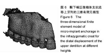
2.1 三维有限元模型的建立 成功建立颧下嵴区微植体支抗远移上牙列三维有限元模型,由于双侧牙列对称,特简化模型,以颚骨中线将模型均分为两半,取右上为研究对象,共获节点43 682个、单元66 273个,模型体网格划分均匀、大小适当,模型符合解剖学形态。网格换分情况托槽、弓丝、上颌骨、牙齿及牙周膜单元数分别为2 101,409,9 580,30 509,4 936;节点数分别为1 985,196,10 345,31 005,151(图6)。"

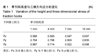
2.2 牵引钩高度与受力情况分析 由表1可知,随着牵引钩高度的增高,垂直方向应力逐渐增加,而与水平分力的变化无关。"

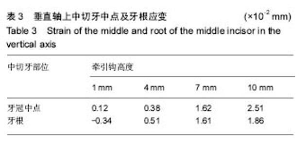
2.3 中切牙应变变化 由表2所知,随着牵引钩高度的增加,矢状轴上中切牙牙冠中点的应变逐渐降低,当牵引钩高度增加到7 mm和10 mm时,变为负值,而牙根应变的变化与前者正好相反,随着牵引钩高度增加,其应变增大,最大值为2.23×10⁻² mm。由表3所知,随着牵引钩高度的增加,垂直轴上中切牙牙冠中点和牙根的应变逐渐增大。依据应变变化推断,中切牙随着牵引钩高度的增加,由顺时针旋转变为逆时针旋转。"

"


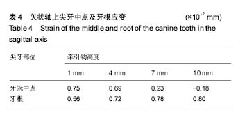
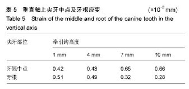
2.4 尖牙应变变化 由表4所知,随着牵引钩高度的增加,矢状轴上牙冠的应变逐渐降低,当牵引钩高度增加到 10 mm时,变为负值,而牙根应变的变化与前者正好相反,随着牵引钩高度增加,其应变增大,最大值为0.8×10⁻² mm。由表5所知,随着牵引钩高度的增加,垂直轴上牙冠中点的应变逐渐增大,而牙根应变逐渐减小。依据应变变化推断,尖牙与侧切牙一样,随着牵引钩高度的增加,由顺时针旋转变为逆时针旋转。"
"

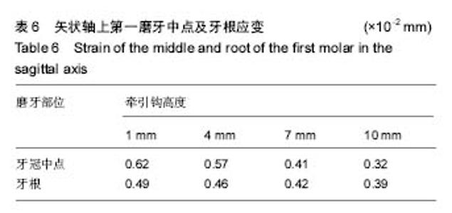
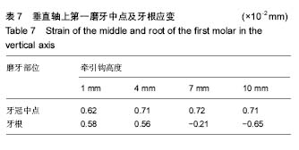
2.5 第一磨牙应变变化 由表6可见,随着牵引钩高度的增加,矢状轴上牙冠中点的应变逐渐降低,而牙根应变变化不大。由表7所知,随着牵引钩高度的增加,垂直轴上牙冠中点的应变变化不大,而牙根应变逐渐减小,当牵引钩高度为10 mm时,依据应变变化推断,尖牙与第一磨牙一样,随着牵引钩高度的增加,由顺时针旋转变为逆时针旋转。"


"
| [1]兰泽栋,林珠,龙红月,等.支抗种植体长度对骨界面应力分布的影响[J].中国口腔种植学杂志,2003,8(3):112-115.[2]Jensen OT,Adams MW,Smith E.Paranasal bone: the prime factor affecting the decision to use transsinus vs zygomatic implants for biomechanical support for immediate function in maxillary dental implant reconstruction.Int J Oral Maxillofac Implants.2014;29(1):e130-138. [3]张扬,张丹,冯翠娟.微小种植体正畸支抗生物力学的三维有限元分析[J].上海口腔医学,2005,14(3):281-283.[4]Lee HS, Choi HM, Choi DS,et al.Bone thickness of the infrazygomatic crest area in skeletal Class III growing patients:A computed tomographic study.Imaging Sci Dent. 2013;43(4):261-266.[5]王震东,王林,倪晓宇,等.不同长度微种植体支抗应力差异的三维有限元研究[J].口腔医学,2005,25(2):96-97.[6]刘慧娟.微种植体支抗后退整个上牙列时平面改变的三维有限元研究[D].中国医科大学,2012.[7]Lee HS,Choi HM,Choi DS,et al.Bone thickness of the infrazygomatic crest area in skeletal Class III growing patients: A computed tomographic study.Am J Orthod Dentofacial Orthop.2012;141(3):345-351. [8]陈立艳,刘志杰,苑芳连,等.颧压槽嵴区骨宽度及皮质骨厚度的CBCT测量分析[J].口腔医学研究,2017,33(6):637-641.[9]Lee NK,Baek SH.Stress and displacement between maxillary protraction with miniplates placed at the infrazygomatic crest and the lateral nasal wall: a 3-dimensional finite element analysis.Am J Orthod Dentofacial Orthop. 2012;141(3): 345-351.[10]张翼,张磊,樊瑜波,等.微植体支抗滑动法内收上颌前牙的三维有限元研究[J].华西口腔医学杂志,2009,27(5):557-560.[11]Hung KF,Ai QY,Fan SC,et al.Measurement of the zygomatic region for the optimal placement of quad zygomatic implants.Clin Implant Dent Relat Res.2017;19(5):841-848.[12]李琦.上颌骨微植体支抗正畸矫治系统数值分析研究[D].重庆医科大学,2007.[13]艾林,丁伟,倪龙兴.有限元法与口腔生物力学[J].临床口腔医学杂志,2005,21(5):318-319.[14]Butterworth CJ,Rogers SN.The zygomatic implant perforated (ZIP) flap: a new technique for combined surgical reconstruction and rapid fixed dental rehabilitation following low-level maxillectomy.Int J Implant Dent.2017;3(1):37. [15]毕振宇,刘阳,黄文华.有限元分析法在口腔生物力学领域的应用[J].解剖科学进展,2009,15(4):427-431.[16]Coppedê A,de Mayo T,de Sá Zamperlini M,et al.Three-year clinical prospective follow-up of extrasinus zygomatic implants for the rehabilitation of the atrophic maxilla.Clin Implant Dent Relat Res.2017;19(5):926-934.[17]车蓓,张昊,钱才梅,等.不同垂直骨面型安氏II类1分类错颌患者颞下颌关节三维形态结构的比较[J].中华口腔医学杂志, 2014, 49(7):399-402.[18][胡丽,林久祥,陈莉莉.基于颈椎骨龄定量分期法的安氏II类I分类错颌畸形患者最佳矫形时机的探讨.[J].临床口腔医学杂志. 2013,29(3):131-134.[19]陈文君,于海涛,古力巴哈买买提力.不同功能矫治安氏II类错颌下颌支抗丧失的对比研究[J].新疆医科大学学报, 2014,37(10): 1366-1368.[20]胡铮,谢雨菲,陆佩珺,等.颧牙槽嵴区域骨密度厚度的锥形束CT测量分析[J].国际口腔医学杂志,2014,41(3):281-285.[21]Billiet T,de-Pauw G,Dermaut L.Location of the centre of resistance of the upper dentition and the nasomaxillary complex:An Experimental study.Eur J Orthod. 2011;23(3): 263-273.[22]Chung Gm,Sung SJ,Lee LJ,et al.Finite element investigation of the center of resistance of the maxillary dentition korean J Orthod.2009;39(1):83-94.[23]Mozzati M,Mortellaro C,Arata V,et al.Rehabilitation with 4 zygomatic implants with a new surgical protocol using ultrasonic technique.J Craniofac Surg.2015,26(3):722-728.[24]Jensen OT, Adams MW, Smith E.Paranasal bone: the prime factor affecting the decision to use transsinus vs zygomatic implants for biomechanical support for immediate function in maxillary dental implant reconstruction.Int J Oral Maxillofac Implants.2014;29(1):e130-138.[25]Lee HS,Choi HM,Choi DS,et al.Bone thickness of the infrazygomatic crest area in skeletal Class III growing patients: A computed tomographic study.Imaging Sci Dent. 2013;43(4): 261-266.[26]Lee NK,Baek SH.Stress and displacement between maxillary protraction with miniplates placed at the infrazygomatic crest and the lateral nasal wall: a 3-dimensional finite element analysis.Am J Orthod Dentofacial Orthop. 2012;141(3): 345-351[27]Ishida Y,Ono T.Nonsurgical treatment of an adult with a skeletal Class II gummy smile using zygomatic temporary anchorage devices and improved superelastic nickel-titanium alloy wires.Am J Orthod Dentofacial Orthop. 2017;152(5): 693-705.[28]Benatto GB,Bueno CRS,Curvello VP,et al.Management of Zygomatic Fixture Complication Case Using Extra-Short Implants.J Craniofac Surg.2017;(8):e797-e799. |
| [1] | Xu Feng, Kang Hui, Wei Tanjun, Xi Jintao. Biomechanical analysis of different fixation methods of pedicle screws for thoracolumbar fracture [J]. Chinese Journal of Tissue Engineering Research, 2021, 25(9): 1313-1317. |
| [2] | Zhang Tongtong, Wang Zhonghua, Wen Jie, Song Yuxin, Liu Lin. Application of three-dimensional printing model in surgical resection and reconstruction of cervical tumor [J]. Chinese Journal of Tissue Engineering Research, 2021, 25(9): 1335-1339. |
| [3] | Chen Xinmin, Li Wenbiao, Xiong Kaikai, Xiong Xiaoyan, Zheng Liqin, Li Musheng, Zheng Yongze, Lin Ziling. Type A3.3 femoral intertrochanteric fracture with augmented proximal femoral nail anti-rotation in the elderly: finite element analysis of the optimal amount of bone cement [J]. Chinese Journal of Tissue Engineering Research, 2021, 25(9): 1404-1409. |
| [4] | Zhou Jihui, Li Xinzhi, Zhou You, Huang Wei, Chen Wenyao. Multiple problems in the selection of implants for patellar fracture [J]. Chinese Journal of Tissue Engineering Research, 2021, 25(9): 1440-1445. |
| [5] | Zeng Yanhua, Hao Yanlei. In vitro culture and purification of Schwann cells: a systematic review [J]. Chinese Journal of Tissue Engineering Research, 2021, 25(7): 1135-1141. |
| [6] | Xu Yulin, Shen Shi, Zhuo Naiqiang, Yang Huilin, Yang Chao, Li Yang, Zhao Heng, Zhao Lu. Biomechanical comparison of three different plate fixation methods for acetabular posterior column fractures in standing and sitting positions [J]. Chinese Journal of Tissue Engineering Research, 2021, 25(6): 826-830. |
| [7] | Cai Qunbin, Zou Xia, Hu Jiantao, Chen Xinmin, Zheng Liqin, Huang Peizhen, Lin Ziling, Jiang Ziwei. Relationship between tip-apex distance and stability of intertrochanteric femoral fractures with proximal femoral anti-rotation nail: a finite element analysis [J]. Chinese Journal of Tissue Engineering Research, 2021, 25(6): 831-836. |
| [8] | Song Chengjie, Chang Hengrui, Shi Mingxin, Meng Xianzhong. Research progress in biomechanical stability of lateral lumbar interbody fusion [J]. Chinese Journal of Tissue Engineering Research, 2021, 25(6): 923-928. |
| [9] | Liu Zhao, Xu Xilin, Shen Yiwei, Zhang Xiaofeng, Lü Hang, Zhao Jun, Wang Zhengchun, Liu Xuzhuo, Wang Haitao. Guiding role and prospect of staging and classification combined collapse prediction method for osteonecrosis of femoral head [J]. Chinese Journal of Tissue Engineering Research, 2021, 25(6): 929-934. |
| [10] | Xie Chongxin, Zhang Lei. Comparison of knee degeneration after anterior cruciate ligament reconstruction with or without remnant preservation [J]. Chinese Journal of Tissue Engineering Research, 2021, 25(5): 735-740. |
| [11] | Xu Dongzi, Zhang Ting, Ouyang Zhaolian. The global competitive situation of cardiac tissue engineering based on patent analysis [J]. Chinese Journal of Tissue Engineering Research, 2021, 25(5): 807-812. |
| [12] | Wu Zijian, Hu Zhaoduan, Xie Youqiong, Wang Feng, Li Jia, Li Bocun, Cai Guowei, Peng Rui. Three-dimensional printing technology and bone tissue engineering research: literature metrology and visual analysis of research hotspots [J]. Chinese Journal of Tissue Engineering Research, 2021, 25(4): 564-569. |
| [13] | Chang Wenliao, Zhao Jie, Sun Xiaoliang, Wang Kun, Wu Guofeng, Zhou Jian, Li Shuxiang, Sun Han. Material selection, theoretical design and biomimetic function of artificial periosteum [J]. Chinese Journal of Tissue Engineering Research, 2021, 25(4): 600-606. |
| [14] | Liu Fei, Cui Yutao, Liu He. Advantages and problems of local antibiotic delivery system in the treatment of osteomyelitis [J]. Chinese Journal of Tissue Engineering Research, 2021, 25(4): 614-620. |
| [15] | Li Xiaozhuang, Duan Hao, Wang Weizhou, Tang Zhihong, Wang Yanghao, He Fei. Application of bone tissue engineering materials in the treatment of bone defect diseases in vivo [J]. Chinese Journal of Tissue Engineering Research, 2021, 25(4): 626-631. |
| Viewed | ||||||
|
Full text |
|
|||||
|
Abstract |
|
|||||