Chinese Journal of Tissue Engineering Research ›› 2018, Vol. 22 ›› Issue (17): 2692-2698.doi: 10.3969/j.issn.2095-4344.0482
Previous Articles Next Articles
Human umbilical cord blood-derived mesenchymal stem cells differentiate into neuron-like cells induced by combination of mouse nerve growth factor and brain-derived neurotrophic factor
Chen Jun, Yang Zi-jin, Li Hong-mei
- Department of Pediatrics, Lianyungang Hospital Affiliated to Xuzhou Medical University (First People’s Hospital of Lianyungang), Lianyungang 222002, Jiangsu Province, China
-
Revised:2017-11-25Online:2018-06-18Published:2018-06-18 -
Contact:Yang Zi-jin, Chief physician, Master’s supervisor, Department of Pediatrics, Lianyungang Hospital Affiliated to Xuzhou Medical University (First People’s Hospital of Lianyungang), Lianyungang 222002, Jiangsu Province, China -
About author:Chen Jun, Master candidate, Department of Pediatrics, Lianyungang Hospital Affiliated to Xuzhou Medical University (First People’s Hospital of Lianyungang), Lianyungang 222002, Jiangsu Province, China -
Supported by:the Key Division Construction of Lianyungang Municipal Science and Technology Department, No. SH1117
CLC Number:
Cite this article
Chen Jun, Yang Zi-jin, Li Hong-mei. Human umbilical cord blood-derived mesenchymal stem cells differentiate into neuron-like cells induced by combination of mouse nerve growth factor and brain-derived neurotrophic factor[J]. Chinese Journal of Tissue Engineering Research, 2018, 22(17): 2692-2698.
share this article
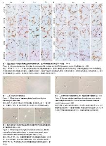
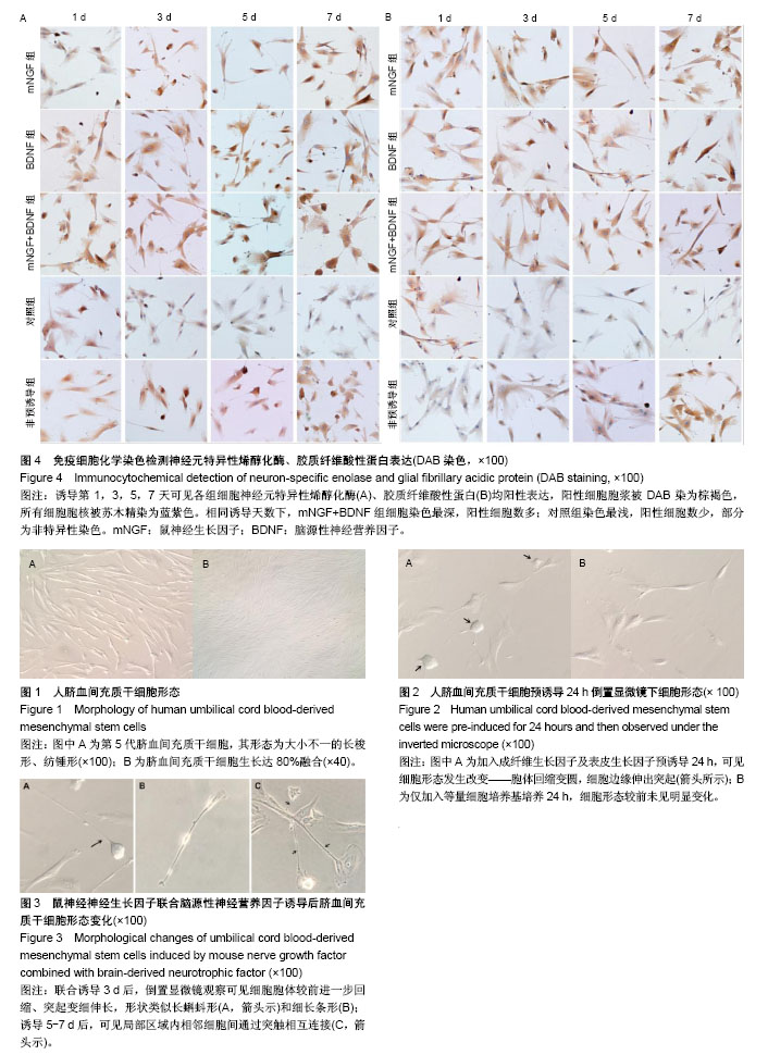
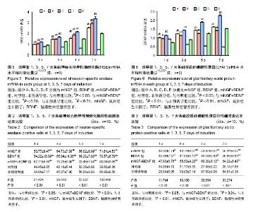
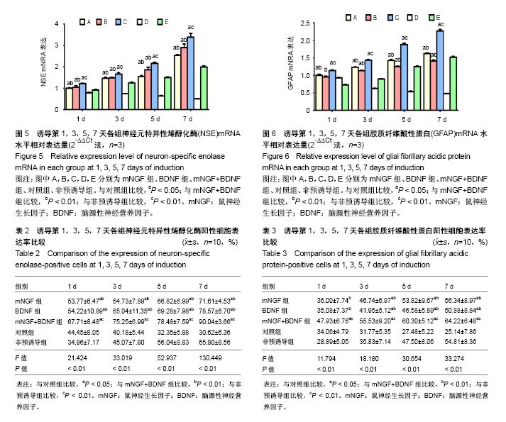
2.1 脐血间充质干细胞的形态特征 倒置显微镜下观察第5代脐血间充质干细胞,其形态为大小不一的长梭形、纺锤形(图1A)。传代培养五六天细胞可达80%融合状态,此时镜下观察可见细胞集落呈漩涡状、放射状生长(图1B)。 2.2 诱导过程中脐血间充质干细胞形态学变化 加入成纤维生长因子及表皮生长因子预诱导24 h后,细胞胞体回缩变圆,细胞边缘伸出单个或多个突起,而未经预诱导的脐血间充质干细胞则未见明显形态变化(图2)。正式诱导3 d后,细胞胞体以胞核为中心进一步回缩,细胞突起逐渐变细伸长,形态类似长蝌蚪形、细长条形,呈类神经元样细胞改变;正式诱导5-7 d后,可见局部区域内相邻细胞间通过突触相互连接。各实验组细胞有类似的形态变化,但联合诱导组的细胞形态改变最为显著(图3)。在7 d诱导时间内所有实验组细胞生长状态均良好,未见明显的细胞脱落、死亡现象。 2.3 免疫细胞化学染色鉴定 对诱导第1,3,5,7天各实验组细胞行免疫细胞化学染色,发现各实验组细胞NSE、GFAP均阳性表达,表明经诱导后脐血间充质干细胞向神经元样细胞和星形胶质细胞分化。对相同诱导天数下各组细胞染色情况进行观察对比,发现mNGF+BDNF组细胞染色最深,NSE、GFAP阳性细胞多于mNGF组、BDNF组,对照组染色最浅,阳性细胞数较少,部分为非特异性染色(图4)。 对比各实验组细胞NSE、GFAP阳性率(表2,3),发现在7 d诱导时间内,mNGF组、BDNF组、mNGF+BDNF组、非预诱导组NSE、GFAP细胞阳性率均呈逐渐上升趋势,而对照组则逐渐下降;在诱导天数相同的情况下,mNGF+BDNF组NSE、GFAP细胞阳性率均高于mNGF组、BDNF组以及对照组,且高于非预诱导组,差异均有显著性意义(P < 0.05)。 2.4 实时荧光定量PCR检测结果 各实验组细胞NSE mRNA和GFAP mRNA在诱导第1,3,5,7天时均有表达,且mNGF组、BDNF组、mNGF+BDNF组、非预诱导组二者表达量均呈上升趋势,在第7天时表达量最高,而对照组表达量则逐渐降低,总体趋势同免疫细胞化学染色结果相一致。相同诱导天数下mNGF+BDNF组NSE mRNA和GFAP mRNA的表达量高于mNGF组、BDNF组以及对照组,差异有显著性意义(P < 0.05)。另外,进行预诱导的mNGF+BDNF组NSE mRNA和GFAP mRNA的表达量明显高于非预诱导组,差异有显著性意义(P < 0.05),见图5,6。"
"
| [1] Singh S, Srivastava A, Srivastava P, et al. Advances in Stem Cell Research- A Ray of Hope in Better Diagnosis and Prognosis in Neurodegenerative Diseases. Front Mol Biosci. 2016;3:72.[2] Mine Y, Tatarishvili J, Oki K, et al. Grafted human neural stem cells enhance several steps of endogenous neurogenesis and improve behavioral recovery after middle cerebral artery occlusion in rats. Neurobiol Dis. 2013;52:191-203.[3] 刘俊华,王大斌,顾教伟,等. 神经干细胞移植治疗脑性瘫痪:神经修复的效果和安全性评估[J]. 中国组织工程研究, 2015,19(19):3032-3036.[4] Ager RR, Davis JL, Agazaryan A, et al. Human neural stem cells improve cognition and promote synaptic growth in two complementary transgenic models of Alzheimer's disease and neuronal loss. Hippocampus. 2015;25(7):813-826.[5] Taran R, Mamidi MK, Singh G, et al. In vitro and in vivo neurogenic potential of mesenchymal stem cells isolated from different sources. J Biosci. 2014;39(1):157-169.[6] Lunn JS, Sakowski SA, Feldman EL. Concise review: Stem cell therapies for amyotrophic lateral sclerosis: recent advances and prospects for the future. Stem Cells. 2014;32(5):1099-1109.[7] Zhu Y, Guan YM, Huang HL, et al. Human umbilical cord blood mesenchymal stem cell transplantation suppresses inflammatory responses and neuronal apoptosis during early stage of focal cerebral ischemia in rabbits. Acta Pharmacol Sin. 2014;35(5):585-591.[8] Wang L, Lu M. Regulation and direction of umbilical cord blood mesenchymal stem cells to adopt neuronal fate. Int J Neurosci. 2014;124(3):149-159.[9] Shahbazi A, Safa M, Alikarami F, et al. Rapid Induction of Neural Differentiation in Human Umbilical Cord Matrix Mesenchymal Stem Cells by cAMP-elevating Agents. Int J Mol Cell Med. 2016;5(3): 167-177.[10] Rafieemehr H, Kheirandish M, Soleimani M. Improving the neuronal differentiation efficiency of umbilical cord blood-derived mesenchymal stem cells cultivated under appropriate conditions. Iran J Basic Med Sci. 2015;18(11):1100-1106.[11] Zeng Y, Rong M, Liu Y, et al. Electrophysiological characterisation of human umbilical cord blood-derived mesenchymal stem cells induced by olfactory ensheathing cell-conditioned medium. Neurochem Res. 2013;38(12):2483-2489.[12] Hei WH, Almansoori AA, Sung MA, et al. Adenovirus vector-mediated ex vivo gene transfer of brain-derived neurotrophic factor (BDNF) tohuman umbilical cord blood-derived mesenchymal stem cells (UCB-MSCs) promotescrush-injured rat sciatic nerve regeneration. Neurosci Lett. 2017;643:111-120.[13] Nan C, Guo L, Zhao Z, et al. Tetramethylpyrazine induces differentiation of human umbilical cord-derived mesenchymal stem cells into neuron-like cells in vitro. Int J Oncol. 2016;48(6):2287-2294.[14] 杨自金,郭佳丽,卢思广,等.人脐血间充质干细胞移植联合神经节苷脂注射治疗脑性瘫痪[J]. 中国组织工程研究, 2016, 20(19):2803-2809.[15] Can A, Balci D. Isolation, culture, and characterization of human umbilical cord stroma-derived mesenchymal stem cells. Methods Mol Biol. 2011;698:51-62.[16] Khoo ML, Shen B, Tao H, et al. Long-term serial passage and neuronal differentiation capability of human bone marrow mesenchymal stem cells. Stem Cells Dev. 2008;17(5):883-896.[17] Duan P, Sun S, Li B, et al. miR-29a modulates neuronal differentiation through targeting REST in mesenchymal stem cells. PLoS One. 2014;9(5):e97684.[18] Zhuang H, Zhang R, Zhang S, et al. Altered expression of microRNAs in the neuronal differentiation of human Wharton's Jelly mesenchymal stem cells. Neurosci Lett. 2015;600:69-74.[19] Paczkowska E, ?uczkowska K, Piecyk K, et al. The influence of BDNF on human umbilical cord blood stem/progenitor cells: implications for stem cell-based therapy of neurodegenerative disorders. Acta Neurobiol Exp (Wars). 2015;75(2):172-191.[20] Takemoto T, Ishihara Y, Ishida A, et al. Neuroprotection elicited by nerve growth factor and brain-derived neurotrophic factor released from astrocytes in response to methylmercury. Environ Toxicol Pharmacol. 2015;40(1):199-205.[21] Shi W, Huang CJ, Xu XD, et al. Transplantation of RADA16-BDNF peptide scaffold with human umbilical cord mesenchymal stem cells forced with CXCR4 and activated astrocytes for repair of traumatic brain injury. Acta Biomater. 2016;45:247-261.[22] Ji HY, Kim MS, Lee MY, et al. GABAergic neuronal differentiation induced by brain-derived neurotrophic factor in human mesenchymal stem cells. Animal Cells & Systems. 2014; 18(1):17-24.[23] Zhao J, Cheng YY, Fan W, et al. Botanical drug puerarin coordinates with nerve growth factor in the regulation of neuronal survival and neuritogenesis via activating ERK1/2 and PI3K/Akt signaling pathways in the neurite extension process. CNS Neurosci Ther. 2015;21(1):61-70.[24] 郝冬荣,厉红,庞保东.鼠神经生长因子治疗小儿面神经炎42例[J]. 中国药业,2014, 23(1):82-84.[25] 许马利,王杨. 鼠神经生长因子治疗新生儿缺氧缺血性脑病的Meta分析[J]. 中国临床药理学杂志,2016, 32(7):652-654.[26] 李巧秀,张俊清,翟红印,等. 鼠神经生长因子运动区注射治疗小儿脑瘫疗效观察[J]. 中国妇幼保健,2015, 30(28):4889-4891.[27] Liu YR1, Liu Q. Meta-analysis of mNGF therapy for peripheral nerve injury: a systematic review. Chin J Traumatol. 2012;15(2):86-91.[28] Salehinejad P, Alitheen NB, Ali AM, et al. Neural differentiation of human umbilical cord matrix-derived mesenchymal cells under special culture conditions. Cytotechnology. 2015;67(3):449-460.[29] Huat TJ, Khan AA, Pati S, et al. IGF-1 enhances cell proliferation and survival during early differentiation of mesenchymal stem cells to neural progenitor-like cells. BMC Neurosci. 2014;15:91.[30] Rafieemehr H, Kheirandish M, Soleimani M. A new two-step induction protocol for neural differentiation of human umbilical cord blood-derived mesenchymal stem cells. Iranian Journal of Blood & Cancer. 2015; 7(2):111-116.[31] Sanalkumar R, Vidyanand S, Lalitha Indulekha C, et al. Neuronal vs. glial fate of embryonic stem cell-derived neural progenitors (ES-NPs) is determined by FGF2/EGF during proliferation. J Mol Neurosci. 2010;42(1):17-27.[32] Salehinejad P, Alitheen NB, Ali AM, et al. Neural differentiation of human umbilical cord matrix-derived mesenchymal cells under special culture conditions. Cytotechnology. 2015;67(3):449-460.[33] Lim JY, Park SI, Kim SM, et al. Neural differentiation of brain-derived neurotrophic factor-expressing human umbilical cord blood-derived mesenchymal stem cells in culture via TrkB-mediated ERK and β-catenin phosphorylation and following transplantation into the developing brain. Cell Transplant. 2011;20(11-12):1855-1866.[34] Lim JY, Park SI, Oh JH, et al. Brain-derived neurotrophic factor stimulates the neural differentiation of human umbilical cord blood-derived mesenchymal stem cells and survival of differentiated cells through MAPK/ERK and PI3K/Akt-dependent signaling pathways. J Neurosci Res. 2008;86(10):2168-2178.[35] Santa-Olalla J, Covarrubias L. Basic fibroblast growth factor promotes epidermal growth factor responsiveness and survival of mesencephalic neural precursor cells. J Neurobiol. 1999;40(1):14-27.[36] Sanchez-Ramos J, Song S, Cardozo-Pelaez F, et al. Adult bone marrow stromal cells differentiate into neural cells in vitro. Exp Neurol. 2000;164(2):247-256. |
| [1] | Lin Qingfan, Xie Yixin, Chen Wanqing, Ye Zhenzhong, Chen Youfang. Human placenta-derived mesenchymal stem cell conditioned medium can upregulate BeWo cell viability and zonula occludens expression under hypoxia [J]. Chinese Journal of Tissue Engineering Research, 2021, 25(在线): 4970-4975. |
| [2] | Pu Rui, Chen Ziyang, Yuan Lingyan. Characteristics and effects of exosomes from different cell sources in cardioprotection [J]. Chinese Journal of Tissue Engineering Research, 2021, 25(在线): 1-. |
| [3] | Zhang Tongtong, Wang Zhonghua, Wen Jie, Song Yuxin, Liu Lin. Application of three-dimensional printing model in surgical resection and reconstruction of cervical tumor [J]. Chinese Journal of Tissue Engineering Research, 2021, 25(9): 1335-1339. |
| [4] | Hou Jingying, Yu Menglei, Guo Tianzhu, Long Huibao, Wu Hao. Hypoxia preconditioning promotes bone marrow mesenchymal stem cells survival and vascularization through the activation of HIF-1α/MALAT1/VEGFA pathway [J]. Chinese Journal of Tissue Engineering Research, 2021, 25(7): 985-990. |
| [5] | Shi Yangyang, Qin Yingfei, Wu Fuling, He Xiao, Zhang Xuejing. Pretreatment of placental mesenchymal stem cells to prevent bronchiolitis in mice [J]. Chinese Journal of Tissue Engineering Research, 2021, 25(7): 991-995. |
| [6] | Liang Xueqi, Guo Lijiao, Chen Hejie, Wu Jie, Sun Yaqi, Xing Zhikun, Zou Hailiang, Chen Xueling, Wu Xiangwei. Alveolar echinococcosis protoscolices inhibits the differentiation of bone marrow mesenchymal stem cells into fibroblasts [J]. Chinese Journal of Tissue Engineering Research, 2021, 25(7): 996-1001. |
| [7] | Fan Quanbao, Luo Huina, Wang Bingyun, Chen Shengfeng, Cui Lianxu, Jiang Wenkang, Zhao Mingming, Wang Jingjing, Luo Dongzhang, Chen Zhisheng, Bai Yinshan, Liu Canying, Zhang Hui. Biological characteristics of canine adipose-derived mesenchymal stem cells cultured in hypoxia [J]. Chinese Journal of Tissue Engineering Research, 2021, 25(7): 1002-1007. |
| [8] | Geng Yao, Yin Zhiliang, Li Xingping, Xiao Dongqin, Hou Weiguang. Role of hsa-miRNA-223-3p in regulating osteogenic differentiation of human bone marrow mesenchymal stem cells [J]. Chinese Journal of Tissue Engineering Research, 2021, 25(7): 1008-1013. |
| [9] | Lun Zhigang, Jin Jing, Wang Tianyan, Li Aimin. Effect of peroxiredoxin 6 on proliferation and differentiation of bone marrow mesenchymal stem cells into neural lineage in vitro [J]. Chinese Journal of Tissue Engineering Research, 2021, 25(7): 1014-1018. |
| [10] | Zhu Xuefen, Huang Cheng, Ding Jian, Dai Yongping, Liu Yuanbing, Le Lixiang, Wang Liangliang, Yang Jiandong. Mechanism of bone marrow mesenchymal stem cells differentiation into functional neurons induced by glial cell line derived neurotrophic factor [J]. Chinese Journal of Tissue Engineering Research, 2021, 25(7): 1019-1025. |
| [11] | Duan Liyun, Cao Xiaocang. Human placenta mesenchymal stem cells-derived extracellular vesicles regulate collagen deposition in intestinal mucosa of mice with colitis [J]. Chinese Journal of Tissue Engineering Research, 2021, 25(7): 1026-1031. |
| [12] | Pei Lili, Sun Guicai, Wang Di. Salvianolic acid B inhibits oxidative damage of bone marrow mesenchymal stem cells and promotes differentiation into cardiomyocytes [J]. Chinese Journal of Tissue Engineering Research, 2021, 25(7): 1032-1036. |
| [13] | Li Cai, Zhao Ting, Tan Ge, Zheng Yulin, Zhang Ruonan, Wu Yan, Tang Junming. Platelet-derived growth factor-BB promotes proliferation, differentiation and migration of skeletal muscle myoblast [J]. Chinese Journal of Tissue Engineering Research, 2021, 25(7): 1050-1055. |
| [14] | Wang Feng, Zhou Liyu, Saijilafu, Qi Shibin, Ma Yanxia, Wei Shanwen. CaMKII-Smad1 promotes axonal regeneration of peripheral nerves [J]. Chinese Journal of Tissue Engineering Research, 2021, 25(7): 1064-1068. |
| [15] | Wang Xianyao, Guan Yalin, Liu Zhongshan. Strategies for improving the therapeutic efficacy of mesenchymal stem cells in the treatment of nonhealing wounds [J]. Chinese Journal of Tissue Engineering Research, 2021, 25(7): 1081-1087. |
| Viewed | ||||||
|
Full text |
|
|||||
|
Abstract |
|
|||||