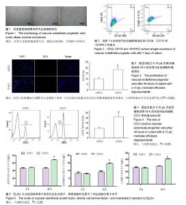
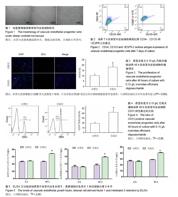
| [1] 李劼慧,王琦,杨秀滨,等. 不完全纠正心肌缺血对接受血管重建术患者长期预后的影响[J]. 中国循环杂志, 2016, 31(11):1051-1055.[2] 倪明科,林琍,宗文霞,等. 心脏康复治疗对缺血性心肌病室性心律失常的影响[J]. 中国康复医学杂志, 2016, 31(7):770-774.[3] 赵庆斌,刘丹丹,周娟. 糖尿病心肌缺血大鼠骨髓干细胞动员及心肌组织细胞因子的表达[J]. 西安交通大学学报:医学版, 2017, 38(1):29-33.[4] 张美华,张伟,盖凌,等. 血管内皮祖细胞在治疗动脉粥样硬化中的作用[J]. 中国心血管杂志, 2012, 17(1):73-75.[5] 张硕,杜怡斌,杜公文,等. 骨髓源性EPCs对脊髓源性NSCs增殖分化的影响[J]. 安徽医科大学学报,2015,50(1):20-24.[6] Loukovaara S, Gucciardo E, Repo P, et al. Indications of lymphatic endothelial differentiation and endothelial progenitor cell activation in the pathology of proliferative diabetic retinopathy. Acta Ophthalmol. 2015;93(6):512-523.[7] Sriram G, Tan JY, Islam I, et al. Efficient differentiation of human embryonic stem cells to arterial and venous endothelial cells under feeder- and serum-free conditions. Stem Cell Res Ther. 2015;6:261.[8] 王宁,周鹏,冯国清,等. 巴戟天糖链对缺氧复氧损伤内皮细胞的血管形成作用研究[J]. 中国药理学通报, 2011, 27(2):281-284.[9] Li YF, Gong ZH, Yang M, et al. Inhibition of the oligosaccharides extracted from Morinda officinalis, a Chinese traditional herbal medicine, on the corticosterone induced apoptosis in PC12 cells. Life Sci. 2003;72(8): 933-942.[10] Li YF, Yuan L, Xu YK, et al. Antistress effect of oligosaccharides extracted from Morinda officinalis in mice and rats. Acta Pharmacol Sin. 2001;22(12):1084-1088.[11] 王宁,周鹏,冯国清,等.巴戟天糖链对缺氧复氧损伤内皮细胞的血管形成作用研究[J]. 中国药理学通报, 2011,27(2):281-284.[12] 杨景靠,冯国清,于爽,等. 巴戟天醇提取物促大鼠缺血心肌治疗性血管生成的实验研究[J]. 中国药理学通报, 2010, 26(3): 367-371.[13] Yang Z, Yi Y, Gao C, et al. Isolation of inulin-type oligosaccharides from Chinese traditional medicine: Morinda officinalis How and their characterization using ESI-MS/MS. J Sep Sci. 2010;33(1):120-125.[14] Chang MS, Kim WN, Yang WM, et al. Cytoprotective effects of Morinda officinalis against hydrogen peroxide-induced oxidative stress in Leydig TM3 cells. Asian J Androl. 2008; 10(4):667-674.[15] Chen DL, Zhang P, Lin L, et al. Protective effect of Bajijiasu against β-amyloid-induced neurotoxicity in PC12 cells. Cell Mol Neurobiol. 2013;33(6):837-850.[16] 马阳,王红.血管内皮祖细胞增殖与迁移的研究进展[J]. 中国医学创新, 2016, 13(11):133-136.[17] 李文华,张群辉,戎浩,等. 内皮祖细胞与高血压病的应用研究进展[J]. 中国组织工程研究, 2016, 20(15):2273-2280.[18] 赵湜,王红祥,李宾公,等.糖尿病周围血管病血管内皮祖细胞数量和功能的改变及其机制探讨[J]. 中国糖尿病杂志, 2007, 15(1): 42-43.[19] 陈君柱,张芙荣,朱建华,等. 他汀类药物对外周血内皮祖细胞数量和功能的影响[J]. 中华心血管病杂志, 2004, 32(4):346-350.[20] 朱军慧,陈君柱,王兴祥,等. 氧化低密度脂蛋白对外周血内皮祖细胞数量和功能的影响[J]. 中国循环杂志, 2004, 19(s1):13-17.[21] 邹连勇,张鸿燕.巴戟天寡糖抗抑郁作用的研究进展[J].中国新药杂志,2012,21(16):1889-1891.[22] Heo JW, Cho JH, Jang JB, et al. Dose Dependent Effect of Morindae officinalis Radix Extract Solution on the Reproductive Capacities in the Mice. Journal of Oriental Obstetrics & Gynecology. 2005;18(3):17-31.[23] Yang X, Zhang YH, Ding CF, et al. Extract from Morindae officinalis against oxidative injury of function to human sperm membrane. Zhongguo Zhong Yao Za Zhi. 2006;31(19): 1614-1617.[24] Qiu ZK, Liu CH, Gao ZW, et al. The inulin-type oligosaccharides extract from morinda officinalis, a traditional Chinese herb, ameliorated behavioral deficits in an animal model of post-traumatic stress disorder. Metab Brain Dis. 2016;31(5):1143-1149.[25] 张中启,袁莉,赵楠,等. 巴戟天醇提取物的抗抑郁作用[J]. 中国药学杂志, 2000, 35(11):739-741.[26] 梁建辉,舒良,罗质璞,等. 巴戟天水提物治疗抑郁症临床疗效初探[J]. 中国中药杂志, 2002, 27(1):75-78.[27] 赖满香,阮志燕,许意平. 补肾中药巴戟天药理作用研究进展[J]. 亚太传统医药, 2017, 13(1):63-64.[28] Yang X, Zhang YH, Ding CF, et al. Extract from Morindae officinalis against oxidative injury of function to human sperm membrane. Zhongguo Zhong Yao Za Zhi. 2006;31(19): 1614-1617.[29] Bae WJ, Lee JM, Lee CH, et al. Effects of Allii Tuberosi Semen, Ginseng Radix Alba and Morindae Officinalis Radix Extract on Reproductive Capacities in Mice. Journal of Oriental Obstetrics & Gynecology. 2008;21(1):16-27.[30] 吴雪晖,罗飞,何清义,等. 血管内皮祖细胞研究进展[J]. 医学研究生学报, 2008, 21(9):981-985.[31] 黄伟剑,肖方毅,张怀勤, 等. 两种内皮祖细胞修复兔颈动脉内膜损伤血管的对比研究[J]. 中华医学杂志, 2007, 87(44): 3143-3147.[32] Zhang HL, Zhang QW, Zhang XQ, et al. Chemical Constituents from the Roots of Morinda officinalis. Chin J Nat Med. 2010; 8(3):192-195.[33] 何传波,李琳,汤凤霞,等.巴戟天多糖的提取及纯化研究[J].食品科学, 2008, 29(8):326-329.[34] 曹志伟. ST段抬高性急性心肌梗死患者循环血管内皮祖细胞及内皮生长因子与心功能的关系[J]. 中国药业, 2016, 25(17): 93-95.[35] 买超平,阎春生,哈小琴. 海拔高度对冠心病患者EPCs数量及功能的研究[J]. 中国循证心血管医学杂志, 2015,7(6):754-757.[36] 吕民,陈丽波,赵冬梅. 自体血管内皮祖细胞移植治疗高肺血流量肺动脉高压[J]. 中华实验外科杂志, 2015, 32(2):370-373.[37] 谢德波,何云,成小凤,等. 外周动脉反应性充血指数在冠心病患者血管内皮功能评价中的临床价值[J]. 第三军医大学学报, 2016, 38(19):2168-2173.[38] 代文静,张军,周敬群,等. 转化生长因子-α在小鼠颈动脉损伤血管修复中的作用[J]. 中国循环杂志, 2015,30(4):384-389. |