Chinese Journal of Tissue Engineering Research ›› 2018, Vol. 22 ›› Issue (1): 32-39.doi: 10.3969/j.issn.2095-4344.0408
Previous Articles Next Articles
Overexpression of stromal cell-derived factor-1 promotes the proliferation and migration of bone marrow mesenchymal stem cells in vitro
Chen Shao-qiang1, Wu Bi-lian2, Wang Shan-shan1, Huang Hai-hui1
- 1Department of Anatomy, Histology and Embryology, Fujian Medical University, Minhou 350122, Fujian Province, China; 2Department of Human Anatomy, Fujian Health College, Minhou 350101, Fujian Province, China
-
Revised:2017-10-09Online:2018-01-08Published:2018-01-08 -
About author:Chen Shao-qiang, M.D., Professor, Department of Anatomy, Histology and Embryology, Fujian Medical University, Minhou 350122, Fujian Province, China -
Supported by:the Natural Science Foundation of Fujian Province, No. 2014J01332
CLC Number:
Cite this article
Chen Shao-qiang, Wu Bi-lian, Wang Shan-shan, Huang Hai-hui. Overexpression of stromal cell-derived factor-1 promotes the proliferation and migration of bone marrow mesenchymal stem cells in vitro[J]. Chinese Journal of Tissue Engineering Research, 2018, 22(1): 32-39.
share this article
Add to citation manager EndNote|Reference Manager|ProCite|BibTeX|RefWorks

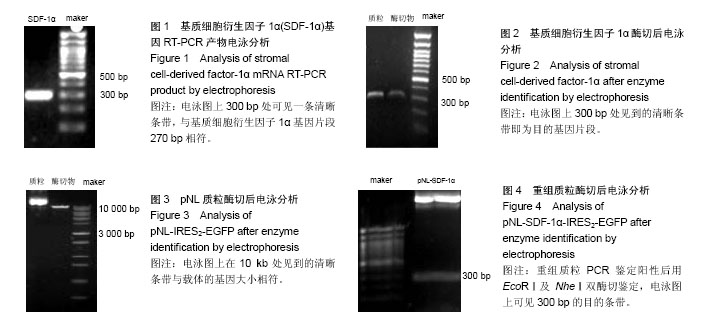
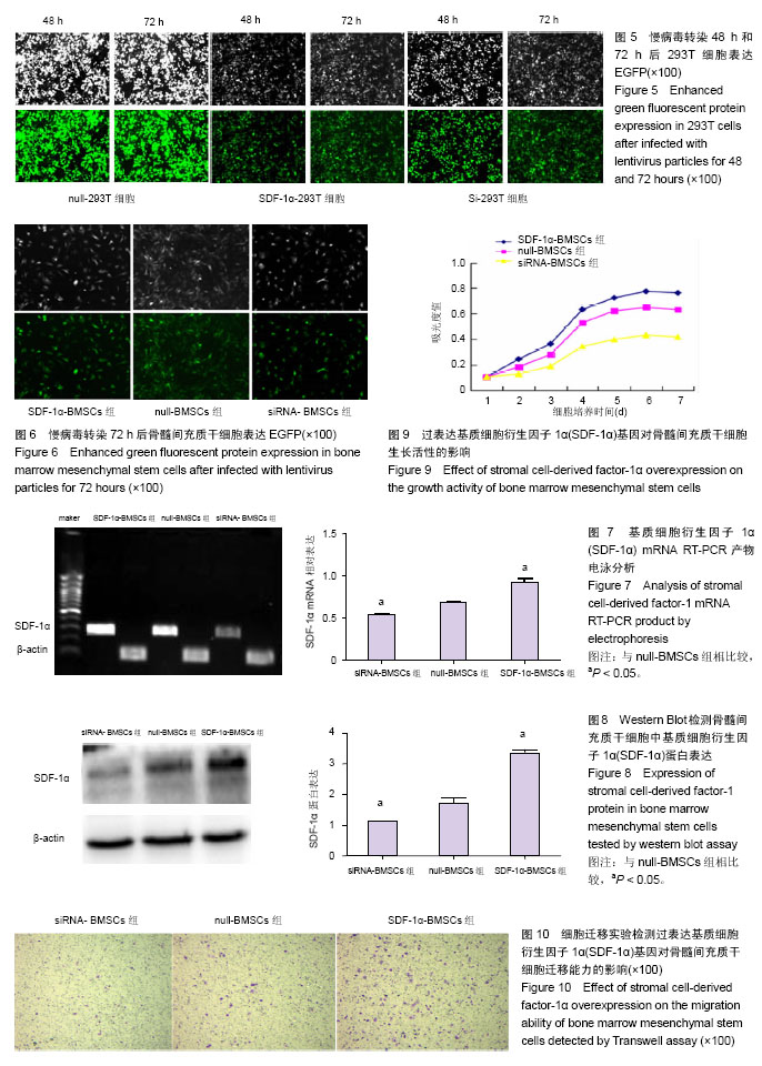
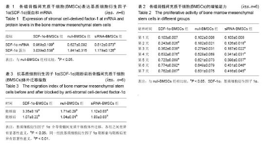
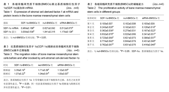
2.1 SDF-1α目的基因的获取结果 所提取大鼠肝脏组织总RNA的A260/A280为1.96,RNA质量浓度为2 168 mg/L。1.0%琼脂糖凝胶电泳后可以清楚地看到3条条带,分别为28s rRNA、18s rRNA及5s rRNA。RT-PCR后行1.0%琼脂糖凝胶电泳检测,电泳图上在300 bp处可见一条明亮的清晰条带,与预期的特异SDF-1α基因片段270 bp相符(图1)。 2.2 过表达SDF-1α质粒的构建与鉴定结果 SDF-1α基因及pNL-IRES2-EGFP质粒用EcoRⅠ及NheⅠ双酶切后,分别行1.0%及0.8%琼脂糖凝胶电泳,电泳图上在300 bp和10 kb处可见两条明亮的清晰条带(图2,3),300 bp处条带即为目的基因片段,10 kb处的条带与载体的基因大小相符。连接产物转化后挑取克隆,进行PCR初步鉴定,电泳图结果显示,在300 bp处可见一条明亮的清晰条带,与预期的特异SDF-1α基因片段270 bp相符。重组质粒PCR鉴定阳性后用EcoRⅠ及NheⅠ双酶切鉴定,电泳图上可见300 bp的目的条带及10 kb的载体条带(图4)。将重组质粒送上海铂尚生物技术有限公司进行测序鉴定,测序结果与SDF-1α基因序列,即从起始密码ATG到终止密码TAA序列完全相同。 2.3 骨髓间充质干细胞的分离、培养和鉴定结果 培养24 h后细胞逐渐贴壁,其他血细胞通过换液逐渐除去,此时贴壁细胞共同特点是呈单核细胞状,细胞形态不一,可有梭形、多角形或星芒状突起,为单个或多个细胞的克隆,细胞增殖迅速。培养12-14 d,细胞90%以上融合,融合细胞都呈梭形,原代可获得(1.0-2.0)×106个骨髓间充质干细胞。体外扩增5代后细胞数约为1×10¹¹个。流式细胞仪检测10份第2代细胞样本,结果显示:CD34、 CD45表达阴性,CD29、CD90表达阳性。 2.4 慢病毒SDF-1α基因载体感染细胞及过表达SDF-1α骨髓间充质干细胞系的建立情况 3种质粒共同转染293T细胞后,可在倒置荧光显微镜下观察到大量293T细胞表达EGFP,EGFP阳性率可达95%以上(图5)。病毒感染骨髓间充质干细胞72 h后,可在倒置荧光显微镜下观察到EGFP表达(图6),表明慢病毒转染载体成功将SDF-1α目的基因转入大鼠骨髓间充质干细胞。 RT-PCR检测结果显示,SDF-1α-BMSCs组SDF-1α mRNA的表达高于null-BMSCs组(P < 0.05),而siRNA-BMSCs组低于null-BMSCs组(P < 0.05),各组骨髓间充质干细胞表达SDF-1α mRNA情况见表1和图7。 Western Blot检测结果显示,SDF-1α-BMSCs组SDF-1α蛋白分子的表达高于null-BMSCs组(P < 0.05),而siRNA- BMSCs组低于null-BMSCs组(P < 0.05),各组骨髓间充质干细胞表达SDF-1α蛋白情况见表1和图8。说明过表达SDF-1α基因能提高骨髓间充质干细胞中SDF-1α的表达,SDF-1α沉默能抑制其表达,成功建立过表达SDF-1α骨髓间充质干细胞系。 2.5 过表达SDF-1α对骨髓间充质干细胞增殖的影响 感染第1天时,各组吸光度值差异无显著性意义(P > 0.05),其余时间点各组吸光度值差异均有显著性意义(P < 0.05)。各组吸光度值见表2和图9,SDF-1α-BMSCs组高于null- BMSCs组(P < 0.05),而siRNA-BMSCs组低于null-BMSCs组(P < 0.05),说明过表达SDF-1α基因能提高骨髓间充质干细胞的增殖能力。 2.6 过表达SDF-1α对骨髓间充质干细胞迁移的影响 在Transwell小室上进行体外迁移实验中,聚碳酸酯膜下表面的迁移细胞用结晶紫染色后呈浅紫色,见图10。图中可见SDF-1α-BMSCs组迁移指数高于null-BMSCs组,而siRNA-BMSCs组低于null-BMSCs组,3者之间差异均有显著性意义(P < 0.01)。SDF-1α-BMSCs组及null-BMSCs组在抗SDF-1α多抗作用后迁移指数明显下降,与抗体阻断前差异有显著性意义(P < 0.05);而siRNA-BMSCs组抗体阻断后与阻断前迁移指数差异无显著性意义,见表3。"

"
"
| [1] Recio AC, Felter CE, Schneider AC, et al. High-voltage electrical stimulation for the management of stage III and IV pressure ulcers among adults with spinal cord injury: demonstration of its utility for recalcitrant wounds below the level of injury. J Spinal Cord Med. 2012;35(1):58-63.[2] Ayatollahi M, Salmani MK, Geramizadeh B, et al. Conditions to improve expansion of human mesenchymal stem cells based on rat samples. World J Stem Cells. 2012;4(1):1-8.[3] Lee Z, Dennis J, Alsberg E, et al. Imaging stem cell differentiation for cell-based tissue repair. Methods Enzymol. 2012;506:247-263.[4] Li TS, Komota T, Ohshima M, et al. TGF-beta induces the differentiation of bone marrow stem cells into immature cardiomyocytes. Biochem Biophys Res Commun. 2008; 366(4): 1074-1080.[5] Matsushima A, Kotobuki N, Tadokoro M, et al. In vivo osteogenic capability of human mesenchymal cells cultured on hydroxyapatite and on beta-tricalcium phosphate. Artif Organs. 2009;33(6):474-481.[6] Croft AP, Przyborski SA. Mesenchymal stem cells expressing neural antigens instruct a neurogenic cell fate on neural stem cells. Exp Neurol. 2009;216(2):329-341.[7] Brazelton TR, Rossi FM, Keshet GI, et al. From marrow to brain: expression of neuronal phenotypes in adult mice. Science. 2000;290(5497):1775-1779.[8] Mezey E, Chandross KJ, Harta G, et al. Turning blood into brain: cells bearing neuronal antigens generated in vivo from bone marrow. Science. 2000;290(5497):1779-1782.[9] Sasaki M, Honmou O, Akiyama Y, et al. Transplantation of an acutely isolated bone marrow fraction repairs demyelinated adult rat spinal cord axons. Glia. 2001;35(1):26-34.[10] Ban DX, Ning GZ, Feng SQ, et al. Combination of activated Schwann cells with bone mesenchymal stem cells: the best cell strategy for repair after spinal cord injury in rats. Regen Med. 2011;6(6):707-720.[11] Zhang YJ, Zhang W, Lin CG, et al. Neurotrophin-3 gene modified mesenchymal stem cells promote remyelination and functional recovery in the demyelinated spinal cord of rats. J Neurol Sci. 2012;313(1-2):64-74.[12] Alexanian AR, Fehlings MG, Zhang Z, et al. Transplanted neurally modified bone marrow-derived mesenchymal stem cells promote tissue protection and locomotor recovery in spinal cord injured rats. Neurorehabil Neural Repair. 2011;25(9):873-880.[13] 陈少强,林建华.移植骨髓间质干细胞在损伤脊髓内向神经元的定向分化[J].解剖学杂志,2012,35(3):282-286.[14] 陈少强,林建华.移植骨髓间质干细胞在损伤脊髓内向少突胶质细胞的定向分化[J].中华创伤骨科杂志,2012,14(9):795-799.[15] Chen S, Wu B, Lin J. Effect of intravenous transplantation of bone marrow mesenchymal stem cells on neurotransmitters and synapsins in rats with spinal cord injury. Neural Regen Res. 2012;7(19):1445-1453.[16] 陈少强,林建华.不同移植时间窗对静脉移植骨髓间质干细胞在大鼠损伤脊髓内存活和迁移的影响[J].解剖学杂志,2009,32(2): 190-194.[17] 陈少强,林建华.大鼠脊髓损伤后炎症反应对静脉移植骨髓基质干细胞存活和迁移的影响[J].中华创伤骨科杂志,2009,11(1):61-65.[18] 陈少强,林建华.静脉移植骨髓间充质干细胞对大鼠脊髓损伤后少突胶质细胞再髓鞘化的影响[J].中华实验外科杂志,2009, 26(1): 134.[19] 陈少强,林建华.羧基荧光素乙酰乙酸对大鼠骨髓间质干细胞体外染色的研究[J].福建医科大学学报,2008,42(6):482-486.[20] 陈少强,林建华.骨髓间充质干细胞移植治疗脊髓损伤的研究[J].中国修复重建外科杂志,2008,21(5):507-511.[21] 贾小力,陈少强.Sofast 介导增强型绿色荧光蛋白基因转染骨髓间质干细胞[J].解剖学杂志,2008,31(5):640-642.[22] 吴碧莲,贾小力,陈少强.静脉注射骨髓间质干细胞可抑制大鼠损伤脊髓水通道蛋白-4的表达[J].解剖学杂志,2008,31(5):691-693.[23] 陈少强,吴碧莲,贾小力,等.骨髓基质干细胞旁分泌作用促进大鼠损伤脊髓的血管新生[J].中华创伤骨科杂志,2015,17(3):257-261.[24] Liu X, Duan B, Cheng Z, et al. SDF-1/CXCR4 axis modulates bone marrow mesenchymal stem cell apoptosis, migration and cytokine secretion. Protein Cell. 2011;2(10):845-854.[25] 盛瑾,夏宇,许官学,等.SDF-1/CXCR4轴在MSCs移植促进SD大鼠急性心肌梗死心脏功能恢复中的作用[J].中华医学杂志,2015, 95(18):1421-1424.[26] 陈军,张凯伦,杜心灵,等.SDF-1/CXCR4 轴介导大鼠骨髓间充质干细胞向心肌梗死组织迁移的体外实验研究[J].华中科技大学学报, 2008,27(6):745-748.[27] 孙立影,韩明子.基质细胞衍生因子-1对骨髓间充质干细胞的趋化作用[J].世界华人消化杂志,2008,17(9):992-997.[28] Zhao X, Qian D, Wu N, et al. The spleen recruits endothelial progenitor cell via SDF-1/CXCR4 axis in mice. J Recept Signal Transduct Res. 2010;30(4):246-254.[29] Yu J, Li M, Qu Z, et al. SDF-1/CXCR4-mediated migration of transplanted bone marrow stromal cells toward areas of heart myocardial infarction through activation of PI3K/Akt.J Cardiovasc Pharmacol. 2010;55(5):496-505.[30] Theiss HD, Vallaster M, Rischpler C, et al. Dual stem cell therapy after myocardial infarction acts specifically by enhanced homing via the SDF-1/CXCR4 axis. Stem Cell Res. 2011;7(3): 244-255. [31] Moriya C, Shioda T, Tashiro K, et al. Large quantity production with extreme convenience of human SDF-1alpha and SDF-1beta by a Sendai virus vector. FEBS Lett. 1998;425(1): 105-111.[32] Yu L, Cecil J, Peng SB, et al. Identification and expression of novel isoforms of human stromal cell-derived factor 1. Gene. 2006;374:174-179.[33] Lu D, Li Y, Wang L, et al. Intraarterial administration of marrow stromal cells in a rat model of traumatic brain injury. J Neurotrauma. 2001;18(8):813-819.[34] Chen J, Li Y, Wang L, et al. Therapeutic benefit of intracerebral transplantation of bone marrow stromal cells after cerebral ischemia in rats. J Neurol Sci. 2001;189(1-2):49-57.[35] Mahmood A, Lu D, Wang L, et al. Treatment of traumatic brain injury in female rats with intravenous administration of bone marrow stromal cells. Neurosurgery. 2001;49(5):1196-1203.[36] 李妍,杜红延,李红卫.慢病毒载体及其在RNA干扰技术中的应用与发展[J].分子诊断与治疗杂志,2013,5(1):55-58.[37] Meloche S, Pouysségur J. The ERK1/2 mitogen-activated protein kinase pathway as a master regulator of the G1- to S-phase transition. Oncogene. 2007;26(22):3227-3239.[38] Yoon S, Seger R. The extracellular signal-regulated kinase: multiple substrates regulate diverse cellular functions. Growth Factors. 2006;24(1):21-44.[39] Zhang W, Liu HT. MAPK signal pathways in the regulation of cell proliferation in mammalian cells. Cell Res. 2002;12(1):9-18.[40] 黄晓佳,李摇静,许摇潇.SDF-1促进原代培养大鼠星形胶质细胞增殖的作用[J].中国药理学通报,2014,30(9):1219-1224.[41] 李明峰,乔建林,曾今宇.SDF -1/CXCR4信号通路在造血干细胞归巢中作用的研究[J].国际输血及血液病杂志,2016,39(4):338-340.[42] He H, Zhao ZH, Han FS, et al. Activation of protein kinase C ε enhanced movement ability and paracrine function of rat bone marrow mesenchymal stem cells partly at least independent of SDF-1/CXCR4 axis and PI3K/AKT pathway. Int J Clin Exp Med. 2015;8(1):188-202. |
| [1] | Pu Rui, Chen Ziyang, Yuan Lingyan. Characteristics and effects of exosomes from different cell sources in cardioprotection [J]. Chinese Journal of Tissue Engineering Research, 2021, 25(在线): 1-. |
| [2] | Lin Qingfan, Xie Yixin, Chen Wanqing, Ye Zhenzhong, Chen Youfang. Human placenta-derived mesenchymal stem cell conditioned medium can upregulate BeWo cell viability and zonula occludens expression under hypoxia [J]. Chinese Journal of Tissue Engineering Research, 2021, 25(在线): 4970-4975. |
| [3] | Zhang Tongtong, Wang Zhonghua, Wen Jie, Song Yuxin, Liu Lin. Application of three-dimensional printing model in surgical resection and reconstruction of cervical tumor [J]. Chinese Journal of Tissue Engineering Research, 2021, 25(9): 1335-1339. |
| [4] | Hou Jingying, Yu Menglei, Guo Tianzhu, Long Huibao, Wu Hao. Hypoxia preconditioning promotes bone marrow mesenchymal stem cells survival and vascularization through the activation of HIF-1α/MALAT1/VEGFA pathway [J]. Chinese Journal of Tissue Engineering Research, 2021, 25(7): 985-990. |
| [5] | Shi Yangyang, Qin Yingfei, Wu Fuling, He Xiao, Zhang Xuejing. Pretreatment of placental mesenchymal stem cells to prevent bronchiolitis in mice [J]. Chinese Journal of Tissue Engineering Research, 2021, 25(7): 991-995. |
| [6] | Liang Xueqi, Guo Lijiao, Chen Hejie, Wu Jie, Sun Yaqi, Xing Zhikun, Zou Hailiang, Chen Xueling, Wu Xiangwei. Alveolar echinococcosis protoscolices inhibits the differentiation of bone marrow mesenchymal stem cells into fibroblasts [J]. Chinese Journal of Tissue Engineering Research, 2021, 25(7): 996-1001. |
| [7] | Fan Quanbao, Luo Huina, Wang Bingyun, Chen Shengfeng, Cui Lianxu, Jiang Wenkang, Zhao Mingming, Wang Jingjing, Luo Dongzhang, Chen Zhisheng, Bai Yinshan, Liu Canying, Zhang Hui. Biological characteristics of canine adipose-derived mesenchymal stem cells cultured in hypoxia [J]. Chinese Journal of Tissue Engineering Research, 2021, 25(7): 1002-1007. |
| [8] | Geng Yao, Yin Zhiliang, Li Xingping, Xiao Dongqin, Hou Weiguang. Role of hsa-miRNA-223-3p in regulating osteogenic differentiation of human bone marrow mesenchymal stem cells [J]. Chinese Journal of Tissue Engineering Research, 2021, 25(7): 1008-1013. |
| [9] | Lun Zhigang, Jin Jing, Wang Tianyan, Li Aimin. Effect of peroxiredoxin 6 on proliferation and differentiation of bone marrow mesenchymal stem cells into neural lineage in vitro [J]. Chinese Journal of Tissue Engineering Research, 2021, 25(7): 1014-1018. |
| [10] | Zhu Xuefen, Huang Cheng, Ding Jian, Dai Yongping, Liu Yuanbing, Le Lixiang, Wang Liangliang, Yang Jiandong. Mechanism of bone marrow mesenchymal stem cells differentiation into functional neurons induced by glial cell line derived neurotrophic factor [J]. Chinese Journal of Tissue Engineering Research, 2021, 25(7): 1019-1025. |
| [11] | Duan Liyun, Cao Xiaocang. Human placenta mesenchymal stem cells-derived extracellular vesicles regulate collagen deposition in intestinal mucosa of mice with colitis [J]. Chinese Journal of Tissue Engineering Research, 2021, 25(7): 1026-1031. |
| [12] | Pei Lili, Sun Guicai, Wang Di. Salvianolic acid B inhibits oxidative damage of bone marrow mesenchymal stem cells and promotes differentiation into cardiomyocytes [J]. Chinese Journal of Tissue Engineering Research, 2021, 25(7): 1032-1036. |
| [13] | Li Cai, Zhao Ting, Tan Ge, Zheng Yulin, Zhang Ruonan, Wu Yan, Tang Junming. Platelet-derived growth factor-BB promotes proliferation, differentiation and migration of skeletal muscle myoblast [J]. Chinese Journal of Tissue Engineering Research, 2021, 25(7): 1050-1055. |
| [14] | Liu Cong, Liu Su. Molecular mechanism of miR-17-5p regulation of hypoxia inducible factor-1α mediated adipocyte differentiation and angiogenesis [J]. Chinese Journal of Tissue Engineering Research, 2021, 25(7): 1069-1074. |
| [15] | Wang Xianyao, Guan Yalin, Liu Zhongshan. Strategies for improving the therapeutic efficacy of mesenchymal stem cells in the treatment of nonhealing wounds [J]. Chinese Journal of Tissue Engineering Research, 2021, 25(7): 1081-1087. |
| Viewed | ||||||
|
Full text |
|
|||||
|
Abstract |
|
|||||