Chinese Journal of Tissue Engineering Research ›› 2018, Vol. 22 ›› Issue (31): 4943-4948.doi: 10.3969/j.issn.2095-4344.0392
Previous Articles Next Articles
Effect of two-level anterior cervical discectomy and fusion on cervical sagittal balance
Sun Bai-han1, Liu Yong-tao2, Liu Meng1, Guo Kai-jin2, Huang Dong2, Xin Bing2
- 1School of Graduate, Xuzhou Medical University, Xuzhou 221000, Jiangsu Province, China; 2Department of Orthopedics, the Affiliated Hospital of Xuzhou Medical University, Xuzhou 221000, Jiangsu Province, China
-
Online:2018-11-08Published:2018-11-08 -
Contact:Xin Bing, Chief physician, Master’s supervisor, Department of Orthopedics, the Affiliated Hospital of Xuzhou Medical University, Xuzhou 221000, Jiangsu Province, China -
About author:Sun Bai-han, Master candidate, School of Graduate, Xuzhou Medical University, Xuzhou 221000, Jiangsu Province, China
CLC Number:
Cite this article
Sun Bai-han, Liu Yong-tao, Liu Meng, Guo Kai-jin, Huang Dong, Xin Bing. Effect of two-level anterior cervical discectomy and fusion on cervical sagittal balance[J]. Chinese Journal of Tissue Engineering Research, 2018, 22(31): 4943-4948.
share this article
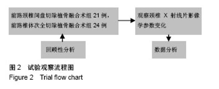
2.1 影像学相关参数评价 两组一般资料具有可比性,差异无显著性意义(表1)。术前两组各影像学参数相比差异无显著性意义(P > 0.05,表2)。试验观察流程图见图2。"

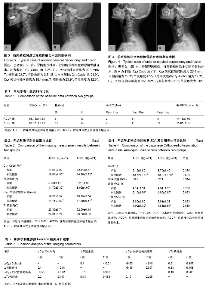
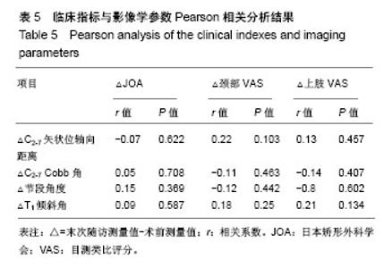
ACDF组:末次随访时C2-7 Cobb角由术前(11.04±7.98)°增加到(18.01±4.85)°,节段角度由(5.29±3.41)°增至(11.72± 3.32)°,C2-7矢状位轴向距离由(19.35±6.84) mm降至(14.16± 3.59) mm,各影像学参数较术前相比差异有显著性意义 (P < 0.05,表2)。T1倾斜角末次随访时(24.34±6.24)°较术前(23.36±6.74)°相比差异无显著性意义(P > 0.05,表2)。 ACCF组:末次随访时C2-7Cobb角由术前(12.14± 5.47)°增加到(14.90±2.72)°,节段角度由(6.28±4.36)°增加到(8.66± 4.69)°,C2-7矢状位轴向距离由(20.88±5.54) mm降至(17.70± 6.67) mm,各影像学参数较术前相比差异有显著性意义(P < 0.05,表2)。T1倾斜角末次随访时(22.99±6.14)°较术前(23.58±6.09)°相比差异无显著性意义(P > 0.05,表2)。 末次随访时两组间比较:ACDF组C2-7 Cobb角(18.01± 4.85)°、节段角度(11.72±3.32)°,明显高于ACCF组(14.90± 2.72)°、(8.66±4.69)°,两者相比差异有显著性意义(P < 0.05,表2)。ACDF组C2-7 矢状位轴向距离(14.16±3.59) mm明显低于ACCF组(17.70±6.67) mm,两者相比差异有显著性意义(P > 0.05,表2)。ACDF组与ACCF组T1倾斜角比较差异无显著性意义(P > 0.05,表2)。典型病例见图3,4。 两组影像学参数Pearson分析结果示:C2-7矢状位轴向距离与C2-7 Cobb角呈负相关(r=-0.55,P < 0.05,表3);C2-7 Cobb角与节段角度呈正相关(r=0.40,P < 0.05,表3)。 2.2 临床功能评价 ACDF组:在末次随访时,JOA评分由(9.19±1.86)分增至(13.62±1.71)分,JOA改善率为58.7%,颈部目测类比评分由(4.14±1.31)分降至(2.10±1.04)分,上肢目测类比评分由(3.80±1.80)降至(1.90±1.30)分,各临床指标较术前相比差异有显著性意义(P < 0.05,表4)。 ACCF组:在末次随访时,JOA评分由(8.75±1.45分增至(13.87±1.39)分,JOA改善率为62.1%,颈部目测类比评分由(4.04±1.20)分降至(1.95±0.80)分,上肢目测类比评分由(3.91±1.38降至(1.83±1.04)分,各临床指标较术前相比差异有显著性意义(P < 0.05,表4)。 在末次随访时,ACDF组和ACCF组两组间JOA评分、JOA改善率、颈部VAS、上肢VAS相比,差异均无显著性意义(P > 0.05,表4)。 没有影像学参数显示与临床指标具有相关性(P > 0.05,表5)。 "
"

| [1] Smith JS, Lafage V, Ryan DJ, et al. Association of myelopathy scores with cervical sagittal balance and normalized spinal cord volume: analysis of 56 preoperative cases from the AOSpine North America Myelopathy study. Spine. 2013;38(1):161-70.[2] Cho JH, Ha JK, Kim DG, et al. Does preoperative T1 slope affect radiological and functional outcomes following cervical laminoplasty? Spine. 2014;39(26):E1575.[3] Hyun SJ, Kim KJ, Jahng TA, et al. Relationship between T1 slope and cervical alignment following multilevel posterior cervical fusion surgery: impact of T1 slope minus cervical lordosis. Spine. 2016;41(7):E396.[4] Kim TH, Lee SY, Kim YC, et al. T1 slope as a predictor of kyphotic alignment change after laminoplasty in patients with cervical myelopathy. Spine. 2013;38(16):E992.[5] Angevine PD, Arons RR, Mccormick PC. National and regional rates and variation of cervical discectomy with and without anterior fusion, 1990-1999. Spine. 2003;28(9):931-939.[6] Wen ZQ, Du JY, Ling ZH, et al. Anterior cervical discectomy and fusion versus anterior cervical corpectomy and fusion in the treatment of multilevel cervical spondylotic myelopathy: systematic review and a meta-analysis. Ther Clini Risk Manag. 2015;11(3):161-70.[7] Liu T, Yang HL, Xu YZ, et al. ACDF with the PCB cage-plate system versus laminoplasty for multilevel cervical spondylotic myelopathy. Clin Spine Surg. 2011;24(24):213-220.[8] Carrier CS, Bono CM, Lebl DR. Evidence-based analysis of adjacent segment degeneration and disease after ACDF: a systematic review. Spine J. 2013;13(10):1370-1378.[9] Roguski M, Benzel EC, Curran JN, et al. Postoperative cervical sagittal imbalance negatively affects outcomes after surgery for cervical spondylotic myelopathy. Spine. 2014;39(25):2070-2077.[10] Weng C, Wang J, Tuchman A, et al. The influence of T1 slope on the cervical sagittal balance in degenerative cervical spine: an analysis using kinematic MRI. Spine. 2015;41(3):185.[11] Ames CP, Smith JS, Scheer JK, et al. Impact of spinopelvic alignment on decision making in deformity surgery in adults: a review. J Neurosurg Spine. 2012;16(6):547.[12] Tang JA, Scheer JK, Smith JS, et al. The impact of standing regional cervical sagittal alignment on outcomes in posterior cervical fusion surgery. Neurosurgery. 2012;71(3):662.[13] Jr NJ, Sherk HH. Biomechanical evaluation of the extensor musculature of the cervical spine. Spine. 1988;13(1):9-11.[14] Katsuura A, Hukuda S, Saruhashi Y, et al. Kyphotic malalignment after anterior cervical fusion is one of the factors promoting the degenerative process in adjacent intervertebral levels. Euro Spine J. 2001;10(4):320.[15] Knott PT, Mardjetko SM, Techy F. The use of the T1 sagittal angle in predicting overall sagittal balance of the spine. Spine J. 2010;10(11): 994-998.[16] Ono A, Tonosaki Y, Numasawa T, et al. The relationship between the anatomy of the nuchal ligament and postoperative axial pain after cervical laminoplasty: cadaver and clinical study. Spine. 2012;37(26): 1607-1613.[17] Schwab F, Patel A, Ungar B, et al. Adult spinal deformity-postoperative standing imbalance: how much can you tolerate? An overview of key parameters in assessing alignment and planning corrective surgery. Spine. 2010;35(25):2224.[18] Lee SH, Kim KT, Seo EM, et al. The influence of thoracic inlet alignment on the craniocervical sagittal balance in asymptomatic adults. J Spinal Disord Tech. 2012;25(2):E41.[19] Jun HS, Kim JH, Ahn JH, et al. T1 slope and degenerative cervical spondylolisthesis. Spine. 2015;40(4):220-226.[20] Villavicencio AT, Babuska JM, Ashton A, et al. Prospective, randomized, double-blind clinical study evaluating the correlation of clinical outcomes and cervical sagittal alignment. Neurosurgery. 2011;68(5):1309-1316.[21] Lee CK, Shin DA, Yi S, et al. Correlation between cervical spine sagittal alignment and clinical outcome after cervical laminoplasty for ossification of the posterior longitudinal ligament. J Neurosurg Spine. 2016;24(1):100-107.[22] Taemin O, Scheer JK, Robert E, et al. Cervical compensatory alignment changes following correction of adult thoracic deformity: a multicenter experience in 57 patients with a 2-year follow-up. J Neurosurg Spine. 2015;22(6):658-665.[23] Denaro V, Longo UG, Berton A, et al. Cervical spondylotic myelopathy: the relevance of the spinal cord back shift after posterior multilevel decompression. A systematic review. Eur Spine J. 2015;7(7):832-841.[24] Mummaneni PV, Kaiser MG, Matz PG, et al. Cervical surgical techniques for the treatment of cervical spondylotic myelopathy. J Neurosurg Spine. 2009;11(2):130.[25] Gaffney CJ, Spiker WR. Treating multilevel (three or four level) cervical myelopathy with ACDF or ACCF. Semin Spine Surg. 2014;26(3):122-127.[26] Barsa P, Suchomel P. Factors affecting sagittal malalignment due to cage subsidence in standalone cage assisted anterior cervical fusion. Eur Spine J. 2007;16(9):1395.[27] Tomé-Bermejo F, Morales-Valencia JA, Moreno-Pérez J, et al. Long-term changes in sagittal alignment and its clinical implications after cervical interbody fusion cage subsidence for degenerative cervical disc disease. A prospective study with standalone lordotic tantalum cages. J Spinal Disord Tech. 2015;30(5):E648.[28] 张伟,陈德玉,杨立利,等.两种颈椎前路减压融合术治疗脊髓型颈椎病的临床疗效分析[J].中国矫形外科杂志,2011,19(3):181-183.[29] Sakai K, Yoshii T, Hirai T, et al. Impact of the surgical treatment for degenerative cervical myelopathy on the preoperative cervical sagittal balance: a review of prospective comparative cohort between anterior decompression with fusion and laminoplasty. Eur Spine J. 2017;26(1): 104-112.[30] Oh MC, Zhang HY, Park JY, et al. Two-level anterior cervical discectomy versus one-level corpectomy in cervical spondylotic myelopathy. Spine. 2009;34(7):692-696. |
| [1] | Zhang Tongtong, Wang Zhonghua, Wen Jie, Song Yuxin, Liu Lin. Application of three-dimensional printing model in surgical resection and reconstruction of cervical tumor [J]. Chinese Journal of Tissue Engineering Research, 2021, 25(9): 1335-1339. |
| [2] | Zeng Yanhua, Hao Yanlei. In vitro culture and purification of Schwann cells: a systematic review [J]. Chinese Journal of Tissue Engineering Research, 2021, 25(7): 1135-1141. |
| [3] | Xu Dongzi, Zhang Ting, Ouyang Zhaolian. The global competitive situation of cardiac tissue engineering based on patent analysis [J]. Chinese Journal of Tissue Engineering Research, 2021, 25(5): 807-812. |
| [4] | Wu Zijian, Hu Zhaoduan, Xie Youqiong, Wang Feng, Li Jia, Li Bocun, Cai Guowei, Peng Rui. Three-dimensional printing technology and bone tissue engineering research: literature metrology and visual analysis of research hotspots [J]. Chinese Journal of Tissue Engineering Research, 2021, 25(4): 564-569. |
| [5] | Chang Wenliao, Zhao Jie, Sun Xiaoliang, Wang Kun, Wu Guofeng, Zhou Jian, Li Shuxiang, Sun Han. Material selection, theoretical design and biomimetic function of artificial periosteum [J]. Chinese Journal of Tissue Engineering Research, 2021, 25(4): 600-606. |
| [6] | Liu Fei, Cui Yutao, Liu He. Advantages and problems of local antibiotic delivery system in the treatment of osteomyelitis [J]. Chinese Journal of Tissue Engineering Research, 2021, 25(4): 614-620. |
| [7] | Li Xiaozhuang, Duan Hao, Wang Weizhou, Tang Zhihong, Wang Yanghao, He Fei. Application of bone tissue engineering materials in the treatment of bone defect diseases in vivo [J]. Chinese Journal of Tissue Engineering Research, 2021, 25(4): 626-631. |
| [8] | Zhang Zhenkun, Li Zhe, Li Ya, Wang Yingying, Wang Yaping, Zhou Xinkui, Ma Shanshan, Guan Fangxia. Application of alginate based hydrogels/dressings in wound healing: sustained, dynamic and sequential release [J]. Chinese Journal of Tissue Engineering Research, 2021, 25(4): 638-643. |
| [9] | Chen Jiana, Qiu Yanling, Nie Minhai, Liu Xuqian. Tissue engineering scaffolds in repairing oral and maxillofacial soft tissue defects [J]. Chinese Journal of Tissue Engineering Research, 2021, 25(4): 644-650. |
| [10] | Xing Hao, Zhang Yonghong, Wang Dong. Advantages and disadvantages of repairing large-segment bone defect [J]. Chinese Journal of Tissue Engineering Research, 2021, 25(3): 426-430. |
| [11] | Chen Siqi, Xian Debin, Xu Rongsheng, Qin Zhongjie, Zhang Lei, Xia Delin. Effects of bone marrow mesenchymal stem cells and human umbilical vein endothelial cells combined with hydroxyapatite-tricalcium phosphate scaffolds on early angiogenesis in skull defect repair in rats [J]. Chinese Journal of Tissue Engineering Research, 2021, 25(22): 3458-3465. |
| [12] | Wang Hao, Chen Mingxue, Li Junkang, Luo Xujiang, Peng Liqing, Li Huo, Huang Bo, Tian Guangzhao, Liu Shuyun, Sui Xiang, Huang Jingxiang, Guo Quanyi, Lu Xiaobo. Decellularized porcine skin matrix for tissue-engineered meniscus scaffold [J]. Chinese Journal of Tissue Engineering Research, 2021, 25(22): 3473-3478. |
| [13] | Mo Jianling, He Shaoru, Feng Bowen, Jian Minqiao, Zhang Xiaohui, Liu Caisheng, Liang Yijing, Liu Yumei, Chen Liang, Zhou Haiyu, Liu Yanhui. Forming prevascularized cell sheets and the expression of angiogenesis-related factors [J]. Chinese Journal of Tissue Engineering Research, 2021, 25(22): 3479-3486. |
| [14] | Liu Chang, Li Datong, Liu Yuan, Kong Lingbo, Guo Rui, Yang Lixue, Hao Dingjun, He Baorong. Poor efficacy after vertebral augmentation surgery of acute symptomatic thoracolumbar osteoporotic compression fracture: relationship with bone cement, bone mineral density, and adjacent fractures [J]. Chinese Journal of Tissue Engineering Research, 2021, 25(22): 3510-3516. |
| [15] | Liu Liyong, Zhou Lei. Research and development status and development trend of hydrogel in tissue engineering based on patent information [J]. Chinese Journal of Tissue Engineering Research, 2021, 25(22): 3527-3533. |
| Viewed | ||||||
|
Full text |
|
|||||
|
Abstract |
|
|||||