Chinese Journal of Tissue Engineering Research ›› 2018, Vol. 22 ›› Issue (31): 4975-4981.doi: 10.3969/j.issn.2095-4344.0336
Previous Articles Next Articles
Biomechanical characteristics of tibio-femoral joint under different flexion angles by finite element analysis
Wang Jun-ran1, Du Wei-jin1, Wang Chang-jiang2, Guo Yuan1, Chen Wei-yi1
- 1Institute of Applied Mechanics and Biomedical Engineering, Taiyuan University of Technology, Taiyuan 030024, Shanxi Province, China; 2Department of Engineering and Design, University of Sussex, Brighton, UK, BNI 9RH
-
Online:2018-11-08Published:2018-11-08 -
Contact:Chen Wei-yi, PhD, Professor, Institute of Applied Mechanics and Biomedical Engineering, Taiyuan University of Technology, Taiyuan 030024, Shanxi Province, China -
About author:Wang Jun-ran, Master, Institute of Applied Mechanics and Biomedical Engineering, Taiyuan University of Technology, Taiyuan 030024, Shanxi Province, China -
Supported by:the National Natural Science Foundation of China, No. 11472185 and 11772214
CLC Number:
Cite this article
Wang Jun-ran, Du Wei-jin, Wang Chang-jiang, Guo Yuan, Chen Wei-yi. Biomechanical characteristics of tibio-femoral joint under different flexion angles by finite element analysis [J]. Chinese Journal of Tissue Engineering Research, 2018, 22(31): 4975-4981.
share this article

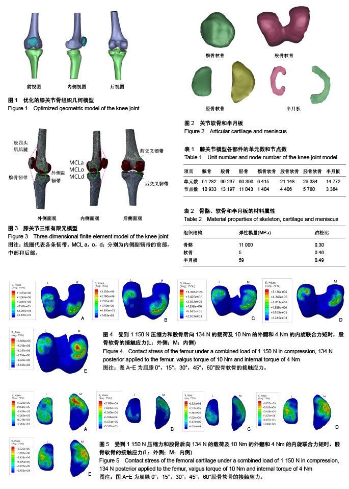
2.1 三维有限元模型的鉴定 实验构建了成年男性正常膝关节的三维有限元模型,包括髌骨、股骨、胫骨、关节软骨、内外侧半月板、髌韧带(PL)、股四头肌(QT)、前后交叉韧带(ACL,PCL)和内外侧副韧带(MCL,LCL)。模型的结构和相对位置均符合正常的膝关节生理构造,共包含200 043个单元数和50 127个节点。 2.2 膝关节在不同屈曲角度受力的力学特性 在屈曲0°,受到1 150 N的压缩力及股骨后向134 N的载荷时,实验得到的前后、内外方向上的位移分别为4.25,1.39 mm;内外旋和内外翻角度分别为3.6°和0.66°。在相同载荷条件下,Song等[27]得到的结果分别为4.3,0.39 mm,1.9°和0.09°;Peña等[28]得到的结果分别为4.75 mm、0.56 mm、1.6°和0.76°。通过实验结果与其他作者的结果比较,可说明此模型的有效性[27-29]。 膝关节在屈曲0°到60°,施加1 150 N的压缩力及股骨后向134 N的载荷时,胫股关节间的相对位移,以前后位移为主,屈曲0°时位移最小,为4.25 mm;屈曲30°时位移最大,为6.93 mm。其他方向的相对位移较小,且旋转角度变化不大,见表4。"

"

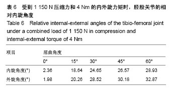
膝关节在屈曲0°-60°,受到1 150 N的压缩力及10 Nm的内、外翻力矩时,主要研究股骨相对胫骨产生的内、外翻。Wascher等[30]对胫骨施加15 Nm内、外翻的力矩,在10°-20°的屈膝过程中,得到其内、外翻的范围是 7.7°-9.2°。Markolf等[31]采用29 Nm的力矩,得到在屈膝0°-90°,内、外翻角度为2°-8°。实验由于没有包括肌肉和皮肤等组织,所以使用10 Nm的内、外翻力矩,膝关节在屈曲0°-60°,得到内、外翻活动范围为3.54°-6.12°(表5)。可见膝关节内、外翻运动相对稳定,角度变化较小。"

膝关节在屈曲0°-60°运动中,受到1 150 N的压缩力及4 Nm的内、外旋力矩时,主要研究股骨相对胫骨产生的内、外旋。Wascher等[30]研究得出当膝关节微屈10°-20°时,内、外旋的活动范围为31.6° -37.6°。Markolf等[31]采用4 Nm的旋转力矩时,研究得出当膝关节屈膝超过20°时,其内、外旋角度均可达到25°。实验也使用4 Nm的内旋力矩,测得在0°-60°屈膝过程中,膝关节内旋的范围为2.36°-28.93°,外旋的范围为1.98°-32.87°(表6)。可见在0°-60°屈膝过程中内、外旋角度随屈曲角度的增大逐渐增大,但外旋的活动范围略大于内旋范围。"

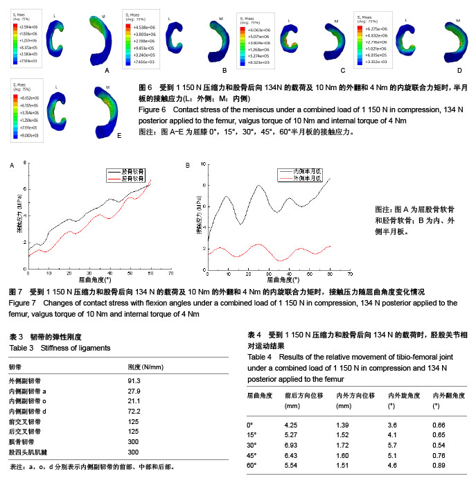
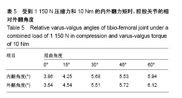
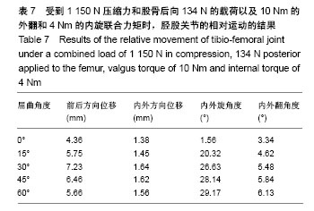
为了更准确地模拟人体膝关节运动,在0°-60°屈膝运动中,施加1 150 N压缩力和股骨后向134 N的载荷以及 10 Nm的外翻和4 Nm的内旋联合力矩,得到如下数据(表7)。内旋和外翻的角度随屈膝角度的增大而增大,其活动范围分别为1.56°-29.17°和 3.34°-6.13°;胫股关节的前后位移,在屈曲0°时位移最小,为4.36 mm,30°时最大,为7.23 mm,则其前后位移范围是4.36-7.23 mm。 为了更好的分析胫股关节间的力学特性,给出了膝关节在0°-60°受到1 150 N压缩力和股骨后向134 N的载荷及10 Nm的外翻和4 Nm的内旋联合力矩时,胫股关节接触应力云图(图4-6)和各关节面接触应力变化曲线(图7)。 由图4-7可知,股骨软骨、胫骨软骨和半月板在0°的最大接触应力分别为1.45,1.03和2.59 MPa;而60°的最大接触应力分别为6.41,6.73和8.65 MPa。膝关节在0°-60°的屈曲运动中,股骨软骨和胫骨软骨的接触应力随着屈膝角度的增加而增大,且接触区域逐渐向后侧移动。在此过程中,内、外侧半月板接触应力的变化不同,内侧半月板的接触应力的波动较大,而与之相反,外侧半月板的接触应力相对稳定,且内侧半月板的接触应力大于外侧半月板的应力值。膝关节在0°-30°的屈膝阶段,胫股关节的接触区域主要发生在胫骨平台和半月板的前侧,且接触面积相对较小;而从30°-60°屈膝阶段,接触区域逐渐转移到胫骨平台和半月板的中央,接触面积也随之变大。"
| [1] 贾焕英.膝关节软骨损伤磁共振扫描序列临床应用价值的研究[J].中国药物与临床,2017,17(6):826-827.[2] 林昊,张余,李国安.人工全膝关节研究新进展[J].医用生物力学, 2012,27(2):115-121.[3] 李春娥.膝关节有限元模型研究进展[J].科技经济导刊,2017, 1(9):158.[4] Tanska P, Mononen ME, Korhonen RK. A multi-scale finite element model for investigation of chondrocyte mechanics in normal and medial meniscectomy human knee joint during walking. J Biomech. 2015;48(8):1397-1406.[5] Naghibi BH, Janssen D, Van GS, et al. The influence of ligament modelling strategies on the predictive capability of finite element models of the human knee joint. J Biomech. 2017;65(5):1-11.[6] Kang KT, Koh YG, Son J, et al. Finite element analysis of the biomechanical effects of 3 posterolateral corner reconstruction techniques for the knee joint. Arthroscopy. 2017;33(8):1537-1550.[7] Woiczinski M, Steinbrück A, Weber P, et al. Development and validation of a weight-bearing finite element model for total knee replacement. Comput Methods Biomech Biomed Engin. 2016;19(10):1033.[8] Ali AA, Shalhoub SS, Cyr AJ, et al. Validation of predicted patellofemoral mechanics in a finite element model of the healthy and cruciate-deficient knee. J Biomech. 2016;49(2): 302-309.[9] Dong R, Liu Y, Zhang X, et al. The evaluation of the role of medial collateral ligament maintaining knee stability by a finite element analysis. J Orthop Surg Res. 2017;12(1):64-71.[10] Rybicki EF, Simonen FA, Jr WE. On the mathematical analysis of stress in the human femur. J Biomech. 1972; 5(2): 203-215.[11] Wismans J, Veldpaus F, Janssen J, et al. A three-dimensional mathematical model of the knee-joint. J Biomech. 1980;13(8): 677-679.[12] Bendjaballah MZ, Shirazi A. Biomechanics of the human knee joint in compression: reconstruction, mesh generation and finite element analysis. Knee. 1995;2(2):69-79.[13] Jilani A, Shirazi-Adl A, Bendjaballah MZ. Biomechanics of human tibio-femoral joint in axial rotation. Knee. 1997;4(4): 203-213.[14] Caruntu DI, Hefzy MS. 3-D anatomically based dynamic modeling of the human knee to include tibio-femoral and patello-femoral joints. J Biomech Eng. 2004;126(1):44-53.[15] Zielinska B, Donahue TL. 3D finite element model of meniscectomy: changes in joint contact behavior. J Biomech Eng. 2006;128(1):115-123.[16] 姜华亮,华锦明,许新忠,等.正常人膝关节三维有限元模型的建立[J].苏州大学学报:医学版,2008,28(3):421-422.[17] Baldwin MA, Clary CW, Fitzpatrick CK, et al. Dynamic finite element knee simulation for evaluation of knee replacement mechanics. J Biomech. 2012;45(3):474-483.[18] 鲍春雨,郭宝川,孟庆华.人体膝关节有限元模型建立及其有效性验证[J].应用力学学报,2017,34(3):559-563.[19] 陈文栋,杨光.不同载荷条件下半月板动态仿真生物力学分析[J].中国组织工程研究,2017,21(11):1742-1747.[20] Manda K, Ryd L, Eriksson A. Finite element simulations of a focal knee resurfacing implant applied to localized cartilage defects in a sheep model. J Biomech. 2011;44(5):794-801.[21] 崔晓倩,王辅忠,张慧春.膝关节股骨远端软骨硬化前后力学性能分析[J].医用生物力学,2015,30(1):25-29.[22] Rao C, Fitzpatrick CK. A statistical finite element model of the knee accounting for shape and alignment variability. Medical Engineering&Physics. 2013,35(10):1450-1456.[23] 万超,郝智秀,温诗铸.前交叉韧带力学特性差异对膝关节有限元仿真结果的影响[J].医用生物力学,2012,27(4):375-380.[24] Donzelli PS, Spilker RL, Ateshian GA, et al. Contact analysis of biphasic transversely isotropic cartilage layers and correlations with tissue failure. J Biomech. 1999;32(10): 1037-1047.[25] Marouane H, Shiraziadl A, Hashemi J. Quantification of the role of tibial posterior slope in knee joint mechanics and ACL force in simulated gait. J Biomech. 2015;48(10):1899-905.[26] 吴辉群,严培培,耿兴云,等.基于Mimics软件虚拟人膝关节三维图像融合实验[J].中国组织工程研究,2011,15(48):8943-8946.[27] Song Y, Debski RE, Musahl V, et al. A three -dimensional finite element model of the human anterior cruciate ligament: a computational analysis with experimental validation. J Biomech. 2004;37(3):383- 390.[28] Peña AE, Calvo B, Martã¬Nez MA, et al. A three-dimensional finite element analysis of the combined behavior of ligaments and menisci in the healthy human knee joint. J Biomech. 2006;39(9):1686-1701.[29] Suggs J, Wang C, Li G. The effect of graft stiffness on knee joint biomechanics after ACL reconstruction-a 3D computational simulation. Clin Biomech. 2003;18(1):35-43.[30] Wascher DC, Markolf KL, Shapiro MS,et al. Direct in vitro measurement of forces in the cruciate ligaments. Part I: the effect of multiplane loading in the intact knee. J Bone Joint Surg Am. 1993;75(3):377-386.[31] Morkolf K. Stiffness and laxity of the knee-the contributions of the supporting structures. J Bone Joint Surg A. 1976;58(5): 583-594.[32] Naghibi H, Khoshgoftar M, Sprengers A, et al. A comparison between dynamic implicit and explicit finite element simulations of the native knee joint. Med Eng Phys. 2016; 10(38):1123-1130.[33] 王光达,张祚福,齐晓军,等.膝关节三维有限元模型的建立及生物力学分析[J].中国组织工程研究,2010,14(52):9702-9705. |
| [1] | Huang Dengcheng, Wang Zhike, Cao Xuewei. Comparison of the short-term efficacy of extracorporeal shock wave therapy for middle-aged and elderly knee osteoarthritis: a meta-analysis [J]. Chinese Journal of Tissue Engineering Research, 2021, 25(9): 1471-1476. |
| [2] | Xu Feng, Kang Hui, Wei Tanjun, Xi Jintao. Biomechanical analysis of different fixation methods of pedicle screws for thoracolumbar fracture [J]. Chinese Journal of Tissue Engineering Research, 2021, 25(9): 1313-1317. |
| [3] | Chen Xinmin, Li Wenbiao, Xiong Kaikai, Xiong Xiaoyan, Zheng Liqin, Li Musheng, Zheng Yongze, Lin Ziling. Type A3.3 femoral intertrochanteric fracture with augmented proximal femoral nail anti-rotation in the elderly: finite element analysis of the optimal amount of bone cement [J]. Chinese Journal of Tissue Engineering Research, 2021, 25(9): 1404-1409. |
| [4] | Zhou Jihui, Li Xinzhi, Zhou You, Huang Wei, Chen Wenyao. Multiple problems in the selection of implants for patellar fracture [J]. Chinese Journal of Tissue Engineering Research, 2021, 25(9): 1440-1445. |
| [5] | Wu Xun, Meng Juanhong, Zhang Jianyun, Wang Liang. Concentrated growth factors in the repair of a full-thickness condylar cartilage defect in a rabbit [J]. Chinese Journal of Tissue Engineering Research, 2021, 25(8): 1166-1171. |
| [6] | Li Jiacheng, Liang Xuezhen, Liu Jinbao, Xu Bo, Li Gang. Differential mRNA expression profile and competitive endogenous RNA regulatory network in osteoarthritis [J]. Chinese Journal of Tissue Engineering Research, 2021, 25(8): 1212-1217. |
| [7] | Geng Qiudong, Ge Haiya, Wang Heming, Li Nan. Role and mechanism of Guilu Erxianjiao in treatment of osteoarthritis based on network pharmacology [J]. Chinese Journal of Tissue Engineering Research, 2021, 25(8): 1229-1236. |
| [8] | Zhong Hehe, Sun Pengpeng, Sang Peng, Wu Shuhong, Liu Yi. Evaluation of knee stability after simulated reconstruction of the core ligament of the posterolateral complex [J]. Chinese Journal of Tissue Engineering Research, 2021, 25(6): 821-825. |
| [9] | Xu Yulin, Shen Shi, Zhuo Naiqiang, Yang Huilin, Yang Chao, Li Yang, Zhao Heng, Zhao Lu. Biomechanical comparison of three different plate fixation methods for acetabular posterior column fractures in standing and sitting positions [J]. Chinese Journal of Tissue Engineering Research, 2021, 25(6): 826-830. |
| [10] | Cai Qunbin, Zou Xia, Hu Jiantao, Chen Xinmin, Zheng Liqin, Huang Peizhen, Lin Ziling, Jiang Ziwei. Relationship between tip-apex distance and stability of intertrochanteric femoral fractures with proximal femoral anti-rotation nail: a finite element analysis [J]. Chinese Journal of Tissue Engineering Research, 2021, 25(6): 831-836. |
| [11] | Liu Shaohua, Zhou Guanming, Chen Xicong, Xiao Keming, Cai Jian, Liu Xiaofang. Influence of anterior cruciate ligament defect on the mid-term outcome of fixed-bearing unicompartmental knee arthroplasty [J]. Chinese Journal of Tissue Engineering Research, 2021, 25(6): 860-865. |
| [12] | Song Chengjie, Chang Hengrui, Shi Mingxin, Meng Xianzhong. Research progress in biomechanical stability of lateral lumbar interbody fusion [J]. Chinese Journal of Tissue Engineering Research, 2021, 25(6): 923-928. |
| [13] | Liu Zhao, Xu Xilin, Shen Yiwei, Zhang Xiaofeng, Lü Hang, Zhao Jun, Wang Zhengchun, Liu Xuzhuo, Wang Haitao. Guiding role and prospect of staging and classification combined collapse prediction method for osteonecrosis of femoral head [J]. Chinese Journal of Tissue Engineering Research, 2021, 25(6): 929-934. |
| [14] | Kong Lingbao, Lü Xin. Effect of implant selection and approach on support in the operation of posterolateral tibial plateau fractures [J]. Chinese Journal of Tissue Engineering Research, 2021, 25(6): 942-947. |
| [15] | Huang Dengcheng, Wang Zhike, Cao Xuewei. Intravenous, topical tranexamic acid alone or their combination in total knee arthroplasty: a meta-analysis of randomized controlled trials [J]. Chinese Journal of Tissue Engineering Research, 2021, 25(6): 948-956. |
| Viewed | ||||||
|
Full text |
|
|||||
|
Abstract |
|
|||||