Chinese Journal of Tissue Engineering Research ›› 2026, Vol. 30 ›› Issue (24): 6196-6206.doi: 10.12307/2026.293
Previous Articles Next Articles
Molecular dynamic characteristics of rat gastrocnemius muscle under acute and short-term exercise intervention during the subacute phase of spinal cord injury
Wei Xinyi1, Zheng Yan2, Chen Qian3, Ren Jiajia1, Li Jian4
- 1School of Physical Education and Health, East China Normal University, Shanghai 200241, China; 2School of Physical Education, Soochow University, Suzhou 215021, Jiangsu Province, China; 3School of Physical Education, Xi’an Shiyou University, Xi’an 710065, Shaanxi Province, China; 4Peking University Health Science Center, Beijing 100191, China
-
Received:2025-07-19Revised:2025-12-18Online:2026-08-28Published:2026-01-29 -
Contact:Li Jian, MD, Assistant researcher, Peking University Health Science Center, Beijing 100191, China -
About author:Wei Xinyi, MS, School of Physical Education and Health, East China Normal University, Shanghai 200241, China
CLC Number:
Cite this article
Wei Xinyi, Zheng Yan, Chen Qian, Ren Jiajia, Li Jian. Molecular dynamic characteristics of rat gastrocnemius muscle under acute and short-term exercise intervention during the subacute phase of spinal cord injury[J]. Chinese Journal of Tissue Engineering Research, 2026, 30(24): 6196-6206.
share this article
Add to citation manager EndNote|Reference Manager|ProCite|BibTeX|RefWorks
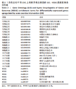
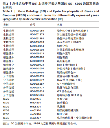
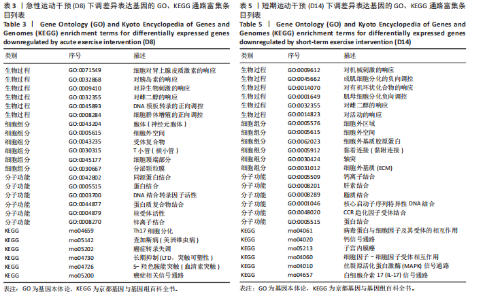
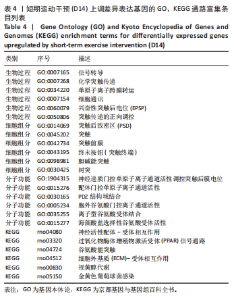
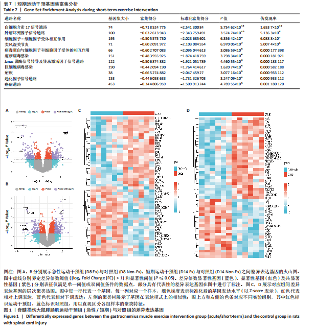
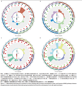
2.1 微阵列数据信息与差异表达基因筛选 在GSE45550数据集下载大鼠腓肠肌数据,主要采用纽约大学机械脊髓损伤装置(NYU-MASCIS)方法对大鼠进行中等挫伤脊髓损伤造模,造模后第1天记为D1,随后进行正常饲养,研究将动物随机分为4组(每组6只),在术后第8天和第14天分别进行取样,以评估不同运动训练周期对脊髓损伤的影响。急性运动对照组(D8 Non-ex)和短期运动对照组(D14 Non-ex)仅接受手术处理,不进行运动干预;急性运动干预组(D8 Ex)在术后第7天进行2次20 min跑台训练,第8天进行1次20 min跑台训练;短期运动干预组(D14 Ex)在术后第7-14天间共训练5 d,每天训练2次,每次20 min。分别于第8天和第14天采集样本进行分析,具体干预方式及分组见表1。然后对GSE45550数据集中急性运动干预组及其对照组(D8 Ex、D8 Non-Ex),短期运动干预组及其对照组(D14 Ex、D14 Non-Ex)进行差异基因分析,并进行可视化处理。在火山图中,对急性运动干预组及其对照组(D8 Non-Ex与Ex)大鼠腓肠肌表达基因进行对比,发现Hrh2、Plet1、Sele等106个基因上调,RGD1305928、Adcy10、Ugt2b1等97个基因下调,见图1A。对短期运动干预组及其对照组(D14 Ex与D14 Non-Ex)大鼠腓肠肌进行差异基因分析,发现Tas2r123、Ca10等138个基因上调,Pcp4、Col19a1、Eif2s3y、Myh4等105个基因下调,见图1B。热图展示了急性干预和短期运动干预与对照组基因样本间的表达差异。层次聚类分析揭示了急性运动干预组(D8 Ex)和对照组(D8 Non-Ex)之间显著的转录差异,基因表达呈现明显的上调与下调模式,其中Sele、Slit2和Areg等基因在急性运动干预组中表达水平较高,而Calcb、Rnf180和Kcne1等基因表达水平较低,见图1C。类似地,在短期运动干预后,运动组与对照组基因表达的聚类模式依然清晰,表明运动对基因转录的影响在时间维度上持续存在,Klk8、Ctsr和Fcgbp等基因在短期运动干预组中表达上调,而Gabrb2、Tacr2和Pcp4等基因表达水平下调,见图1D。这些结果表明,急性与短期运动干预诱导的基因表达变化具有显著时序性,不同时间点均呈现特异性分子特征。 2.2 差异表达基因功能及KEGG富集分析 采用GO和KEGG通路富集分析,以探讨基因在生物学功能中的分布和富集情况。GO分析包括3个主要分类:生物学过程、细胞组成和分子功能。KEGG分析用于识别显著富集的代谢及信号通路。为进一步分析生物学过程、细胞组成、分子功能及KEGG通路中最显著上调或下调的前6名基因,分别进行可视化处理,以揭示在各自通路中的潜在作用和关联性。富集分析采用超几何检验方法计算P值,并进行多重假设检验校正,筛选显著性水平P < 0.05的通路,结果通过circlize R包进行可视化,绘制GO和KEGG富集圈图,从外至内分别表示分类、基因数、基因重叠情况及富集分数,基因数的颜色深度表征-log10(P值)。基因重叠情况中红色、绿色分别代表上调、下调基因。GSE45550数据集中,脊髓损伤后的急性运动干预(D8)使大鼠腓肠肌上调的差异表达基因显著富集在染色体分离(Chromosome segregation)、肾上腺素能受体信号通路(Adrenergic receptor signaling pathway)、染色体分离的正向调控(Positive regulation of chromosome segregation)等生物学过程;细胞外空间(Extracellular space)、细胞表面(Cell surface)、凝缩染色体(Condensed chromosome)等细胞组成;微管运动蛋白活性(Microtubule motor activity)、DNA结合(DNA binding)、ATP水解活性(ATP hydrolysis activity)等分子功能;以及胃酸分泌(Gastric acid secretion)、运动蛋白(Motor proteins)、乳腺癌(Breast cancer)等信号通路,见图2A。下调的差异表达基因主要富集于肾上腺类固醇刺激的细胞反应(Cellular response to dexamethasone stimulus)、胰岛素响应(Response to insulin)、外源性刺激(Response to xenobiotic stimulus)等生物学过程;细胞外空间(Extracellular space)、细胞表面(Cell surface)、受体复合物(Receptor complex)等细胞组成;同源蛋白结合(Identical protein binding)、蛋白结合(Protein binding)、DNA结合转录因子活性(DNA-binding transcription factor activity)等分子功能;以及Th17细胞分化(Th17 cell differentiation)、查加斯病(Chagas disease)、癌症转录失调(Transcriptional misregulation in cancer)等信号通路,见图2B。 脊髓损伤后的短期运动干预(D14)使大鼠腓肠肌上调的差异表达基因显著富集在信号转导(Signal transduction)、化学突触传递(Chemical synaptic transmission)、单原子离子跨膜运输(Monoatomic ion transmembrane transport)等生物学过程;突触后致密物(Postsynaptic density)、突触(Synapse)、突触前膜(Presynaptic membrane)等细胞组成;神经递质门控的单原子离子通道活性(Transmitter-gated monoatomic ion channel activity)、配体门控单原子离子通道活性(Ligand-gated monoatomic ion channel activity)、PDZ结构域结合(PDZ domain binding)等分子功能;以及神经活性配体-受体相互作用(Neuroactive ligand-receptor interaction)、PPAR信号通路(PPAR signaling pathway)、谷氨酸能突触(Glutamatergic synapse)等信号通路,见图2C。而下调的差异表达基因主要富集于对机械刺激的响应(Response to mechanical stimulus)、肌母细胞分化负向调控(Negative regulation of myoblast differentiation)、对有机环状化合物的响应(Response to organic cyclic compound)等生物学过程;细胞外区域(Extracellular region)、细胞外空间(Extracellular space)、细胞外基质胶原蛋白(Collagen-containing extracellular matrix)等细胞组成;钙离子结合(Calcium ion binding)、肝素结合(Heparin binding)、脂质结合(Lipid binding)等分子功能;以及病毒蛋白与细胞因子及其受体的相互作用(Viral protein interaction with cytokine and cytokine receptor)、钙信号通路(Calcium signaling pathway)、子宫内膜癌(Endometrial cancer)等信号通路,见图2D。GO及KEGG中序号及术语名称见表2-5。"
"
"
"
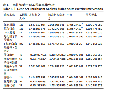

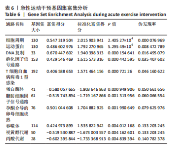
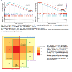
2.3 基因集富集分析 脊髓损伤后亚急性期的急性运动干预组呈现显著的生物学通路调控。如表6所示,排名前10的通路中,细胞周期(NES=2.02,FDR=7.70×10?5)、DNA复制(NES=1.94,FDR=1.65×10?2)及运动蛋白相关通路(NES=1.79,FDR=8.47×10?3)呈现显著正向富集,见图3A,提示大鼠脊髓损伤后亚急性期急性运动干预可能主要促进腓肠肌内细胞再生。短期运动干预组差异基因变化则体现为炎症信号通路的系统性抑制,见表7,白细胞介素17信号通路(NES=-2.54,FDR=1.65×10?10)、肿瘤坏死因子信号通路(NES=-2.34,FDR=5.14×10?8)及细胞因子-受体互作通路(NES=-2.02,FDR=6.09×10?7)均显著负向富集,见图3B。 2.4 急性与短期运动干预后腓肠肌差异基因对比 对比急性运动干预(D8 Ex),短期运动干预(D14 Ex)后两组大鼠腓肠肌上调及下调基因是否有交集,绘制韦恩图。其中,颜色深浅表示差异"
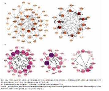
基因不同组别的重叠次数。韦恩图可观察到不同组别之间的重叠与差异。急性运动干预上调基因与短期运动干预上调基因交集有3个基因表现出一致的上调模式,为Ctsr、Zfyve28、Nkx3-1。 急性运动干预下调基因与短期运动干预下调基因之间的交集为Tacr2。急性运动干预上调基因与短期运动干预下调基因之间也存在相互作用,LOC102550254基因在急性运动干预组表现为高表达,而在短期运动干预组则转为低表达。最后,急性运动干预下调基因与短期运动干预上调基因之间的交集为4个基因,分别为Rdh12、Naalad1、LOC10254892、LOC102552654,见图4。 2.5 差异表达基因蛋白互作网络分析 将筛选得到的差异基因上传至STRING在线数据库中,并使用Cytoscape软件构建蛋白-蛋白互作网络。度中心性由蛋白质节点颜色表现,颜色越深表示度中心性越高,即与其他蛋白互相作用越强。在GSE45550数据集中,急性运动干预组观察到2个中心模块,第1个模块中互作较强的蛋白为Sox10、Pou4f1、Fgf6、Shh、IL13、Kng1、Neurod1、Rbp3,与神经、免疫细胞发育密切联系;第2个模块中,核心相互作用蛋白包括Esco2、Top2a、Aspm、Ska1、Smc2、Uhrf1、Ncapg2、Kif18b、Melk、Cenpe、Brca1,与细胞"

合成高度相关,见图5A。短期运动干预组观察到3个中心模块,第1个模块度中心性较高的蛋白包括Comp、Serpinb7、Fmod、Gadd45a、Bglap、Myc、Hapln1、Adamts1、II2ra、Tnf、IL11、Adamts4、Pparα,调控细胞进程;第2个模块主要互作蛋白包括Grik2、Neto1、Cacng8、Sv2a、Dlgap3、Opn4、Oxt、Gria2、Drd1,与神经信号调控、突触传递相关;第3个模块主要包括Sycn、Cyp2a1、Cyp26a1、Pla2g2c等,参与代谢、氧化反应进程,见图5B。"

"

| [1] 刘帅祎,赵晓璇,李奇,等.中央模式发生器在脊髓损伤后下肢运动恢复中作用的研究进展[J].中国脊柱脊髓杂志,2024,34(10):1092-1098. [2] RYBAK IA, SHEVTSOVA NA, MARKIN SN, et al. Operation regimes of spinal circuits controlling locomotion and the role of supraspinal drives and sensory feedback. Elife. 2024;13:RP98841. [3] MINASSIAN K, BAYART A, LACKNER P, et al. Rare phenomena of central rhythm and pattern generation in a case of complete spinal cord injury. Nat Commun. 2023;14(1):3276. [4] YU P, ZHANG W, LIU Y, et al. The effects and potential mechanisms of locomotor training on improvements of functional recovery after spinal cord injury. Int Rev Neurobiol. 2019;147:199-217. [5] LOY K, BAREYRE FM. Rehabilitation following spinal cord injury: how animal models can help our understanding of exercise-induced neuroplasticity. Neural Regen Res. 2019;14(3):405-412. [6] ZENG CW, ZHANG CL. Neuronal regeneration after injury: a new perspective on gene therapy. Front Neurosci. 2023;17:1181816. [7] HU X, XU W, REN Y, et al. Spinal cord injury: molecular mechanisms and therapeutic interventions. Signal Transduct Target Ther. 2023;8(1):245. [8] 潘璐,谭波涛,虞乐华,等.运动训练影响脊髓损伤后功能恢复机制的研究进展[J].中国康复医学杂志,2020,35(12):1537-1541. [9] KAUR S, ARUMUGAM N, CHHABRA H. A Systematic Review: Exercise based approaches to activate central pattern generator in spinal cord injury survivors. Int J Neurol Phys Ther. 2024;10:8-15. [10] HORNBY TG, REISMAN DS, WARD IG, et al. Clinical Practice Guideline to Improve Locomotor Function Following Chronic Stroke, Incomplete Spinal Cord Injury, and Brain Injury. J Neurol Phys Ther. 2020;44(1):49-100. [11] BILCHAK JN, CARON G, CÔTÉ MP. Exercise-Induced Plasticity in Signaling Pathways Involved in Motor Recovery after Spinal Cord Injury. Int J Mol Sci. 2021;22(9):4858. [12] GOLDSHMIT Y, LYTHGO N, GALEA MP, et al. Treadmill training after spinal cord hemisection in mice promotes axonal sprouting and synapse formation and improves motor recovery. J Neurotrauma. 2008;25(5):449-465. [13] ZHANG H, LIU Y, ZHOU K, et al. Restoring Sensorimotor Function Through Neuromodulation After Spinal Cord Injury: Progress and Remaining Challenges. Front Neurosci. 2021;15:749465. [14] CHANG W, PEDRONI A, BERTUZZI M, et al. Locomotion dependent neuron-glia interactions control neurogenesis and regeneration in the adult zebrafish spinal cord. Nat Commun. 2021;12(1):4857. [15] KAZIM SF, BOWERS CA, COLE CD, et al. Corticospinal Motor Circuit Plasticity After Spinal Cord Injury: Harnessing Neuroplasticity to Improve Functional Outcomes. Mol Neurobiol. 2021;58(11):5494-5516. [16] WANG H, LIU NK, ZHANG YP, et al. Treadmill training induced lumbar motoneuron dendritic plasticity and behavior recovery in adult rats after a thoracic contusive spinal cord injury. Exp Neurol. 2015;271:368-378. [17] YU JI, SEO TB. The effects of weight- and non-weight-bearing exercise on corticospinal axon sprouting, regeneration-related proteins and functional recovery after spinal cord contusion. J Exerc Rehabil. 2024;20(6):213-219. [18] ERSCHBAMER MK, PHAM TM, ZWART MC, et al. Neither environmental enrichment nor voluntary wheel running enhances recovery from incomplete spinal cord injury in rats. Exp Neurol. 2006;201(1):154-164. [19] LEWIS NE, TABARESTANI TQ, CELLINI BR, et al. Effect of Acute Physical Interventions on Pathophysiology and Recovery After Spinal Cord Injury: A Comprehensive Review of the Literature. Neurospine. 2022;19(3):671-686. [20] MIGLIORINI F, COCCONI F, SCHÄFER L, et al. Pharmacological management of secondary chronic spinal cord injury: a systematic review. Br Med Bull. 2024; 151(1):49-68. [21] ANJUM A, YAZID MD, FAUZI DAUD M, et al. Spinal Cord Injury: Pathophysiology, Multimolecular Interactions, and Underlying Recovery Mechanisms. Int J Mol Sci. 2020;21(20):7533. [22] ALIZADEH A, DYCK SM, KARIMI-ABDOLREZAEE S. Traumatic Spinal Cord Injury: An Overview of Pathophysiology, Models and Acute Injury Mechanisms. Front Neurol. 2019;10:282. [23] HOU J, NELSON R, MOHAMMAD N, et al. Effect of Simultaneous Combined Treadmill Training and Magnetic Stimulation on Spasticity and Gait Impairments after Cervical Spinal Cord Injury. J Neurotrauma. 2020;37(18):1999-2013. [24] LI X, LI Q, LI C, et al. Effect of high-intensity exercise training on functional recovery after spinal cord injury. Front Neurol. 2025;16:1442004. [25] HE LW, GUO XJ, ZHAO C, et al. Rehabilitation Training after Spinal Cord Injury Affects Brain Structure and Function: From Mechanisms to Methods. Biomedicines. 2023;12(1):41. [26] BALIGAND C, CHEN YW, YE F, et al. Transcriptional Pathways Associated with Skeletal Muscle Changes after Spinal Cord Injury and Treadmill Locomotor Training. Biomed Res Int. 2015;2015:387090. [27] 郭小军,常佳琪,何乐玮,等.硬膜外脊髓电刺激在脊髓损伤后下肢运动恢复的研究进展[J].中国康复医学杂志,2024,39(6):898-904. [28] LYAKHOVETSKII V, MERKULYEVA N, GORSKII O, et al. Simultaneous bidirectional hindlimb locomotion in decerebrate cats. Sci Rep. 2021;11(1):3252. [29] TOMÀS J, CILLEROS-MAÑÉ V, JUST-BORRÀS L, et al. Brain-derived neurotrophic factor signaling in the neuromuscular junction during developmental axonal competition and synapse elimination. Neural Regen Res. 2025;20(2):394-401. [30] CHAN WS, NG CF, PANG BPS, et al. Exercise-induced BDNF promotes PPARδ-dependent reprogramming of lipid metabolism in skeletal muscle during exercise recovery. Sci Signal. 2024;17(828):eadh2783. [31] SANDROW-FEINBERG HR, IZZI J, SHUMSKY JS, et al. Forced exercise as a rehabilitation strategy after unilateral cervical spinal cord contusion injury. J Neurotrauma. 2009;26(5):721-731. [32] ASANO K, NAKAMURA T, FUNAKOSHI K. Early mobilization in spinal cord injury promotes changes in microglial dynamics and recovery of motor function. IBRO Neurosci Rep. 2022;12:366-376. [33] 王子恒,吴霜.脊髓损伤后氧化应激相关基因及分子机制:基于GEO数据库的数据分析及验证[J].中国组织工程研究,2025,29(32):6893-6904. [34] 于浩洋,祝英文,李亚楠,等.跑台运动小鼠骨组织外泌体微小RNA表达谱及其生物信息学分析[J].医用生物力学,2024,39(S1):612. [35] 章森,刘文彬,夏杰,等.运动改善ASMT基因敲除小鼠抑郁行为的海马蛋白质组学机制[J].上海体育大学学报,2024,48(3):36-48. [36] HENRY M, COXE RC, BARRY A, et al. A research protocol to study the critical time window for rehabilitation after incomplete spinal cord injury: early vs. late locomotor training. BMC Neurol. 2024;24(1):482. [37] MUN S, HAN K, HYUN JK. The Time Sequence of Gene Expression Changes after Spinal Cord Injury. Cells. 2022;11(14):2236. [38] MATSON KJE, RUSS DE, KATHE C, et al. Single cell atlas of spinal cord injury in mice reveals a pro-regenerative signature in spinocerebellar neurons. Nat Commun. 2022;13(1):5628. [39] LI C, WU Z, ZHOU L, et al. Temporal and spatial cellular and molecular pathological alterations with single-cell resolution in the adult spinal cord after injury. Signal Transduct Target Ther. 2022;7(1):65. [40] CHIO JCT, WANG J, SURENDRAN V, et al. Delayed administration of high dose human immunoglobulin G enhances recovery after traumatic cervical spinal cord injury by modulation of neuroinflammation and protection of the blood spinal cord barrier. Neurobiol Dis. 2021;148:105187. [41] DAVIS JQ, LAMBERT S, BENNETT V. Molecular composition of the node of Ranvier: identification of ankyrin-binding cell adhesion molecules neurofascin (mucin+/third FNIII domain-) and NrCAM at nodal axon segments. J Cell Biol. 1996;135(5):1355-1367. [42] LAMBERT S, DAVIS JQ, BENNETT V. Morphogenesis of the node of Ranvier: co-clusters of ankyrin and ankyrin-binding integral proteins define early developmental intermediates. J Neurosci. 1997;17(18):7025-7036. [43] ESHED-EISENBACH Y, DEVAUX J, VAINSHTEIN A, et al. Precise Spatiotemporal Control of Nodal Na+ Channel Clustering by Bone Morphogenetic Protein-1/Tolloid-like Proteinases. Neuron. 2020;106(5):806-815.e6. [44] RASBAND MN, PELES E. Mechanisms of node of Ranvier assembly. Nat Rev Neurosci. 2021;22(1):7-20. [45] TSAYTLER P, BLAESS G, SCHOLZE-WITTLER M, et al. Early neural specification of stem cells is mediated by a set of SOX2-dependent neural-associated enhancers. Stem Cell Reports. 2024;19(5):618-628. [46] LIU JA, TAI A, HONG J, et al. Fbxo9 functions downstream of Sox10 to determine neuron-glial fate choice in the dorsal root ganglia through Neurog2 destabilization. Proc Natl Acad Sci U S A. 2020;117(8):4199-4210. [47] FARIOLI-VECCHIOLI S, MATTERA A, MICHELI L, et al. Running rescues defective adult neurogenesis by shortening the length of the cell cycle of neural stem and progenitor cells. Stem Cells. 2014;32(7):1968-1982. [48] KRUPPA AJ, BUSS F. Motor proteins at the mitochondria-cytoskeleton interface. J Cell Sci. 2021;134(7):jcs226084. [49] ONG KH, LAI HY, SUN DP, et al. Prognostic Significance of DNA Topoisomerase II Alpha (TOP2A) in Cholangiocarcinoma. Front Biosci (Landmark Ed). 2023; 28(4):75. [50] TCHAKARSKA G, SOLA B. The double dealing of cyclin D1. Cell Cycle. 2020;19(2): 163-178. [51] CRASKE B, WELBURN JPI. Leaving no-one behind: how CENP-E facilitates chromosome alignment. Essays Biochem. 2020;64(2):313-324. [52] GRAVES LY, KEANE KF, TAYLOR JY, et al. Subacute and Chronic Spinal Cord Injury: A Scoping Review of Epigenetics and Secondary Health Conditions. Epigenet Insights. 2023;16:25168657231205679. [53] ZHAO YN, LI JM, CHEN CX, et al. Effect on intensity of treadmill running on learning, memory and expressions of cell cycle-related proteins in rats with cerebral ischemia. Oncotarget. 2017;8(25):40633-40642. [54] PEHAR M, HEWITT M, WAGNER A, et al. Histamine stimulates human microglia to alter cellular prion protein expression via the HRH2 histamine receptor. Sci Rep. 2024;14(1):25519. [55] CARTHY E, ELLENDER T. Histamine, Neuroinflammation and Neurodevelopment: A Review. Front Neurosci. 2021;15:680214. [56] 於来康,吕媛媛,顾博雅,等.有氧运动对APP/PS1小鼠海马突触超微结构和功能的可塑性作用[J].中国体育科技,2023,59(2):18-24. [57] 戴雯,金晖,彼末一之.目标设置在不同肌肉强度的短期运动训练中对大脑运动皮层突触可塑性影响研究[J].中国体育科技,2021,57(9):46-54. [58] HELLENBRAND DJ, QUINN CM, PIPER ZJ, et al. Inflammation after spinal cord injury: a review of the critical timeline of signaling cues and cellular infiltration. J Neuroinflammation. 2021;18(1):284. [59] XU X, TALIFU Z, ZHANG CJ, et al. Mechanism of skeletal muscle atrophy after spinal cord injury: A narrative review. Front Nutr. 2023;10:1099143. [60] DING Y, CHEN Q. Recent advances on signaling pathways and their inhibitors in spinal cord injury. Biomed Pharmacother. 2024;176:116938. [61] SUN D, LI W, DING D, et al. IL-17a promotes hepatocellular carcinoma by increasing FAP expression in hepatic stellate cells via activation of the STAT3 signaling pathway. Cell Death Discov. 2024;10(1):230. [62] ZHAO Y, URBONAVICIUTE V, XU B, et al. Cartilage Oligomeric Matrix Protein Induced Arthritis-A New Model for Rheumatoid Arthritis in the C57BL/6 Mouse. Front Immunol. 2021;12:631249. [63] GRABACKA M, PIERZCHALSKA M, REISS K. Peroxisome proliferator activated receptor α ligands as anticancer drugs targeting mitochondrial metabolism. Curr Pharm Biotechnol. 2013;14(3):342-356. [64] STRAND E, LYSNE V, GRINNA ML, et al. Short-Term Activation of Peroxisome Proliferator-Activated Receptors α and γ Induces Tissue-Specific Effects on Lipid Metabolism and Fatty Acid Composition in Male Wistar Rats. PPAR Res. 2019;2019:8047627. [65] TAYLOR DF, BISHOP DJ. Transcription Factor Movement and Exercise-Induced Mitochondrial Biogenesis in Human Skeletal Muscle: Current Knowledge and Future Perspectives. Int J Mol Sci. 2022;23(3):1517. [66] TANG Y, WANG X, HUANG M, et al. Sports training improves motor function after spinal cord injury by regulating microtubule dynamics. Biochim Biophys Acta Mol Basis Dis. 2025;1871(3):167587. |
| [1] | Sun Yaotian, Xu Kai, Wang Peiyun. Potential mechanisms by which exercise regulates iron metabolism in immune inflammatory diseases [J]. Chinese Journal of Tissue Engineering Research, 2026, 30(6): 1486-1498. |
| [2] | Cao Xinyan, Yu Zifu, Leng Xiaoxuan, Gao Shiai, Chen Jinhui, Liu Xihua. Effect of repetitive transcranial magnetic stimulation and transcranial direct current stimulation on motor function and gait in children with cerebral palsy: a network meta-analysis [J]. Chinese Journal of Tissue Engineering Research, 2026, 30(6): 1539-1548. |
| [3] | Lyu Guoqing, Aizimaitijiang·Rouzi, Xiong Daohai. Irisin inhibits ferroptosis in human articular chondrocytes: roles and mechanisms [J]. Chinese Journal of Tissue Engineering Research, 2026, 30(6): 1359-1367. |
| [4] | Liu Kexin, , Hao Kaimin, Zhuang Wenyue, , Li Zhengyi. Autophagy-related gene expression in pulmonary fibrosis models: bioinformatic analysis and experimental validation [J]. Chinese Journal of Tissue Engineering Research, 2026, 30(5): 1129-1138. |
| [5] | Chen Qiang, Wu Wenjuan, Jiang Shuhua, Huang Da. Physical exercise improves physical function in burn patients: a systematic review and meta-analysis [J]. Chinese Journal of Tissue Engineering Research, 2026, 30(5): 1269-1281. |
| [6] | Zhou Jian, Zhang Tao, Zhou Weili, Zhao Xingcheng, Wang Jun, Shen Jie, Qian Li, Lu Ming. Effects of resistance training on quadriceps mass and knee joint function in patients with osteoporosis and sarcopenia [J]. Chinese Journal of Tissue Engineering Research, 2026, 30(5): 1081-1088. |
| [7] | Yin Yongcheng, Zhao Xiangrui, Yang Zhijie, Li Zheng, Li Fang, Ning Bin. Effect and mechanism of peroxiredoxin 1 in microglial inflammation after spinal cord injury [J]. Chinese Journal of Tissue Engineering Research, 2026, 30(5): 1106-1113. |
| [8] | Liao Guangtao, Feng Ziyu, Fu Xiaoyong, Zhao Qinglan, Chen Chao, Hong Jinsong. Subtalar arthroereisis for treatment of pediatric flexible flatfoot: relationship between radiographic indicators and clinical efficacy [J]. Chinese Journal of Tissue Engineering Research, 2026, 30(3): 661-670. |
| [9] | Wang Feifei, Wang Zhennan. Scientometric deconstruction of developmental dynamics in upper-limb rehabilitation robotics: evidence network analysis via CiteSpace [J]. Chinese Journal of Tissue Engineering Research, 2026, 30(24): 6400-6409. |
| [10] | Zheng Peng, Jia Xiaoning, Tao Jingwei, Fan Xiao. Tetramethylpyrazine improves iron metabolism disorders in a rat model of spinal cord injury via the Keap-1/Nrf2 signaling pathway [J]. Chinese Journal of Tissue Engineering Research, 2026, 30(23): 6134-6141. |
| [11] | Li Zijing, Chen Xuwu, Ouyang Xinye, Wang Maoyuan. Mitophagy impairment mediated muscular atrophy: insights from the Drosophila model [J]. Chinese Journal of Tissue Engineering Research, 2026, 30(23): 5897-5905. |
| [12] | Zhou Wen, Yang Hongwei. Molecular mechanism and natural drug screening for ferroptosis-targeted therapy in rheumatoid arthritis [J]. Chinese Journal of Tissue Engineering Research, 2026, 30(23): 6051-6061. |
| [13] | Liao Guibin, Wu Yixuan, Tang Jing, Huang Jinke, Wang Jun, Yan Ziqi, Liu Shujun, Zhang Haiyan. Shared genetic basis and causal relationship between nutrition, nutritional status and inflammatory bowel disease [J]. Chinese Journal of Tissue Engineering Research, 2026, 30(22): 5876-5885. |
| [14] | Hu Xiaoyong, Song Qianhua, Yang Zhaoying, Tang Rui, Li Hongjian. Potential mechanism by which iroquois homeobox 3 regulates the browning of perivascular adipose tissue in vascular injury [J]. Chinese Journal of Tissue Engineering Research, 2026, 30(22): 5671-5681. |
| [15] | Tang Cen, Hu Wanqin. Establishing a diagnostic model for recurrent spontaneous abortion based on the levels of autophagy-related genes in the endometrium [J]. Chinese Journal of Tissue Engineering Research, 2026, 30(22): 5728-5738. |
| Viewed | ||||||
|
Full text |
|
|||||
|
Abstract |
|
|||||