Chinese Journal of Tissue Engineering Research ›› 2025, Vol. 29 ›› Issue (23): 4956-4964.doi: 10.12307/2025.509
Previous Articles Next Articles
Effects of sodium arsenite on lipid metabolism in human hepatocytes and regulatory factors
Tian Zhenli1, Zhang Xiaoxu1, Fang Xingyan2, Xie Tingting1
- 1Center for Clinical Laboratory, Affiliated Hospital of Guizhou Medical University, School of Clinical Laboratory Science of Guizhou Medical University, Guiyang 550004, Guizhou Province, China; 2Guizhou Maotai Hospital, Renhuai 564501, Guizhou Province, China
-
Received:2024-03-21Accepted:2024-06-27Online:2025-08-18Published:2024-09-29 -
Contact:Xie Tingting, MD, Associate professor, Associate chief technician, Center for Clinical Laboratory, Affiliated Hospital of Guizhou Medical University, School of Clinical Laboratory Science of Guizhou Medical University, Guiyang 550004, Guizhou Province, China -
About author:Tian Zhenli, Master candidate, Junior technician, Center for Clinical Laboratory, Affiliated Hospital of Guizhou Medical University, School of Clinical Laboratory Science of Guizhou Medical University, Guiyang 550004, Guizhou Province, China -
Supported by:National Natural Science Foundation of China, No. 81560514 (to XTT); 2022 Doctoral Research Initiation Fund Project of Affiliated Hospital of Guizhou Medical University, No. gyfybsky-2022-33 (to XTT)
CLC Number:
Cite this article
Tian Zhenli, Zhang Xiaoxu, Fang Xingyan, Xie Tingting. Effects of sodium arsenite on lipid metabolism in human hepatocytes and regulatory factors[J]. Chinese Journal of Tissue Engineering Research, 2025, 29(23): 4956-4964.
share this article
Add to citation manager EndNote|Reference Manager|ProCite|BibTeX|RefWorks
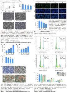
2.1 NaAsO2抑制MIHA细胞活性且呈浓度依赖性 根据CCK-8结果计算IC50值,确定NaAsO2处理时间为48 h,见图1A。与对照组(0 μmol/L NaAsO2)相比,10-30 μmol/L NaAsO2组细胞活性均显著降低(P < 0.05),且随着NaAsO2浓度的增加,MIHA细胞活性逐渐降低,见图1B,呈剂量-效应关系。 2.2 NaAsO2导致MIHA细胞形态改变 倒置显微镜观察显示,对照组(0 μmol/L NaAsO2)细胞状态良好,胞膜光滑完整,胞质丰富饱满,呈多边形以铺路石样排列紧密,见图1C。与对照组相比,实验组随着NaAsO2浓度的增加,胞膜逐渐模糊不清,细胞逐渐失去正常形态,胞质减少,空泡逐渐增多;细胞融合率逐渐降低,间隙增宽,破碎裂解及脱落漂浮的细胞越来越多。 2.3 NaAsO2引起MIHA细胞脂质代谢紊乱 与对照组(0 μmol/LNaAsO2)比较,10-30 μmol/L NaAsO2组细胞上清总胆固醇、三酰甘油水平显著增高(P < 0.05),见图2A,B,且呈剂量-效应关系。与之相反,10-30 μmol/L NaAsO2组细胞上清总胆汁酸水平显著降低(P < 0.05),见图2C,呈剂量-效应关系。 2.4 NaAsO2引起MIHA细胞脂肪变性 油红O染色检测各组MIHA细胞内脂质含量及分布,结果显示,对照组(0 μmol/L NaAsO2)细胞结构正常,胞内无脂滴形成,见图2D。与对照组相比,10-30 μmol/L NaAsO2组细胞肿胀,多数细胞胞浆内脂肪空泡形成,脂滴大量形成;细胞结构明显异常。 2.5 NaAsO2阻滞细胞增殖 0,10,20,30 μmol/L NaAsO2组细胞增殖率分别为(35.71±0.39)%,(30.84±0.57)%,(24.90±0.26)%,(19.73±0.77)%,与对照组(0 μmol/L NaAsO2)相比,10,20,30 μmol/L NaAsO2组细胞增殖率显著降低(P < 0.05),见图3,且随着NaAsO2浓度的增加,细胞增殖抑制愈加明显,呈剂量-效应关系。 2.6 NaAsO2诱导细胞周期停滞 0,10,20,30 μmol/L NaAsO2组细胞S期细胞比例分别为(15.21±0.36)%,(19.19±0.18)%,(25.21±1.03)%,(28.63±0.87)%,G2/M期细胞比例分别为(6.08±0.30)%,(11.02±0.29)%,(13.21±0.40)%,(16.81±0.72)%,与对照组(0 μmol/L NaAsO2)相比,10,20,30 μmol/L NaAsO2组周期被阻滞在S期及G2/M期的细胞比例显著增加(P < 0.05),且随着NaAsO2浓度的增加,细胞S期及G2/M期阻滞引起细胞增殖抑制愈加明显,呈剂量-效应关系,见图4。"

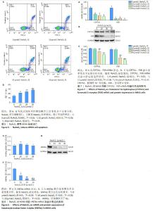
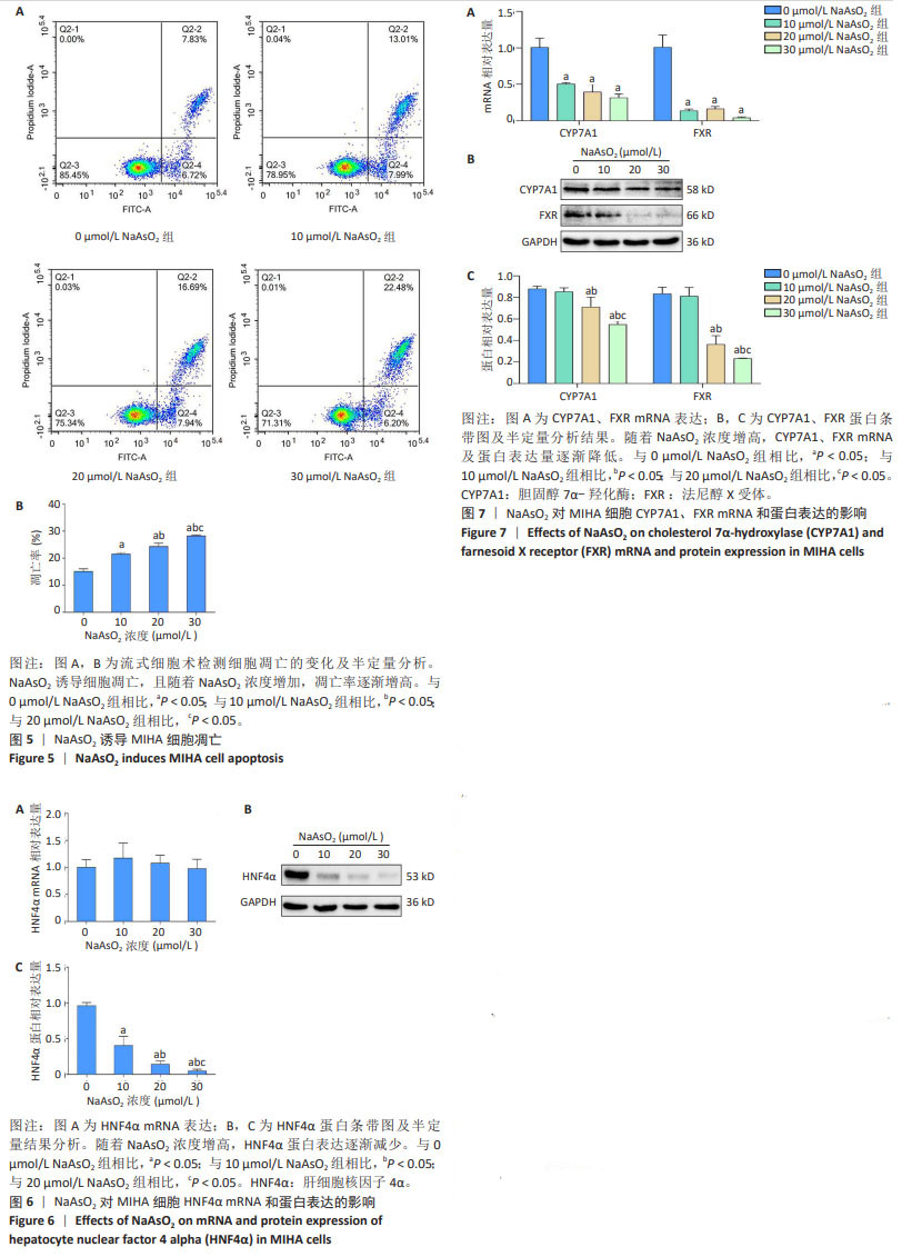
2.7 NaAsO2诱导细胞凋亡 0,10,20,30 μmol/L NaAsO2组细胞凋亡率分别为(15.14±0.57)%,(21.56±0.31)%,(24.34±0.68)%,(28.23±0.23)%,与对照组(0 μmol/L NaAsO2)相比,10,20,30 μmol/L NaAsO2组细胞凋亡率显著增加(P < 0.05),且随着 NaAsO2浓度的增加,细胞凋亡率增加愈明显,呈剂量-效应关系,见图5。 2.8 NaAsO2导致MIHA细胞HNF4α mRNA水平无变化、蛋白水平明显降低 RT-qPCR结果显示,各组间HNF4α mRNA相对表达量均无统计学差异(P > 0.05),见图6A。 Western blot结果显示,与对照组相比,10-30 μmol/L NaAsO2组HNF4α蛋白相对表达量明显降低,且随着NaAsO2浓度的增加,HNF4α蛋白相对表达量逐渐降低,呈剂量-效应关系,见图6B,C。 2.9 NaAsO2诱导MIHA细胞CYP7A1、FXR mRNA及蛋白水平显著降低 随着NaAsO2浓度逐渐增加,CYP7A1、FXR mRNA及蛋白相对表达量逐渐降低。RT-qPCR结果显示,与对照组相比,10-30 μmol/L NaAsO2组CYP7A1、FXR mRNA相对表达量明显降低,见图7A,差异有显著性意义(P < 0.05);Western blot结果显示,与对照组相比,20,30 μmol/L NaAsO2组CYP7A1及FXR蛋白相对表达量明显降低,见图7B,C,差异有显著性意义(P < 0.05)。"
| 1] PASCAL M, BEAUDEAU P, MEDINA S, et al. Global Change: a Public Health Researcher’s Ethical Responsibility. Curr Environ Health Rep. 2019;6(3):160-166. [2] COHEN SM, ARNOLD LL, BECK BD, et al. Evaluation of the carcinogenicity of inorganic arsenic. Crit Rev Toxicol. 2013;43(9):711-752. [3] CANTONI O, ZITO E, GUIDARELLI A, et al. Mitochondrial ROS, ER Stress, and Nrf2 Crosstalk in the Regulation of Mitochondrial Apoptosis Induced by Arsenite. Antioxidants (Basel). 2022;11(5):1034. [4] GUIDELINES FOR DRINKING-WATER QUALITY. Fourth edition incorporating the first and second addenda [Internet]. Geneva: World Health Organization, 2022. [5] RO SH, BAE J, JANG Y, et al. Arsenic Toxicity on Metabolism and Autophagy in Adipose and Muscle Tissues. Antioxidants (Basel). 2022; 11(4):689. [6] GANIE SY, JAVAID D, HAJAM YA, et al. Arsenic toxicity: sources, pathophysiology and mechanism. Toxicol Res (Camb). 2023;13(1): tfad111. [7] SUN J, WU L, WU M, et al. Non-coding RNA therapeutics: Towards a new candidate for arsenic-induced liver disease. Chem Biol Interact. 2023;382:110626. [8] LIANG J, LI L, LI L, et al. Lipid metabolism reprogramming in head and neck cancer. Front Oncol. 2023;13:1271505. [9] XU K, XIA P, GONGYE X, et al. A novel lncRNA RP11-386G11.10 reprograms lipid metabolism to promote hepatocellular carcinoma progression. Mol Metab. 2022;63:101540. [10] YOUNOSSI ZM, GOLABI P, PRICE JK, et al. The Global Epidemiology of Nonalcoholic Fatty Liver Disease and Nonalcoholic Steatohepatitis Among Patients With Type 2 Diabetes. Clin Gastroenterol Hepatol. 2024:S1542-3565(24)00287-8. doi: 10.1016/j.cgh.2024.03.006. [11] MEDDA N, DE SK, MAITI S. Different mechanisms of arsenic related signaling in cellular proliferation, apoptosis and neo-plastic transformation. Ecotoxicol Environ Saf. 2021;208:111752. [12] RENU K, CHAKRABORTY R, MYAKALA H, et al. Molecular mechanism of heavy metals (Lead, Chromium, Arsenic, Mercury, Nickel and Cadmium) - induced hepatotoxicity - A review. Chemosphere. 2021;271:129735. [13] RADI SH, VEMURI K, MARTINEZ-LOMELI J, et al. HNF4α isoforms: the fraternal twin master regulators of liver function. Front Endocrinol (Lausanne). 2023;14:1226173. [14] CHIANG JYL, FERRELL JM. Discovery of farnesoid X receptor and its role in bile acid metabolism. Mol Cell Endocrinol. 2022;548:111618. [15] CHAMBERS KF, DAY PE, ABOUFARRAG HT, et al. Polyphenol Effects on Cholesterol Metabolism via Bile Acid Biosynthesis, CYP7A1: A Review. Nutrients. 2019;11(11):2588. [16] KIR S, ZHANG Y, GERARD RD, et al. Nuclear receptors HNF4α and LRH-1 cooperate in regulating Cyp7a1 in vivo. J Biol Chem. 2012;287(49): 41334-41341. [17] 王甜,赵哲仪,穆银贵,等.亚砷酸钠所致L-02人肝细胞损伤与p14ARF表达下调及MDM2、p53表达增加有关[J].细胞与分子免疫学杂志,2020,36(6):507-512. [18] THYMIAKOU E, OTHMAN A, HORNEMANN T, et al. Defects in High Density Lipoprotein metabolism and hepatic steatosis in mice with liver-specific ablation of Hepatocyte Nuclear Factor 4A. Metabolism. 2020;110:154307. [19] GIRISA S, HENAMAYEE S, PARAMA D, et al. Targeting Farnesoid X receptor (FXR) for developing novel therapeutics against cancer. Mol Biomed. 2021;2(1):21. [20] SCHROEDER F, JOLLY CA, CHO TH, et al. Fatty acid binding protein isoforms: structure and function. Chem Phys Lipids. 1998;92(1):1-25. [21] AFOLABI OK, WUSU AD, OGUNRINOLA OO, et al. Arsenic-induced dyslipidemia in male albino rats: comparison between trivalent and pentavalent inorganic arsenic in drinking water. BMC Pharmacol Toxicol. 2015;16:15. [22] KUO CC, SU PH, SUN CW, et al. Early-life arsenic exposure promotes atherogenic lipid metabolism in adolescence: A 15-year birth cohort follow-up study in central Taiwan. Environ Int. 2018;118:97-105. [23] GARCIAFIGUEROA DY, KLEI LR, AMBROSIO F, et al. Arsenic-stimulated lipolysis and adipose remodeling is mediated by G-protein-coupled receptors. Toxicol Sci. 2013;134(2):335-344. [24] EVANS RM, MANGELSDORF DJ. Nuclear Receptors, RXR, and the Big Bang. Cell. 2014;157(1):255-266. [25] HERRERA-PULIDO JA, BOISVERT FM, Boudreau F. Hepatocyte nuclear factor 4α multiple isoforms, their functions, and their interactomes. Proteomics. 2023;23(13-14):e2200372. [26] HUCK I, MORRIS EM, THYFAULT J, et al. Hepatocyte-Specific Hepatocyte Nuclear Factor 4 Alpha (HNF4) Deletion Decreases Resting Energy Expenditure by Disrupting Lipid and Carbohydrate Homeostasis. Gene Expr. 2021;20(3):157-168. [27] XU Y, ZHOU Z, KANG X, et al. Mettl3-mediated mRNA m6A modification controls postnatal liver development by modulating the transcription factor Hnf4a. Nat Commun. 2022;13(1):4555. [28] ZHU C, DONG B, SUN L, et al. Cell Sources and Influencing Factors of Liver Regeneration: A Review. Med Sci Monit. 2020;26:e929129. [29] HAIDAR Z, FATEMA K, SHOILY SS, et al. Disease-associated metabolic pathways affected by heavy metals and metalloid. Toxicol Rep. 2023; 10:554-570. [30] KHAN AN, SINGH R, BHATTACHARYA A, et al. Glucogallin Attenuates RAW 264.7 Cells from Arsenic Trioxide Induced Toxicity via the NF-ҡB/NLRP3 Pathway. Molecules. 2022;27(16):5263. [31] NING BF, DING J, LIU J, et al. Hepatocyte nuclear factor 4α-nuclear factor-κB feedback circuit modulates liver cancer progression. Hepatology. 2014;60(5):1607-1619. [32] RENU K, SARAVANAN A, ELANGOVAN A, et al. An appraisal on molecular and biochemical signalling cascades during arsenic-induced hepatotoxicity. Life Sci. 2020;260:118438. [33] PASTORET A, MARCOS R, SAMPAYO-REYES A, et al. Inhibition of hepatocyte nuclear factor 1 and 4 alpha (HNF1α and HNF4α) as a mechanism of arsenic carcinogenesis. Arch Toxicol. 2013;87(6): 1001-1012. [34] 赵哲仪,王正蓉,方兴艳,等.亚砷酸钠对AML12肝细胞氧化应激、凋亡损伤及Hippo信号通路的影响[J].中国组织工程研究, 2022,26(26):4186-4191. [35] CAI WY, LIN LY, HAO H, et al. Yes-associated protein/TEA domain family member and hepatocyte nuclear factor 4-alpha (HNF4α) repress reciprocally to regulate hepatocarcinogenesis in rats and mice. Hepatology. 2017;65(4):1206-1221. [36] BIAGIONI F, CROCI O, SBERNA S, et al. Decoding YAP dependent transcription in the liver. Nucleic Acids Res. 2022;50(14):7959-7971. [37] TVETER KM, MEZHIBOVSKY E, WU Y, et al. Bile acid metabolism and signaling: Emerging pharmacological targets of dietary polyphenols. Pharmacol Ther. 2023;248:108457. [38] FAROOQUI N, ELHENCE A, SHALIMAR. A Current Understanding of Bile Acids in Chronic Liver Disease. J Clin Exp Hepatol. 2022;12(1):155-173. [39] 徐懂,尹美君,王钰铭,等.FXR-CYP7A1轴在胆汁淤积性肝病中的调控研究进展[J].中西医结合肝病杂志,2021,31(5):470-473. [40] GUTHRIE G, VONDEROHE C, BURRIN D. Fibroblast growth factor 15/19 expression, regulation, and function: An overview. Mol Cell Endocrinol. 2022;548:111617. [41] SONG Y, XU C, SHAO S, et al. Thyroid-stimulating hormone regulates hepatic bile acid homeostasis via SREBP-2/HNF-4α/CYP7A1 axis. J Hepatol. 2015;62(5):1171-1179. [42] JIN B, LI H, ZHANG H, et al. Effects of carnosic acid on arsenic-induced liver injury in mice: A comparative transcriptomics analysis. J Trace Elem Med Biol. 2022;71:126953. [43] LI J, GUO C, LIU Y, et al. Chronic arsenic exposure-provoked biotoxicity involved in liver-microbiota-gut axis disruption in chickens based on multi-omics technologies. J Adv Res. 2024:S2090-1232(24)00032-8. doi: 10.1016/j.jare.2024.01.019. [44] YANG Y, CHI L, LIU CW, et al. Chronic Arsenic Exposure Perturbs Gut Microbiota and Bile Acid Homeostasis in Mice. Chem Res Toxicol. 2023;36(7):1037-1043. [45] YANG Y, HSIAO YC, LIU CW, et al. The Role of the Nuclear Receptor FXR in Arsenic-Induced Glucose Intolerance in Mice. Toxics. 2023; 11(10):833. [46] RIBAS GS, VARGAS CR. Evidence that Oxidative Disbalance and Mitochondrial Dysfunction are Involved in the Pathophysiology of Fatty Acid Oxidation Disorders. Cell Mol Neurobiol. 2022;42(3):521-532. [47] HAGA S, YIMIN, OZAKI M. Relevance of FXR-p62/SQSTM1 pathway for survival and protection of mouse hepatocytes and liver, especially with steatosis. BMC Gastroenterol. 2017;17(1):9. [48] LV R, ZHU M, CHEN K, et al. Z-Guggulsterone Induces Apoptosis in Gastric Cancer Cells through the Intrinsic Mitochondria-Dependent Pathway. ScientificWorldJournal. 2021;2021:3152304. [49] KIM DH, PARK JS, CHOI HI, et al. The critical role of FXR is associated with the regulation of autophagy and apoptosis in the progression of AKI to CKD. Cell Death Dis. 2021;12(4):320. [50] FUJINO T, MARUKO-OHTAKE A, OHTAKE Y, et al. Farnesoid X receptor knockdown provides significant growth inhibition in hepatocellular carcinoma cells while it does not interfere with the proliferation of primary human hepatocyte-derived cells. J Toxicol Sci. 2015;40(4): 501-508. [51] QIAO P, LI S, ZHANG H, et al. Farnesoid X receptor inhibits proliferation of human colorectal cancer cells via the miR‑135A1/CCNG2 signaling pathway. Oncol Rep. 2018;40(4):2067-2078. [52] 赵哲仪,王正蓉,方兴艳,等.亚砷酸钠对小鼠肝细胞AML12损伤的作用机制[J].贵州医科大学学报,2022,47(1):1-6. |
| [1] | Lan Shuangli, Xiang Feifan, Deng Guanghui, Xiao Yukun, Yang Yunkang, Liang Jie. Naringin inhibits iron deposition and cell apoptosis in bone tissue of osteoporotic rats [J]. Chinese Journal of Tissue Engineering Research, 2025, 29(5): 888-898. |
| [2] | Lin Shuqian, Zhao Xilong, Gao Jing, Pan Xinghua, Li Zian, Ruan Guangping. Comparison of biological characteristics of mouse bone marrow mesenchymal stem cells after interference and overexpression of telomere Cajal body protein-1 [J]. Chinese Journal of Tissue Engineering Research, 2025, 29(31): 6616-6624. |
| [3] | Zhu Liuhui, Zhang Xinyue, Zhu Zhouhai, Yang Xinglong, Guan Ying, Liu Bin. Coiled-coil-helix-coiled-coil-helix domain-containing 2 inhibits apoptosis of Parkinson’ s disease SH-SY5Y cells by promoting mitochondrial autophagy [J]. Chinese Journal of Tissue Engineering Research, 2025, 29(25): 5403-5413. |
| [4] | Yu Peng, Meng Dongfang, Li Huiying, Liu Hongfei, He Zike. Pinoresinol diglucoside activates the Wnt/beta-catenin signaling pathway to protect osteoblasts [J]. Chinese Journal of Tissue Engineering Research, 2025, 29(2): 339-346. |
| [5] | Zhang Jiaqi, Liu Yanhong, Liang Huiting, Zhou Jingjing, Wang Yawen, Xu Jingyu, Li Yushuang, Lei Lijian, Hu Xiaoqin. Bioinformatics analysis and functional verification of hsa-miR-3202 in osteoarthritic chondrocytes [J]. Chinese Journal of Tissue Engineering Research, 2025, 29(12): 2458-2465. |
| [6] | Fu Jiaqi, Chen Xiubao, Cui Xing, Chen Zetao . Mechanism by which Angelica sinensis polysaccharide regulates bone marrow hematopoietic microenvironment for aplastic anemia [J]. Chinese Journal of Tissue Engineering Research, 2025, 29(1): 44-51. |
| [7] | Chen Zepeng, Hou Yonghui, Chen Shudong, Hou Yu, Lin Dingkun. Tauroursodeoxycholic acid treats spinal cord injury by reducing apoptosis of spinal cord neurons under glucose and oxygen deprivation [J]. Chinese Journal of Tissue Engineering Research, 2024, 28(4): 528-534. |
| [8] | Zhao Yiting, Zhang Yuxiang, Ma Jie, He Xuejiao. Vascular endothelial growth factor 165/bone morphogenetic protein improves osteoblast injury under hypoxic and reoxygenated conditions [J]. Chinese Journal of Tissue Engineering Research, 2024, 28(35): 5669-5674. |
| [9] | Zhou Minghan, Zhang Hui, Zheng Xianbo, Xu Wuji. Significance of PI3K/Akt/HIF-1α signaling pathway expression in nucleus pulposus cells at different oxygen concentrations in delaying intervertebral disc degeneration [J]. Chinese Journal of Tissue Engineering Research, 2024, 28(28): 4491-4497. |
| [10] | Duan Yanzhe, Hua Jianlin, Ding Zhibin, Jiang Nan, Song Lijuan, Yan Yuqing, Ma Cungen. Visual analysis of the effect of apoptosis on ischemic stroke [J]. Chinese Journal of Tissue Engineering Research, 2024, 28(26): 4145-4150. |
| [11] | Ma Wanli, Yang Hongsheng, Qu Bo, Zhang Zhengdong, Gong Kai, Lin Yanshui. Mechanisms by which baicalein protects against steriod-induced osteonecrosis of the femoral head in rats [J]. Chinese Journal of Tissue Engineering Research, 2024, 28(23): 3661-3668. |
| [12] | Zuo Jun, Ma Shaolin. Mechanism of beta-sitosterol on hypertrophic scar fibroblasts: an analysis based on network pharmacology [J]. Chinese Journal of Tissue Engineering Research, 2024, 28(2): 216-223. |
| [13] | Chen Simin, Hu Yingjun, Yan Wenrui, Ji Le, Shao Mengli, Sun Ze, Zheng Hongxing, Qi Shanshan. Establishment and evaluation of a streptozotocin-induced diabetic encephalopathy rat model [J]. Chinese Journal of Tissue Engineering Research, 2024, 28(2): 237-241. |
| [14] | Fan Zhihong, Zhang Xian, Li Chao. Wnt signaling pathway in intervertebral disc degeneration [J]. Chinese Journal of Tissue Engineering Research, 2024, 28(12): 1950-1955. |
| [15] | Ma Suilu, He Zhijun, Liu Tao, Li Yan, He Yuanxu, He Bo, Wang Weiwei, Wei Xiaotao. Traditional Chinese medicine monomer in the prevention and treatment of flap necrosis by regulating “autophagy” [J]. Chinese Journal of Tissue Engineering Research, 2024, 28(1): 153-158. |
| Viewed | ||||||
|
Full text |
|
|||||
|
Abstract |
|
|||||