Chinese Journal of Tissue Engineering Research ›› 2024, Vol. 28 ›› Issue (32): 5122-5129.doi: 10.12307/2024.510
Previous Articles Next Articles
Autophagy, ferroptosis-related targets and renal function progression in patients with chronic kidney disease: bioinformatics analysis and experimental verification
Chen Guanting1, Zhang Linqi2, Wang Xixi2, Chen Xu2
- 1The First Clinical Medical College of Henan University of Chinese Medicine, Zhengzhou 450003, Henan Province, China; 2The First Affiliated Hospital of Henan University of Chinese Medicine, Zhengzhou 450003, Henan Province, China
-
Received:2023-03-29Accepted:2023-10-10Online:2024-11-18Published:2023-12-28 -
Contact:Zhang Linqi, Professor, Chief physician, Doctoral supervisor, The First Affiliated Hospital of Henan University of Chinese Medicine, Zhengzhou 450003, Henan Province, China -
About author:Chen Guanting, MD candidate, The First Clinical Medical College of Henan University of Chinese Medicine, Zhengzhou 450003, Henan Province, China -
Supported by:National Natural Science Foundation of China (General Program), No. 81973806 (to ZLQ); Special Project for Scientific Research on Traditional Chinese Medicine in Henan Province, No. 2019ZYZD05 (to ZLQ)
CLC Number:
Cite this article
Chen Guanting, Zhang Linqi, Wang Xixi, Chen Xu. Autophagy, ferroptosis-related targets and renal function progression in patients with chronic kidney disease: bioinformatics analysis and experimental verification[J]. Chinese Journal of Tissue Engineering Research, 2024, 28(32): 5122-5129.
share this article
Add to citation manager EndNote|Reference Manager|ProCite|BibTeX|RefWorks
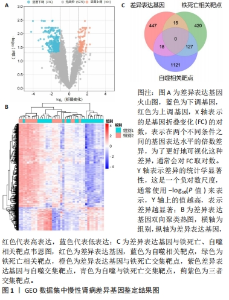
2.1 GSE137570中差异表达基因鉴定结果 实验通过OMIM,GENECARD,FerrDb及HAMdb数据库获取铁死亡相关靶点562个,自噬相关靶点1 266个;通过Networkanalyst数据库对GSE137570数据集进行分析,队列1(n=24)获得685个差异基因,队列2(n=17)获得789个差异基因,合并筛选后共计获取480个差异基因,其中表达上调基因104个,表达下调基因376个。将差异基因与铁死亡、自噬相关靶点取交集,分别获得铁死亡相关靶点15个,自噬相关靶点18个,共有靶点0个,结果如图1所示。"
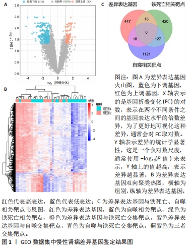
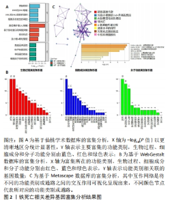
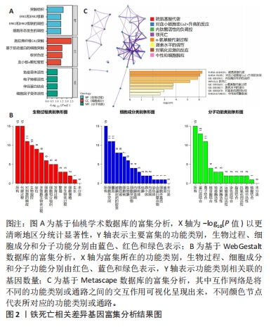
2.2 富集分析结果 实验通过仙桃学术、Metascape、WebGestalt数据库对铁死亡及自噬相关差异基因进行富集分析,GO-BP表明,铁死亡相关差异基因主要涉及对生物体内内肽酶活性、激素水平、能量代谢过程以及ERK1/2级联信号通路的调控,自噬相关差异基因主要涉及生物体内细胞形态、代谢过程及对刺激的响应等方面的调控;GO-CC方面,铁死亡相关差异基因主要涉及施拉弗纤维CA1突触、基于肌动蛋白的细胞突触、细胞外空间及囊泡等,自噬相关差异基因主要涉及生物膜、内膜系统、微体腔室、波尔体基质;GO-MF方面,铁死亡相关差异基因主要涉及蛋白质结合、离子结合、免疫受体活性、电子转移活性,自噬相关差异基因主要涉及氧化还原酶活性、外肽酶活性、蛋白质结合、离子结合等;Wiki,Reactome及KEGG等富集结果表明,铁死亡相关差异基因主要涉及硫氨基酸代谢、血小板胞浆Ca2+升高及中性粒细胞脱粒等生物学过程及铁死亡信号通路,与细胞代谢、免疫响应、细胞生存和细胞死亡等多个方面息息相关;自噬相关差异基因主要富集于血小板脱颗粒、RAC3 GTP酶循环、细胞外基质降解、中性粒细胞脱颗粒以及受体酪氨酸激酶的信号传导,这些过程可能在免疫反应、细胞迁移、组织修复和炎症等生理和病理情况中扮演重要角色。结果如图2,3所示。"
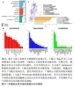
"

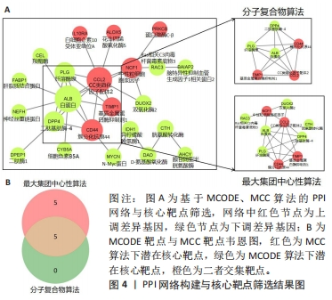
2.3 蛋白互作网络构建 将铁死亡及自噬相关差异基因导入STRING 11.5数据库进行分析,获取PPI网络信息并导入Cytoscape 3.9.1软件进行可视化分析;运用分子复合物检测(molecular complex detection,MCODE)及CytoHubba筛选PPI网络中关键基因,其中MCODE插件旨在庞大的网络中根据节点与边的关系,使用定点加权(Vertex-Weighting)方案发现网络中局部高密度区域并寻找出关键的子网络和基因;CytoHubba插件则是根据节点在网络中的属性进行排名,共有11种拓扑方法,其中以最大集团中心性(maximal clique centrality,MCC)算法精准度最高,结果显示二者高度相似,选取其交集为该实验的潜在核心靶点,其中自噬相关靶点为簇分化抗原44(cluster of differentiation 44,CD44)、白蛋白(albumin,ALB)、纤溶酶原(plasminogen,PLG)、CC类趋化因子配体2(chemokine C-C motif ligand 2,CCL2),铁死亡相关靶点为基质金属蛋白酶抑制剂1(tissue Inhibitor of metalloproteinases 1,TIMP1)、二肽基肽酶4(dipeptidyl peptidase-4,DPP4),结果如图4所示。"


2.4 免疫浸润分析结果 实验根据Timer 2.0数据库中Cibersort算法[12],对不同组别人群的肾组织进行组间免疫浸润细胞相对比例差异分析,并通过WPS CORREL函数计算差异表达细胞与核心靶点之间的相关性。结果如图5所示,队列1两组患者肾组织中休息状态髓样树突细胞(Myeloid dendritic cell resting)具有显著差异性(P < 0.05),其核心靶点CD44,TIMP1,CCL2与其呈中等正相关,ALB与其呈中等负相关;队列2两组患者肾组织中记忆休息状态CD4+记忆性T细胞(T cell CD4+ memory resting),休息状态自然杀伤细胞(NK cell resting),激活状态自然杀伤细胞(NK cell activated),单核细胞(Monocyte)具有显著差异性(P < 0.05),其核心靶点CD44,TIMP1与休息状态自然杀伤细胞呈强正相关,CD44,TIMP1,CCL2与激活状态自然杀伤细胞呈强负相关,DPP4与激活状态自然杀伤细胞呈强正相关。"
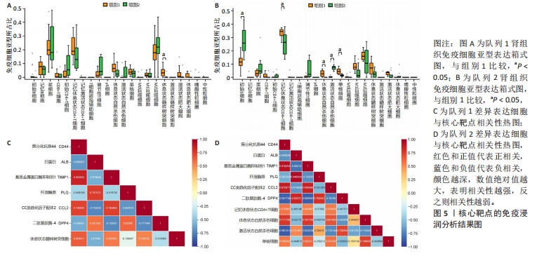
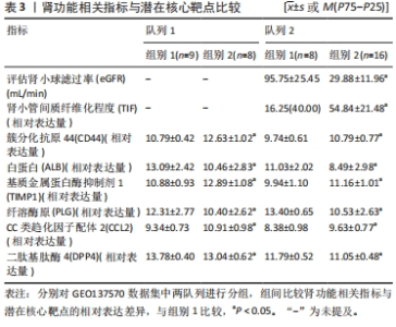
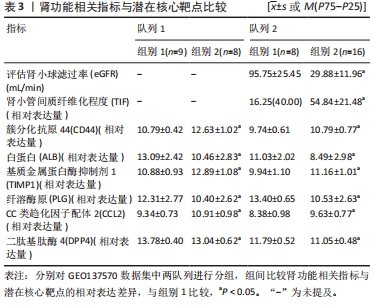
2.5 肾功能相关指标与潜在核心靶点比较结果 根据GSE137570数据集纳入样本进行分析,如表3所示,队列2中组别2 慢性肾病患者eGFR显著低于组别1,TIF显著高于组别1(P < 0.05);两队列中组别2 慢性肾病患者肾组织CD44,CCL2及TIMP1表达均显著高于组别1,ALB,PLG及DPP4表达均显著低于组别1(P < 0.05)。"
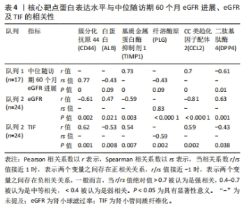
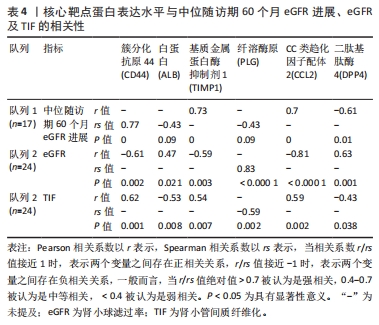
2.6 潜在核心靶点与肾损伤严重程度的相关性 将该实验获取潜在核心靶点分别与队列1中位随访期60个月eGFR进展及队列2患者eGFR及肾小管间质纤维化(renal tubulointerstitial fibrosis,TIF)程度做相关性分析,结果见表4。相关性分析显示,在队列1中CD44,TIMP1,CCL2与eGFR进展呈正相关(P < 0.05),ALB,PLG,DPP4与其呈负相关(P < 0.05);队列2中ALB,PLG,DPP4与eGFR呈正相关(P < 0.05),与TIF呈负相关(P < 0.05),CD44,TIMP1,CCL2与eGFR呈负相关(P < 0.05),与TIF呈正相关(P < 0.05)。"
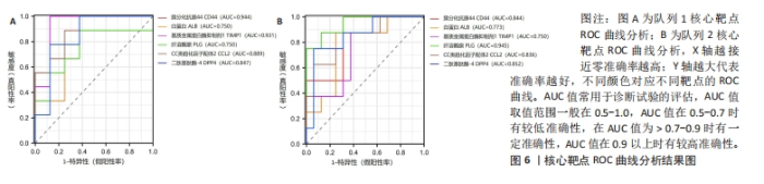
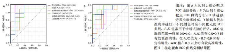
2.7 核心靶点预测慢性肾病患者肾功能进展的效能 通过对GSE137570数据集中核心靶点进行ROC曲线分析,结果如图6所示,CD44,ALB,TIMP1,PLG,CCL2及DPP4可较好地预测肾功能进展,在队列1中,CD44,ALB,TIMP1,PLG,CCL2及DPP4 AUC值分别为0.844(95%CI:0.679-1.000)、0.773(95%CI:0.569-0.977)、0.75(95%CI:0.543-0.957)、0.945(95%CI:0.855-1.000)、0.836(95%CI:0.657-1.000)、0.852(95%CI:0.680-1.000);在队列2中,其AUC值分别为0.944(95%CI:0.827-1.000)、0.750(95%CI:0.482-1.000)、0.931(95%CI:0.788-1.000)、0.750(95%CI:0.495-1.000)、0.733(95%CI:0.568-0.878)、0.639(95%CI:0.827-1.000)。"

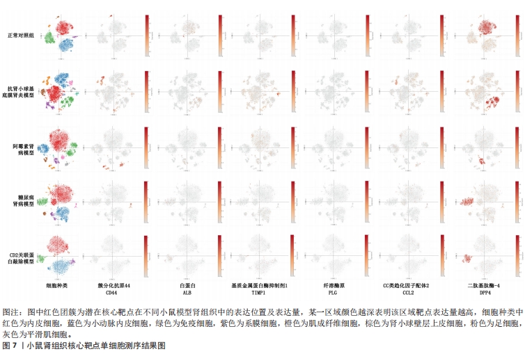
2.8 单细胞测序验证结果 实验利用Single Cell Portal数据库对实验所筛选潜在核心靶点进行单细胞测序分析,如图7所示,与对照组小鼠相比,抗肾小球基底膜肾炎小鼠模型及阿霉素肾病小鼠模型肾组织中CD44,TIMP1,CCL2表达量显著升高,ALB,DPP4表达显著降低,PLG无显著变化;糖尿病肾病小鼠模型肾组织中CCL2表达量显著升高,ALB,DPP4表达显著降低,CD44,TIMP1,PLG无显著变化;CD2关联蛋白敲除小鼠模型肾组织中CD44,ALB,CCL2,DPP4表达显著降低,TIMP1表达量显著升高,PLG无显著变化。除PLG外,各模型组小鼠肾组织靶点基因表达与该实验结果基本一致。"
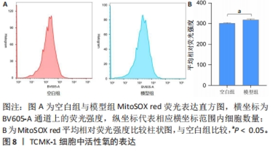
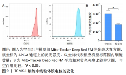
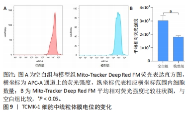
2.9 流式细胞术检测结果 MitoSOX red及Mito-Tracker Deep Red FM是一种荧光染色探针,具有线粒体特异性,主要定位于线粒体内。MitoSOX red与线粒体内产生的活性氧发生反应,一般情况下,MitoSOX red荧光强度与活性氧的表达量呈正相关关系,即活性氧水平升高,MitoSOX的荧光强度也会增加;Mito-Tracker Deep Red FM因其本身具有正电荷,这个正电荷使得Mito-Tracker Deep Red FM在细胞外部的负电势区域更容易进入细胞内,而在细胞内具有负电荷的线粒体内膜更容易吸引和累积Mito-Tracker Deep Red FM,这个积累是与线粒体膜电位的高低成正比的,因此,当线粒体膜电位较高时,Mito-Tracker Deep Red FM分子在线粒体内膜上的积累也相对较高。结果如图8,9所示,与空白组比较,模型组TCMK1细胞内活性氧荧光强度显著增加(P < 0.05),线粒体膜电位显著降低(P < 0.05)。"

"
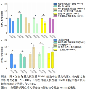
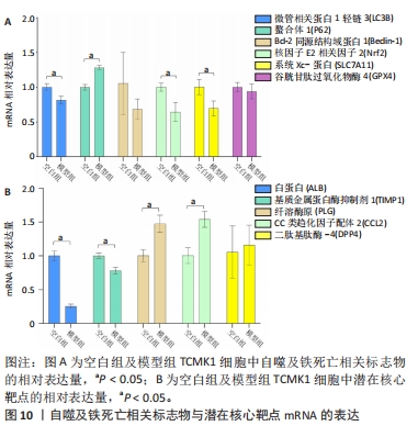
2.10 RT-PCR检测结果 对于自噬及铁死亡表型的验证,结果如图10A所示,与空白组比较,模型组中微管相关蛋白1轻链3(microtubule-associated protein1light chain3,LC3B)、核因子E2相关因子2(nuclear factor erythroid 2-related factor 2,Nrf2)、系统Xc-蛋白(solute carrier family 7 member 11,SLC7A11)mRNA表达显著降低(P < 0.05),螯合体1(sequestosome 1,SQSTM1/P62)mRNA表达显著增高(P < 0.05),Bcl-2同源结构域蛋白1(Beclin-1)及谷胱甘肽过氧化物酶4(glutathione peroxidase 4,GPX4)mRNA表达有所降低,但无显著性;潜在核心靶点验证方面,结果如图10B所示,与空白组比较,模型组中ALB,PLG mRNA表达显著降低(P < 0.05),TIMP1,CCL2 mRNA表达显著升高(P < 0.05),CD44 mRNA表达有所升高,但无显著性。DPP4未检出。"
| [1] 陈欠欠,朱凤阁,陈香美.姜黄素治疗肾间质纤维化分子机制研究进展[J].中草药,2023,54(3):966-975. [2] HUMPHREYS BD. Mechanisms of renal fibrosis. Annu Rev Physiol. 2018;80(1): 309-326. [3] JIANG X, STOCKWELL BR, CONRAD M. Ferroptosis: mechanisms, biology and role in disease. Nat Rev Mol Cell Biol. 2021;22(4):266-282. [4] OTASEVIC V, VUCETIC M, GRIGOROV I, et al. Ferroptosis in different pathological contexts seen through the eyes of mitochondria. Oxid Med Cell Longev. 2021; 2021(6):5537330. [5] WANG J, LIU Y, WANG Y, et al. The cross-link between ferroptosis and kidney diseases. Oxid Med Cell Longev. 2021;2021:6654887. [6] 李磊,夏煜琦,程帆.铁死亡在肾纤维化中的作用研究进展[J].疑难病杂志, 2022,21(8):872-876. [7] CAO W, LI J, YANG K, et al. An overview of autophagy: mechanism, regulation and research progress. Bull Cancer. 2021;108(3):304-322. [8] CHOI EM. Autophagy in kidney disease. Annu Rev Physiol. 2020,82(1):297-322. [9] LIU J, LIU Y, WANG Y, et al. TMEM164 is a new determinant of autophagy-dependent ferroptosis. Autophagy. 2023;19(3):945-956. [10] CHUNG JJ, GOLDSTEIN L, Chen YJ, et al. Single-cell transcriptome profiling of the kidney glomerulus identifies key cell types and reactions to injury. J Am Soc Nephrol. 2020;31(10):2341-2354. [11] PENG Z, GUO HY, LI YQ, et al. The Smad3-dependent microRNA let-7i-5p promoted renal fibrosis in mice with unilateral ureteral obstruction. Front Physiol. 2022;13:937878. [12] KANG K, XIE F, MAO J, et al. Significance of tumor mutation burden in immune infiltration and prognosis in cutaneous melanoma. Front Oncol. 2020;10:573141. [13] SORICE M. Crosstalk of autophagy and apoptosis. Cells. 2022;11(9):1479. [14] 黄衍恒,叶霖,黄小荣,等.肾脏固有细胞自噬对肾纤维化作用的研究进展[J].中华肾脏病杂志,2022,38(3):247-253. [15] PRERNA K, DUBEY VK. Beclin1-mediated interplay between autophagy and apoptosis: new understanding. Int J Biol Macromol. 2022;204:258-273. [16] PENA-MARTINE C, RICKMAN AD, HECKMANN BL. Beyond autophagy: LC3-associated phagocytosis and endocytosis. Sci Adv. 2022;28;8(43):eabn1702. [17] VARGAS JNS, HAMASAKI M, KAWABATA T, et al. The mechanisms and roles of selective autophagy in mammals. Nat Rev Mol Cell Biol. 2023;24(3):167-185. [18] ZHANG B, CHEN X, RU F, et al. Liproxstatin-1 attenuates unilateral ureteral obstruction-induced renal fibrosis by inhibiting renal tubular epithelial cells ferroptosis. Cell Death Dis. 2021;12(9):843-843. [19] GE MH, TIAN H, MAO L, et al. Zinc attenuates ferroptosis and promotes functional recovery in contusion spinal cord injury by activating Nrf2/GPX4 defense pathway. CNS Neurosci Ther. 2021;27(9):1023-1040. [20] LI MY, DAI XH, YU XP, et al. Scalp acupuncture protects against neuronal ferroptosis by activating the p62-Keap1-Nrf2 pathway in rat models of intracranial haemorrhage. J Mol Neurosci. 2022;72(1):82-96. [21] 米海潮,史敏,崔芳.铁自噬与铁死亡及其相关疾病[J].中国生物化学与分子生物学报,2022,38(9):1133-1140. [22] JIN T, CHEN C. Umbelliferone delays the progression of diabetic nephropathy by inhibiting ferroptosis through activation of the Nrf-2/HO-1 pathway. Food Chem Toxicol. 2022;163:112892. [23] WANG J, WANG Y, LIU Y, et al. Ferroptosis, a new target for treatment of renal injury and fibrosis in a 5/6 nephrectomy-induced CKD rat model. Cell Death Discov. 2022;8(1):127-127. [24] ZHOU L, XUE X, HOU Q, et al. Targeting ferroptosis attenuates interstitial inflammation and kidney fibrosis. Kidney Dis (Basel). 2022;8(1):57-71. [25] 张波,汝峰,陈湘,等.自噬通过抑制上皮间质转化减轻梗阻性肾病肾纤维化[J].中南大学学报(医学版),2021,46(6):601-608. [26] 毛稳,夏琳颖,刘贤莉,等.三七皂苷通过抑制自噬减轻TGF-β1诱导的肾小管上皮细胞损伤[J].中国实验方剂学杂志,2022,28(20):86-91. [27] RUBY M, GIFFORD CC, PANDEY R, et al. Autophagy as a therapeutic target for chronic kidney disease and the roles of TGF-β1 in autophagy and kidney fibrosis. Cells. 2023;12(3):412. [28] DJUDJAJ S, BOOR P. Cellular and molecular mechanisms of kidney fibrosis. Mol Aspects Med. 2019;65:16-36. [29] HOU W, XIE Y, SONG X, et.al. Autophagy promotes ferroptosis by degradation of ferritin. Autophagy. 2016;12(8):1425-1428. [30] LI J, CAO F, YIN HL, et al. Ferroptosis: past, present and future. Cell Death Dis. 2020;11(2):88. [31] ZHANG L, YANG P, CHEN J, et al. CD44 connects autophagy decline and ageing in the vascular endothelium. Nat Commun. 2023;14(1):5524-5524. [32] BAI RJ, Liu D, Li YS, et al. OPN inhibits autophagy through CD44, integrin and the MAPK pathway in osteoarthritic chondrocytes. Front Endocrinol (Lausanne). 2022;13:919366. [33] 高翔,梅长林.《慢性肾病早期筛查、诊断及防治指南(2022年版)》解读[J].中国实用内科杂志,2022,42(9):735-739. [34] WARD ES, GELINAS D, DREESEN E, et al. Clinical significance of serum albumin and implications of FcRn inhibitor treatment in IgG-mediated autoimmune disorders. Front Immunol. 2022;13:892534. [35] HUANG H, LIN Q, Dai X, et al. Derivation and validation of urinary TIMP-1 for the prediction of acute kidney injury and mortality in critically ill children. J Transl Med. 2022;20(1):102. [36] KOKENY G, NEMETH A, Kopp JB, et al. Susceptibility to kidney fibrosis in mice is associated with early growth response-2 protein and tissue inhibitor of metalloproteinase-1 expression. Kidney Int. 2022;102(2):337-354. [37] WANG L, WANG J, CHEN L. TIMP1 represses sorafenib-triggered ferroptosis in colorectal cancer cells by activating the PI3K/Akt signaling pathway. Immunopharmacol Immunotoxicol. 2023;45(4):419-425. [38] HEISSIG B, SALAMA Y, TAKAHASHI S, et al.The multifaceted role of plasminogen in inflammation. Cell Signal. 2020;75:109761. [39] KERAGALA CB, MEDCALF RL. Plasminogen: an enigmatic zymogen. Blood. 2021; 137(21):2881-2889. [40] TYKHOMYROV AA, NEDZVETSKY VS, AGCA CA, et al. Plasminogen/plasmin affects expression of glycolysis regulator TIGAR and induces autophagy in lung adenocarcinoma A549 cells. Exp Oncol. 2020;42(4):270-276. [41] LIN Z, SHI JL, CHEN M, et al. CCL2: an important cytokine in normal and pathological pregnancies: a review. Front Immunol. 2022;13:1053457. [42] 李秀萍,王玲,王馨,等.CC类趋化因子配体2对结核分枝杆菌感染人单核巨噬细胞THP-1自噬的影响及分子机制[J].临床误诊误治,2021,34(9):101-106. [43] FANG S, TANG H, LI MZ, et al. Identification of the CCL2 PI3K/Akt axis involved in autophagy and apoptosis after spinal cord injury. Metab Brain Dis. 2023;38(4): 1335. [44] 张志威,汪锴,姜长涛.菌源同工酶分析揭示菌源DPP4同工酶是潜在的抗2型糖尿病靶点[J].遗传,2023,45(8):629-631. [45] CUI X, YUN X, SUN M, et al. HMGCL-induced β-hydroxybutyrate production attenuates hepatocellular carcinoma via DPP4-mediated ferroptosis susceptibility. Hepatol Int. 2023;17(2):377-392. |
| [1] | Shen Jiangyong, He Xi, Tang Yuting, Wang Jianjun, Liu Jinyi, Chen Yuanyuan, Wang Xinyi, Liu Tong, Sun Haoyuan. RAS-selective lethal small molecule 3 inhibits the fibrosis of pathological scar fibroblasts [J]. Chinese Journal of Tissue Engineering Research, 2024, 28(8): 1168-1173. |
| [2] | Wang Ji, Zhang Min, Li Wenbo, Yang Zhongya, Zhang Long. Effect of aerobic exercise on glycolipid metabolism, skeletal muscle inflammation and autophagy in type 2 diabetic rats [J]. Chinese Journal of Tissue Engineering Research, 2024, 28(8): 1200-1205. |
| [3] | Zhou Bangyu, Li Jie, Ruan Yushang, Geng Funeng, Li Shaobo. Effects of Periplaneta americana powder on motor function and autophagic protein Beclin-1 in rats undergoing spinal cord hemisection [J]. Chinese Journal of Tissue Engineering Research, 2024, 28(8): 1223-1228. |
| [4] | Sheng Siqi, Xie Lin, Zhao Xiangyu, Jiang Yideng, Wu Kai, Xiong Jiantuan, Yang Anning, Hao Yinju, Jiao Yun. Involvement of miR-144-3p in Cbs+/- mouse hepatocyte autophagy induced by high-methionine diet [J]. Chinese Journal of Tissue Engineering Research, 2024, 28(8): 1289-1294. |
| [5] | Pan Xiaolong, Fan Feiyan, Ying Chunmiao, Liu Feixiang, Zhang Yunke. Effect and mechanism of traditional Chinese medicine on inhibiting the aging of mesenchymal stem cells [J]. Chinese Journal of Tissue Engineering Research, 2024, 28(7): 1091-1098. |
| [6] | Huang Yuxin, Liang Wenzi, Chen Xiuwen, Ni Na, Zhao Yinglin, Lin Changmin. Role of autophagy in hair regeneration [J]. Chinese Journal of Tissue Engineering Research, 2024, 28(7): 1112-1117. |
| [7] | Liu Qiwei, Zhang Junhui, Yang Yuan, Wang Jinjuan. Role and mechanism of umbilical cord mesenchymal stem cells on polycystic ovary syndrome [J]. Chinese Journal of Tissue Engineering Research, 2024, 28(7): 1015-1020. |
| [8] | Bu Xianzhong, Bu Baoxian, Xu Wei, Zhang Chi, Zhang Yisheng, Zhong Yuanming, Li Zhifei, Tang Fubo, Mai Wei, Zhou Jinyan. Analysis of serum differential proteomics in patients with acute cervical spondylotic radiculopathy [J]. Chinese Journal of Tissue Engineering Research, 2024, 28(4): 535-541. |
| [9] | Zhang Yaru, Chen Yanjun, Zhang Xiaodong, Chen Shenghua, Huang Wenhua. Effect of ferroptosis mediated by glutathione peroxidase 4 in the occurrence and progression of synovitis in knee osteoarthritis [J]. Chinese Journal of Tissue Engineering Research, 2024, 28(4): 550-555. |
| [10] | Xue Jingwen, Wang Fangfang, Zhang Xin, Pang Ruifeng, Wang Xiaoye, Ma Xiaoru. Effect of ganoderma spore on mitochondrial autophagy and apoptosis in testicular tissue of diabetic rats [J]. Chinese Journal of Tissue Engineering Research, 2024, 28(4): 562-568. |
| [11] | Yan Binghan, Li Zhichao, Su Hui, Xue Haipeng, Xu Zhanwang, Tan Guoqing. Mechanisms of traditional Chinese medicine monomers in the treatment of osteoarthritis by targeting autophagy [J]. Chinese Journal of Tissue Engineering Research, 2024, 28(4): 627-632. |
| [12] | Lu Xiaoling, Liu Bin, Xu Bin. Bioinformatics identification and validation of genes related to fatty acid metabolism in rheumatoid arthritis [J]. Chinese Journal of Tissue Engineering Research, 2024, 28(32): 5116-5121. |
| [13] | Xu Wenfei, Ming Chunyu, Duan Kan, Yuan Changshen, Guo Jinrong, Hu Qi, Zeng Chao, Mei Qijie. Identification of ferroptosis signature genes in osteoarthritis based on WGCNA and machine learning and experimental validation [J]. Chinese Journal of Tissue Engineering Research, 2024, 28(30): 4909-4914. |
| [14] | Zhai Sheng, Aikeremujiang • Aerken, Zhang Zheng, Rixiati • Paerhati, Hao Feihu. CircCDR1as/miR-7-5p/RAF1 axis promotes autophagy levels in steroid-induced necrosis of the femoral head [J]. Chinese Journal of Tissue Engineering Research, 2024, 28(28): 4455-4460. |
| [15] | Tian Yong, Zhou Ying, Gu Yongxiang, Yang Guohui. Metformin pretreatment induces cardiac autophagy to reduce myocardial injury in septic mice [J]. Chinese Journal of Tissue Engineering Research, 2024, 28(28): 4469-4476. |
| Viewed | ||||||
|
Full text |
|
|||||
|
Abstract |
|
|||||