Chinese Journal of Tissue Engineering Research ›› 2023, Vol. 27 ›› Issue (28): 4480-4486.doi: 10.12307/2023.562
Previous Articles Next Articles
Expression and effect of microRNA-2116-3p in human hypertrophic scar
Tian Wenrong, Zuo Jun, Yu Yang, Qi Yusong, Ai Jiang, Bu Panpan, Zhao Jiaojun, Ma Zhiwei, Li Peipei, Ma Shaolin
- Department of Plastic Surgery, the First Affiliated Hospital of Xinjiang Medical University, Urumqi 830011, Xinjiang Uygur Autonomous Region, China
-
Received:2022-08-01Accepted:2022-08-27Online:2023-10-08Published:2023-01-29 -
Contact:Ma Shaolin, Chief physician, Professor, Department of Plastic Surgery, the First Affiliated Hospital of Xinjiang Medical University, Urumqi 830011, Xinjiang Uygur Autonomous Region, China -
About author:Tian Wenrong, Master, Physician, Department of Plastic Surgery, the First Affiliated Hospital of Xinjiang Medical University, Urumqi 830011, Xinjiang Uygur Autonomous Region, China -
Supported by:National Natural Science Foundation of China, No. 81760345 (to MSL); Scientific Research and Innovation Projects of Xinjiang Uygur Autonomous Region, Nos. XJ2919G202 (to AJ) and XJ2022G174 (to TWR)
CLC Number:
Cite this article
Tian Wenrong, Zuo Jun, Yu Yang, Qi Yusong, Ai Jiang, Bu Panpan, Zhao Jiaojun, Ma Zhiwei, Li Peipei, Ma Shaolin. Expression and effect of microRNA-2116-3p in human hypertrophic scar[J]. Chinese Journal of Tissue Engineering Research, 2023, 27(28): 4480-4486.
share this article
Add to citation manager EndNote|Reference Manager|ProCite|BibTeX|RefWorks

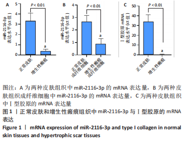
2.1 两种皮肤组织中miR-2116-3p与Ⅰ型胶原的mRNA表达 qRT-PCR检测结果显示,与正常皮肤组织相比,增生性瘢痕组织中miR-2116-3p的mRNA表达量减少(P < 0.01);与正常皮肤成纤维细胞相比,增生性瘢痕组织成纤维细胞中miR-2116-3p的mRNA表达量减少(P < 0.01);与正常皮肤组织相比,增生性瘢痕组织中Ⅰ型胶原的mRNA表达量减少(P < 0.01),见图1。"
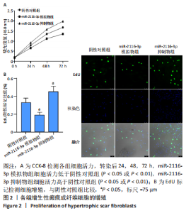
2.2 miR-2116-3p对增生性瘢痕成纤维细胞增殖活力的影响 CCK-8检测显示,转染后0 h,各组细胞活力相近(P > 0.05);转染后24,48,72 h,miR-2116-3p模拟物组细胞活力明显低于阴性对照组(t=5.693,15.541,36.26,P < 0.05或P < 0.01),miR-2116-3p抑制物组细胞活力明显高于阴性对照组(t=7.021,20.782,18.618,P < 0.05或P < 0.01),见图2A。依据EdU试剂盒操作后,用绿色细胞数/蓝色的细胞数的比值进行统计分析,结果显示,与miR-2116-3p阴性对照组相比,miR-2116-3p模拟物组细胞增殖水平显著下降(P < 0.05),miR-2116-3p抑制物组细胞增殖水平显著升高(P < 0.05),见图2B。"

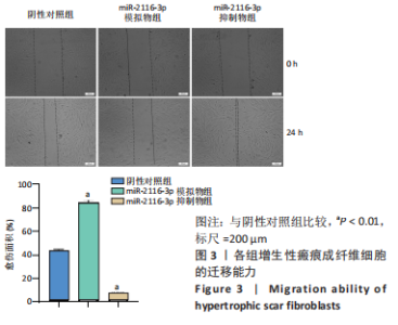
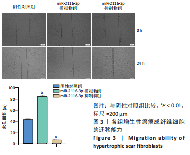
2.3 miR-2116-3p对增生性瘢痕成纤维细胞迁移的影响 转染后24 h,与阴性对照组相比,miR-2116-3p模拟物组细胞迁移面积显著下降(t=25.387,P < 0.01),miR-2116-3p抑制物组细胞迁移面积显著升高(t=42.834,P < 0.01),见图3。"

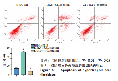
2.4 miR-2116-3p对增生性瘢痕成纤维细胞凋亡的影响 转染后24 h,miR-2116-3p阴性对照组、miR-2116-3p模拟物组、miR-2116-3p抑制物组细胞凋亡率分别为(11.97±1.33)%,(42.03±2.17)%,(7.267±1.65)%,miR-2116-3p模拟物组的细胞凋亡率较阴性对照组显著升高(t=20.432,P < 0.01),miR-2116-3p抑制物组的细胞凋亡率较阴性对照组显著下降(t=3.839,P < 0.05),见图4。"
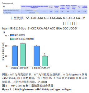
2.5 miR-2116-3p与Ⅰ型胶原的靶向结合 运用 Targetscan预测 miR-2116-3p的下游靶基因,即Ⅰ型胶原,见图5A。双荧光素酶结果示,与miR-2116-3p阴性对照组相比,共转染野生型(WT)Ⅰ型胶原-3′UTR时,miR-2116-3p模拟物组能减弱萤光素酶活性(P < 0.01),共转染突变型(MT)Ⅰ型胶原-3′UTR时,两组萤光素酶活性比较差异无显著性意义(P > 0.05),见图5B。"

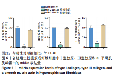
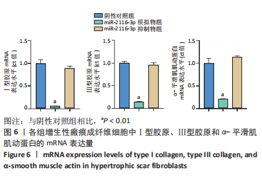
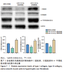
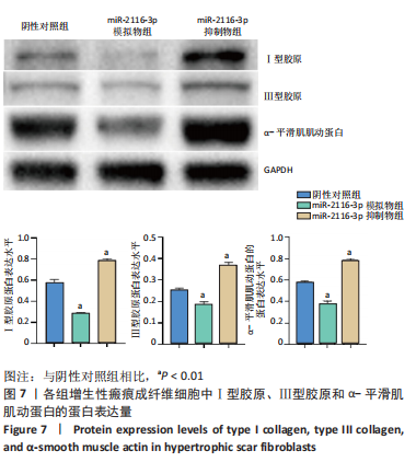
2.6 miR-2116-3p对增生性瘢痕成纤维细胞中相关基因与蛋白表达的影响 qRT-PCR检测结果显示,与阴性对照组相比,miR-2116-3p模拟物组细胞Ⅰ型胶原、Ⅲ型胶原和α-平滑肌肌动蛋白的mRNA表达量显著下降(P < 0.01),miR-2116-3p抑制物组细胞Ⅰ型胶原、Ⅲ型胶原和α-平滑肌肌动蛋白的mRNA表达量明显升高(P < 0.01),见图6。Western blot检测结果显示,与阴性对照组相比,miR-2116-3p模拟物组细胞Ⅰ型胶原、Ⅲ型胶原和α-平滑肌肌动蛋白的蛋白表达量明显降低,miR-2116-3p抑制物组细胞Ⅰ型胶原、Ⅲ型胶原和α-平滑肌肌动蛋白的蛋白表达量升高,见图7。"
"
| [1] FINNERTY C, JESCHKE M, BRANSKI L, et al. Hypertrophic scarring: the greatest unmet challenge after burn injury. Lancet. 2016;388(10052): 1427-1436. [2] LEE H, JANG Y. Recent Understandings of Biology, Prophylaxis and Treatment Strategies for Hypertrophic Scars and Keloids. Int J Mol Sci. 2018;19(3): 711. [3] MCGEARY S, LIN K, SHI C, et al. The biochemical basis of microRNA targeting efficacy. Science. 2019;366(6472):eaav1741. doi: 10.1126/science.aav1741. [4] MENG X, NIKOLIC-PATERSON D, LAN H. TGF-β: the master regulator of fibrosis. Nat Rev Nephrol. 2016;12(6):325-338. [5] WANG X, HE Y, MACKOWIAK B, et al. MicroRNAs as regulators, biomarkers and therapeutic targets in liver diseases. Gut. 2021;70(4): 784-795. [6] WONNACOTT A, DENBY L, JM COWARD R, et al. MicroRNAs and Their Delivery in Diabetic Fibrosis. Adv Drug Deliv Rev. 2022;182:114045. [7] JIN Z. MicroRNA targets and biomarker validation for diabetes-associated cardiac fibrosis. Pharmacol Res. 2021;174:105941. doi: 10.1016/j.phrs.2021.105941. [8] BI S, CHAI L, YUAN X, et al. MicroRNA-98 inhibits the cell proliferation of human hypertrophic scar fibroblasts via targeting Col1A1. Biol Res. 2017;50(1): 22. [9] LI C, ZHU H, BAI W, et al. MiR-10a and miR-181c regulate collagen type I generation in hypertrophic scars by targeting PAI-1 and uPA. FEBS Lett. 2015;589(3):380-389. [10] LI J, LI Y, WANG Y, et al. Overexpression of miR-101 suppresses collagen synthesis by targeting EZH2 in hypertrophic scar fibroblasts. Burns Trauma. 2021;9:tkab038. doi: 10.1093/burnst/tkab038. [11] HUANG X, GU S, LIU C, et al.CD39 + Fibroblasts Enhance Myofibroblast Activation by Promoting IL-11 Secretion in Hypertrophic Scars. J Invest DermatoL. 2022;142(4):1065-1076.e19. [12] ZHANG Z, HUANG X, YANG J, et al. Identification and functional analysis of a three-miRNA ceRNA network in hypertrophic scars. J Transl Med. 2021;19(1):451. [13] GALLANT-BEHM C, PIPER J, LYNCH J, et al. A MicroRNA-29 Mimic (Remlarsen) Represses Extracellular Matrix Expression and Fibroplasia in the Skin. J Invest DermatoL. 2019;139(5):1073-1081. [14] 张紫嫣,徐洪,高学敏,等. microRNA在皮肤纤维化中的研究进展[J].实用皮肤病学杂志,2021,14(2):91-94. [15] HA M, KIM V. Regulation of microRNA biogenesis. Nat Rev Mol Cell Biol. 2014;15(8):509-524. [16] ZHANG Q, GUO B, HUI Q, et al. miR-137 Inhibits Proliferation and Metastasis of Hypertrophic Scar Fibroblasts via Targeting Pleiotrophin. Cell Physiol Biochem. 2018;49(3):985-995. [17] JIANG D, GUO B, LIN F, et al. miR-205 inhibits the development of hypertrophic scars by targeting THBS1. Aging. 2020;12(21):22046-22058. [18] GUO B, HUI Q, XU Z, et al. miR-495 inhibits the growth of fibroblasts in hypertrophic scars. Aging. 2019;11(9):2898-2910. [19] BARONE N, SAFRAN T, VORSTENBOSCH J, et al. Current Advances in Hypertrophic Scar and Keloid Management. Semin Plast Surg. 2021; 35(3):145-152. [20] 郭冰玉,林枫,回蔷,等.微小RNA-627在人增生性瘢痕中的表达及作用[J].中华烧伤杂志,2021,37(4):369-376. [21] 贾本川,郭冰玉,李泰然,等. miR-663靶向抑制TGF-β1表达介导抑制增生性瘢痕皮肤成纤维细胞增殖[J].解剖科学进展,2021,27(2): 218-221,225. [22] 郭冰玉,姜栋文,回蔷,等.微小RNA-205在人增生性瘢痕中的表达及作用[J].中华烧伤杂志,2021,37(2):180-186. [23] XIAO Y. MiR-486-5p inhibits the hyperproliferation and production of collagen in hypertrophic scar fibroblasts IGF1/PI3K/AKT pathway. J Dermatol Treat. 2021;32(8):973-982. [24] ZHANG K, GARNER W, COHEN L, et al. Increased types I and III collagen and transforming growth factor-beta 1 mRNA and protein in hypertrophic burn scar. J Invest Dermatol. 1995;104(5):750-754. [25] CHAI C, TAI I, ZHOU R, et al. MicroRNA-9-5p inhibits proliferation and induces apoptosis of human hypertrophic scar fibroblasts through targeting peroxisome proliferator-activated receptor β. Biol Open. 2020;9(12):bio051904. doi: 10.1242/bio.051904. [26] WU X, LI J, YANG X, et al. miR-155 inhibits the formation of hypertrophic scar fibroblasts by targeting HIF-1α via PI3K/AKT pathway. J Mol Histol. 2018;49(4):377-387. [27] TANG J, YANG J, HU H, et al. miR-211-5p inhibits the proliferation, migration, invasion, and induces apoptosis of human hypertrophic scar fibroblasts by regulating TGFβR2 expression. Ann Transl Med. 2021;9(10):864. [28] ZHANG Z, GAO X, HE Y, et al. MicroRNA-411-3p inhibits bleomycin-induced skin fibrosis by regulating transforming growth factor-β/Smad ubiquitin regulatory factor-2 signalling. J Cell Mol Med. 2021; 25(24):11290-11299. [29] ZHANG T, WANG X, WANG Z, et al. Current potential therapeutic strategies targeting the TGF-β/Smad signaling pathway to attenuate keloid and hypertrophic scar formation. Biomed Pharmacother. 2020; 129:110287. [30] GRAS C, RATUSZNY D, HADAMITZKY C, et al. miR-145 Contributes to Hypertrophic Scarring of the Skin by Inducing Myofibroblast Activity. Mol Med. 2015;21:296-304. [31] GUO L, XU K, YAN H, et al. MicroRNA expression signature and the therapeutic effect of the microRNA‑21 antagomir in hypertrophic scarring. Mol Med Rep. 2017;15(3):1211-1221. [32] LI S, LIU Y, ZHANG T, et al. A Tetrahedral Framework DNA-Based Bioswitchable miRNA Inhibitor Delivery System: Application to Skin Anti-Aging. Adv Mater. 2022;e2204287.doi: 10.1002/adma.202204287. |
| [1] | He Xi, Wan Yu, Tang Yuting, Yang Anning, Wu Kai, Jiao Yun, Bai Zhigang, Jiang Yideng, Shen Jiangyong. Erastin inhibits proliferation of hypertrophic scar fibroblasts [J]. Chinese Journal of Tissue Engineering Research, 2023, 27(在线): 1-. |
| [2] | Ke Weiqiang, Chen Xianghui, Chen Xiaoling, Meng Jie, Ma Yanlin. Rituximab combined with autologous peripheral blood stem cell transplantation in the treatment of diffuse large B-cell lymphoma and the expression of related factors [J]. Chinese Journal of Tissue Engineering Research, 2023, 27(6): 915-920. |
| [3] | Lu Huixiu, Cao Haiyu, Lou Dan, Li Jianying, Liu Hongyuan, Sun Jing. Imiquimod combined with photodynamic therapy for hypertrophic scars: immune response and prognosis [J]. Chinese Journal of Tissue Engineering Research, 2023, 27(5): 690-694. |
| [4] | Hu Xinming, Qiao Yanhua, Wang Xiaofan, Li Linyu, Zhao Bing. Mechanism of long non-coding RNA plasmacytoma variant translocation 1 involved in pelvic organ prolapse [J]. Chinese Journal of Tissue Engineering Research, 2023, 27(5): 669-675. |
| [5] | Deng Moyuan, Peng Kun. Role and regulation of macrophages in biomaterial-mediated fibrosis formation [J]. Chinese Journal of Tissue Engineering Research, 2023, 27(25): 4085-4092. |
| [6] | Zheng Guangji, Wang Qingwei, Lu Junji, Li Yuhong, Chen Fucheng, Zhuang Haishan, Zhong Meilian, Lin Enhui, Pan Yueyu, Luo Qinglu. Different intensities of ultrasound for abdominal skin scar in rabbits [J]. Chinese Journal of Tissue Engineering Research, 2023, 27(23): 3640-3645. |
| [7] | Niu Zihan, Yu Yang, Ai Jiang, Bu Panpan, Li Wenbo, Suriye·Reheman, Ma Shaolin. Total flavonoids of Hippophae rhamnoides L. interfere with the regression of hypertrophic scar tissue blocks in a rabbit ear model [J]. Chinese Journal of Tissue Engineering Research, 2023, 27(2): 258-263. |
| [8] | Zhu Hong, Lin Ziheng, He Rouye, Pan Jinbin, Liu Xiaochuan, He Xiaoling, Zhang Jingying. Antibacterial and hemostatic properties of chitosan collagen sponge [J]. Chinese Journal of Tissue Engineering Research, 2023, 27(16): 2525-2533. |
| [9] | Ma Hongfeng, Ren Qing, Cheng Wanmin, Wang Jiguo, Meng Qingyan. Effects of Sanhuang gel on wound healing, basic fibroblast growth factor and interleukin 17 levels of auricular skin defect in rabbits [J]. Chinese Journal of Tissue Engineering Research, 2023, 27(12): 1842-1847. |
| [10] | Su Meng, Wang Xin, Zhang Jin, Bei Ying, Huang Yu, Zhu Yanzhao, Li Jiali, Wu Yan. Nanocellular vesicles loaded with curcumin promote wound healing in diabetic mice [J]. Chinese Journal of Tissue Engineering Research, 2023, 27(12): 1877-1883. |
| [11] | He Xi, Ma Fang, Wan Yu, Tang Yuting, Ma Shengchao, Jiang Yideng, Shen Jiangyong. miR-382-3p inhibits the proliferation of human hypertrophic scar fibroblasts [J]. Chinese Journal of Tissue Engineering Research, 2023, 27(11): 1758-1764. |
| [12] | Huang Bin, Zheng Jinxu, Zhang Jun. Progress of non-coding RNAs in stem cell abnormality of idiopathic pulmonary fibrosis [J]. Chinese Journal of Tissue Engineering Research, 2023, 27(1): 130-137. |
| [13] | Li Qin, Mao Shuangfa, Li Min, Cheng Jiyan. Protective effect and mechanism of dendrobium on fibroblasts damaged by ultraviolet B [J]. Chinese Journal of Tissue Engineering Research, 2022, 26(8): 1228-1233. |
| [14] | Liu Feng, Peng Yuhuan, Luo Liangping, Wu Benqing. Plant-derived basic fibroblast growth factor maintains the growth and differentiation of human embryonic stem cells [J]. Chinese Journal of Tissue Engineering Research, 2022, 26(7): 1032-1037. |
| [15] | Liu Jin, Li Zhen, Hao Huiqin, Wang Ze, Zhao Caihong, Lu Wenjing. Ermiao san aqueous extract regulates proliferation, migration, and inflammatory factor expression of fibroblast-like synovial cells in collagen-induced arthritis rats [J]. Chinese Journal of Tissue Engineering Research, 2022, 26(5): 688-693. |
| Viewed | ||||||
|
Full text |
|
|||||
|
Abstract |
|
|||||