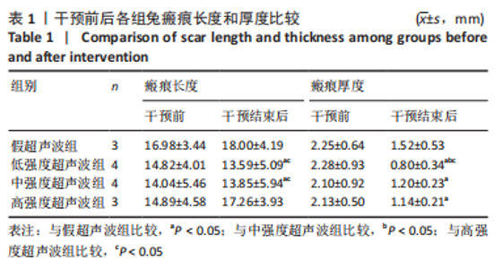
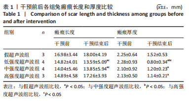
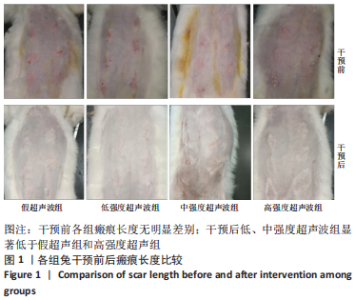
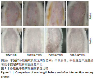
[1] FINNERTY CC, JESCHKE MG, BRANSKI LK, et al. Hypertrophic scarring: the greatest unmet challenge after burn injury. Lancet. 2016;388(10052): 1427-1436.
[2] PETERS SE, JHA B, ROSS M. Rehabilitation following surgery for flexor tendon injuries of the hand. Cochrane Database of Systematic Reviews 2021;1:CD012479.
[3] 魏战杰,颜家琪,杨永刚,等. 温哥华瘢痕量表在曲安奈德治疗甲状腺术后皮肤瘢痕中的应用[J]. 中华实验外科杂志,2022,39(5): 924-927.
[4] WANG ZC, ZHAO WY, CAO Y, et al. The Roles of Inflammation in Keloid and Hypertrophic Scars. Front Immunol. 2020;11:603187.
[5] 郭冰玉,林枫,白泽明,等. 微小RNA-296在兔皮肤瘢痕中的表达及对人成纤维细胞的作用[J]. 中华烧伤杂志,2021,37(8):725-730.
[6] 刘江涛, 曾纯, 欧阳容兰, 等. 人工真皮+负压封闭引流技术+自体刃厚皮片移植联合修复复杂创面的临床应用[J]. 中华损伤与修复杂志(电子版),2020,15(3):215-218.
[7] LEE HJ, JANG YJ. Recent Understandings of Biology, Prophylaxis and Treatment Strategies for Hypertrophic Scars and Keloids. Int J Mol Sci. 2018;19(3):711.
[8] LI Y, ZHANG J, SHI J, et al. Exosomes derived from human adipose mesenchymal stem cells attenuate hypertrophic scar fibrosis by miR-192-5p/IL-17RA/Smad axis. Stem Cell Res Ther. 2021;12(1):221.
[9] ZHANG J, LI Y, BAI X, et al. Recent advances in hypertrophic scar. Histol Histopathol. 2018;33(1):27-39.
[10] VAN DEN TILLAART SA, SCHONEVELD A, PETERS IT, et al. Abdominal scar recurrences of cervical cancer: incidence and characteristics: a case-control study. Int J Gynecol Cancer. 2010;20(6):1031-1040.
[11] XU QL, SONG JH. Characteristics of scar hyperplasia after burn and the rehabilitation treatment in children. Zhonghua Shao Shang Za Zhi. 2018;34(8):509-512.
[12] WANG XQ, LI ZN, WANG QM, et al. Lipid nano-bubble combined with ultrasound for anti-keloids therapy. J Liposome Res. 2018;28(1):5-13.
[13] 谭志,陈俊伟,翁其彪,等. 高压氧联合超声波对病理性瘢痕早期干预的疗效观察[J]. 中华航海医学与高气压医学杂志,2020,27(5): 589-593.
[14] SABET-PEYMAN EJ, WOODWARD JA. Complications using intense ultrasound therapy to treat deep dermal facial skin and subcutaneous tissues. Dermatol Surg. 2014;40(10):1108-1112.
[15] RIAZ HM, ASHRAF CHEEMA S. Paraffin wax bath therapy versus therapeutic ultrasound in management of post burn contractures of small joints of hand. Int J Burns Trauma. 2021;11(3):245-250.
[16] SAKAMOTO M. Effects of Physical Agents on Muscle Healing with a Focus on Animal Model Research. Phys Ther Res. 2021;24(1):1-8.
[17] 贺花, 师书玥, 黄永震, 等. 高校实验室实验动物生物安全管理与规范[J].安徽农业科学,2018,46(31):217-219.
[18] 陈晶, 刘耀宇, 陈友滨. 卡托普利对兔耳皮肤瘢痕影响的实验研究[J]. 中国医药,2017,12(6):925-929.
[19] PANG W, ZHANG Z, ZHANG Y, et al. Extracellular matrix collagen biomarkers levels in patients with chronic thromboembolic pulmonary hypertension. J Thromb Thrombolysis. 2021;52(1):48-58.
[20] Liu H, He Y, Jiang Z, et al. Prodigiosin Alleviates Pulmonary Fibrosis Through Inhibiting miRNA-410 and TGF-β1/ADAMTS-1 Signaling Pathway. Cell Physiol Biochem. 2018;49(2):501-511.
[21] ISSA SF, CHRISTENSEN AF, LINDEGAARD HM, et al. Galectin-3 is Persistently Increased in Early Rheumatoid Arthritis (RA) and Associates with Anti-CCP Seropositivity and MRI Bone Lesions, While Early Fibrosis Markers Correlate with Disease Activity. Scand J Immunol. 2017;86(6):471-478.
[22] SUN P, LU X, ZHANG H, et al. The Efficacy of Drug Injection in the Treatment of Pathological Scar: A Network Meta-analysis. Aesthetic Plast Surg. 2021;45(2):791-805.
[23] 王洪涛,韩军涛,胡大海. 炎症反应在皮肤瘢痕和瘢痕疙瘩形成中的作用及其机制研究进展[J]. 中华烧伤杂志,2021,37(5):490-494.
[24] LEI JA, ZHOU Y, QIN ZL. Research advances on inflammatory responses involved in keloid development. Zhonghua Shao Shang Za Zhi. 2021; 37(6):591-595.
[25] 雷颖, 彭诗绮, 陈红阳, 等. 点阵激光联合脉冲染料激光加多点微量注射曲安奈德对兔耳皮肤瘢痕的影响[J]. 中华整形外科杂志, 2020,36(10):1128-1138.
[26] LI-TSANG CW, LAU JC, CHAN CC. Prevalence of hypertrophic scar formation and its characteristics among the Chinese population. Burns. 2005;31(5):610-616.
[27] LIU J, LIU M, ZHANG S, et al. Intent to have a second child among Chinese women of childbearing age following China’s new universal two-child policy: a cross-sectional study. BMJ Sex Reprod Health. 2019; 46(1):59-66.
[28] 马亦飞. 超声联合检测子宫瘢痕前壁下段厚度对剖宫产再次妊娠女性合理选择分娩方式的指导意义[J]. 中国基层医药,2020,27(19): 2379-2383.
[29] ZHU Z, LI H, ZHANG J. Uterine dehiscence in pregnant with previous caesarean delivery. Ann Med. 2021;53(1):1265-1269.
[30] 谭志, 陈俊伟, 翁其彪, 等. 高压氧联合超声波对病理性瘢痕早期干预的疗效观察[J]. 中华航海医学与高气压医学杂志,2020,27(5): 589-593.
[31] 石海飞, 韩春茂, 胡学庆, 等. 兔耳背植入人工真皮支架模型的应用评价[J]. 浙江医学,2005,27(5):334-336.
[32] NEDELEC B, COUTURE MA, CALVA V, et al. Randomized controlled trial of the immediate and long-term effect of massage on adult postburn scar. Burns. 2019;45(1):128-139.
[33] CONSTANS C, MATEO P, TANTER M, et al. Potential impact of thermal effects during ultrasonic neurostimulation: retrospective numerical estimation of temperature elevation in seven rodent setups. Phys Med Biol. 2018;63(2):025003.
[34] 苏荣家, 王志勇, 刘英开, 等. 变性胶原体外培养模型的建立[J]. 中华创伤杂志,2013,29(4):353-358.
[35] 许庆友, 潘莉, 王月华, 等. 红花对肾间质纤维化实验大鼠肾小管上皮细胞表型转化的抑制作用[J]. 中国老年学杂志,2009,29(11): 1344-1346.
[36] 沈锐, 冯祥生, 宋红梅, 等. 烧伤后皮肤瘢痕患者不同时期血清中PⅢNP的浓度变化及其意义[J]. 国际外科学杂志,2010,37(11): 726-728.
[37] HUANG C, OGAWA R. Keloidal pathophysiology: Current notions. Scars Burn Heal. 2021;7:2059513120980320.
[38] TU L, LIN Z, HUANG Q, et al. USP15 Enhances the Proliferation, Migration, and Collagen Deposition of Hypertrophic Scar–Derived Fibroblasts by Deubiquitinating TGF-βR1 In Vitro. Plast Reconstr Surg. 2021;148(5):1040-1051.
|
