Chinese Journal of Tissue Engineering Research ›› 2023, Vol. 27 ›› Issue (21): 3281-3286.doi: 10.12307/2023.465
Finite element analysis of proximal femoral nail anti-rotation for treating unstable femoral intertrochanteric fracture under different bones with bone cement augmentation
Guo Hao1, Chen Guangxin1, Chen Xiaoqin1, Lian Hongyu2, Liu Xinwei1, Li Zitao2
- 1Mudanjiang Medical University, Mudanjiang 157011, Heilongjiang Province, China; 2Second Department of Orthopedics Surgery, Affiliated Hongqi Hospital, Mudanjiang Medical University, Mudanjiang 157011, Heilongjiang Province, China
-
Received:2022-07-05Accepted:2022-08-03Online:2023-07-28Published:2022-11-23 -
Contact:Li Zitao, Professor, Chief physician, Master’s supervisor, Second Department of Orthopedics Surgery, Affiliated Hongqi Hospital, Mudanjiang Medical University, Mudanjiang 157011, Heilongjiang Province, China -
About author:Guo Hao, Master candidate, Mudanjiang Medical University, Mudanjiang 157011, Heilongjiang Province, China -
Supported by:the Basic Scientific Research Funds of Heilongjiang Provincial Universities, No. 2021-KYYWF-0516 (to LZT)
CLC Number:
Cite this article
Guo Hao, Chen Guangxin, Chen Xiaoqin, Lian Hongyu, Liu Xinwei, Li Zitao. Finite element analysis of proximal femoral nail anti-rotation for treating unstable femoral intertrochanteric fracture under different bones with bone cement augmentation[J]. Chinese Journal of Tissue Engineering Research, 2023, 27(21): 3281-3286.
share this article
Add to citation manager EndNote|Reference Manager|ProCite|BibTeX|RefWorks
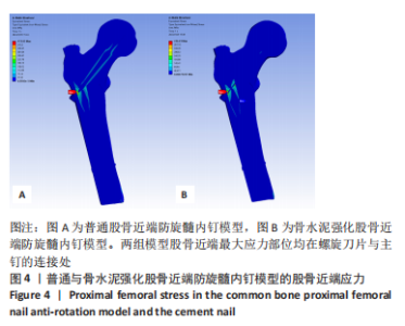
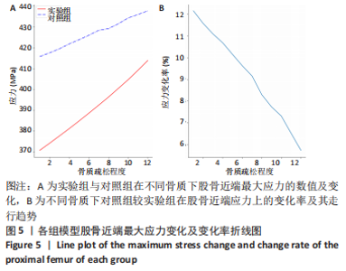
2.1 各组模型股骨近端应力 股骨近端防旋髓内钉对断裂的股骨近端提供主要的支撑作用,通过有限元分析实验模拟计算发现,在对照组与实验组中其股骨近端最大应力部位均在螺旋刀片与主钉的连接处,如图4,并且在相同骨质下对照组股骨近端最大应力值明显高于实验组。实验数据统计将引入变化率的概念,即对照组最大应力值在实验组最大应力值的基础上所发生的变化比例,发现对照组较实验组在最大应力的变化率上随着骨质疏松的程度加重而有所减少,如图5示,从起初对照组较实验组增大了12.2%到最后仅增大了5.74%。"
"
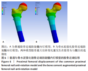
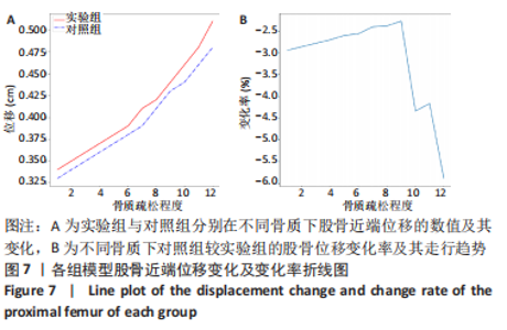
2.2 各组模型股骨近端位移 股骨头表面是股骨主要的受力部位,根据图6可见股骨最大位移变化量发生在股骨头与髋臼的连接处。如图7所示,实验组股骨近端位移量随着骨质疏松程度的加重而随之增高,从起初0.34 cm增加到0.51 cm,且后期股骨位移变化量有明显增长的趋势;对照组在12种骨质下所计算出的位移量均小于实验组固定下的模型,而且在前9种骨质下对照组较实验组股骨近端位移减少的变化率均小于3%,随后伴随骨质疏松程度加重位移降低的变化率明显增大,变化率最多降低了5.89%。"
"

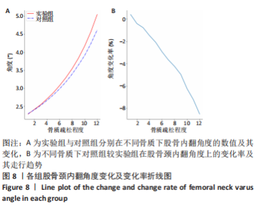
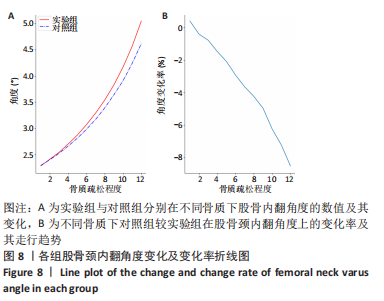
2.3 各组模型股骨颈内翻角度 在ANSYS workbench软件中,在股骨头表面设立点remote A,股骨底面设立点remote B,由于股骨底面约束固定不动,故观察remote A点在x、y、z轴上的变化角度即为股骨内翻的角度。 rotx代表x轴角度变化,roty代表y轴角度变化,rotz代表z轴角度变化。 统计在12种骨质疏松程度下股骨颈内翻角度,如图8A,可见实验组随着骨质疏松程度的加重,股骨颈内翻的角度随之增大,且内翻角度的变化幅度在后期有明显增大的趋势,实验组模型下的股骨颈内翻角度最大可达5.05°;对照组中除第1组正常骨质外,在其余11种骨质中股骨颈内翻角度都较实验组模型低,且随着骨质疏松程度的加重,两组间股骨颈内翻角度降低的变化率明显增加,如图8B,从减少0.41%到减少8.51%。"
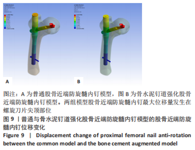
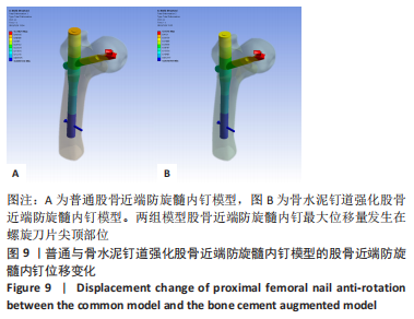
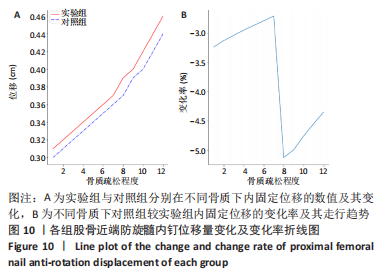
2.4 各组模型股骨近端防旋髓内钉位移 两组模型股骨近端防旋髓内钉最大位移量发生在螺旋刀片尖顶部位,如图9所示。"
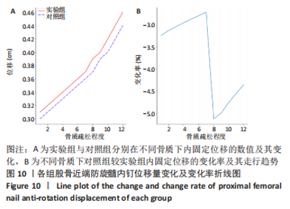
随着骨质疏松程度的加重,实验组模型股骨近端防旋髓内钉螺钉尖端的位移也随之增高,最大可达0.46 cm。在12种骨质分析中发现,对照组模型股骨近端防旋髓内钉螺钉尖端的位移量均小于实验组模型,而且位移变化率在前7种骨质中均小于3.5%,但在后5种骨质中位移变化率明显增大,最大可减少到5.13%,如图10所示。"
| [1] VERONESE N, MAGGI S. Epidemiology and social costs of hip fracture. Injury 2018;49(8):1458-1460. [2] CHANG SM, HOU ZY, HU SJ, et al. Intertrochanteric Femur Fracture Treatment in Asia: What We Know and What the World Can Learn. Orthop Clin North Am. 2020;51(2):189-205. [3] VAN DE REE CLP, DE JONGH MAC, PEETERS CMM, et al. Hip fractures in elderly people:surgery or no surgery?A systematic review and meta-analysis. Geriatr Orthop Surg Rehabil. 2017;8:173-180. [4] 张全,曾勇,舒鑫.不同手术方法治疗老年股骨粗隆间骨折效果及其生物力学研究[J].医用生物力学,2020,35(5):602-607. [5] KASHIGAR A, VINCENT A, GUNTON MJ, et al. Predictors of failure for cephalomedullary nailing of proximal femoral fractures. Bone Joint J. 2014;96-B(8): 1029-1034. [6] LIU JJ, SHAN LC, DENG BY, et al. Reason and treatment of failure of proximal femoral nail antirotation internal fixation for femoral intertrochanteric fractures of senile patients. Genet Mol Res. 2014;13(3):5949-5956. [7] KAMMERLANDER C, ERHART S, DOSHI H, et al. Principles of osteoporotic fracture treatment. Best Pract Res Clin Rheumatol. 2013;27:757-769. [8] 王勇,尤炯鸣,吴银生,等.标准骨水泥强化型与传统股骨近端防旋髓内钉治疗老年骨质疏松性股骨转子间不稳定骨折的疗效比较[J].中华创伤杂志, 2020,36(12):1077-1082. [9] KAMMERLANDER C, DOSHI H, GEBHARD F, et al. Long-term results of the augmented PFNA: a prospective multicenter trial. Arch Orthop Trauma Surg. 2014;134(3):343-349. [10] ZYSSET P, QIN L, LANG T, et al. Clinical Use of Quantitative Computed Tomography-Based Finite Element Analysis of the Hip and Spine in the Management of Osteoporosis in Adults: the 2015 ISCD Official Positions-Part II. J Clin Densitom. 2015;18(3):359-392. [11] SCHILEO E, TADDEI F. Finite Element Assessment of Bone Fragility from Clinical Images. Curr Osteoporos Rep. 2021;19(6):688-698. [12] ZHENG L, CHEN X, ZHENG Y, et al. Cement augmentation of the proximal femoral nail antirotation for the treatment of two intertrochanteric fractures - a comparative finite element study. BMC Musculoskelet Disord. 2021;22(1):1010. [13] 陈心敏,罗斯嘉,夏卓伟,等.钉道强化股骨近端防旋髓内钉治疗老年A3.3型股骨转子间骨折的有限元分析[J].中国组织工程研究,2020,24(27): 4265-4271. [14] HONG CC, NASHI N, MAKANDURA MC, et al.The long and short of cephalomedullary nails in the treatment of osteo poroticpertrochanteric fracture. Singapore Med J. 2017;58(2):85-91. [15] 蔡群斌,邹霞,胡剑涛,等.有限元法分析尖顶距与股骨近端防旋髓内钉固定股骨转子间骨折稳定性的关系[J].中国组织工程研究,2021,25(6):831-836. [16] GAO Z, LV Y, ZHOU F, et al. Risk factors for implant failure after fixation of proximal femoral fractures with fracture of the lateral femoral wall. Injury. 2018;49(2): 315-322. [17] HELWIG P, FAUST G, HINDENLANG U, et al. Biomechanical evaluation of the gliding nail in trochanteric fractures. Z Orthop Ihre Grenzgeb. 2006;144: 594e601. [18] DHANOPIA A, BHARGAVA M. Finite element analysis of human fractured femur bone implantation with PMMA thermoplastic prosthetic plate. Proc Engin. 2017; 173:1658-1665. [19] POLIKEIT A, NOLTE LP, FERGUSON SJ, et al. The effect of cement augmentation on the load transfer in an osteoporotic functional spinal unit (finite-element analysis). Spine (Phila Pa 1976). 2003;28(10):991-996. [20] 朱兴华,宫赫,白雪飞,等.弹性模量与表观密度的分段函数关系用于股骨近端的结构模拟[J].中国生物医学工程学报,2003,22(3):250-257. [21] 李家琼,王冬梅,孙璟川,等.骨水泥对椎体成形术治疗胸腰椎骨质疏松压缩性骨折的生物力学影响[J].医用生物力学,2018,33(1):6-12. [22] SHAO Q, ZHANG Y, SUN GX, et al. Positive or negative anteromedial cortical support of unstable pertrochanteric femoral fractures: A finite element analysis study. Biomed Pharmacother. 2021;138:111473. [23] NUÑO N, AMABILI M, GROPPETTI R, et al. Static coefcient of friction between Ti-6Al-4V and PMMA for cemented hip and knee implants. J Biomed Mater Res. 2002;59(1):191-200. [24] KARAGIANNIS A, PAPAKITSOU E, DRETAKIS K, et al. Mortality rates of patients with a hip fracture in a southwestern district of Greece: ten-year follow-up with reference to the type of fracture. Calcif Tissue Int. 2006;78(2):72-77. [25] ARCHIBECK MJ, CAROTHERS JT, TRIPURANENI KR, et al. Total hip arthroplasty after failed internal fixation of proximal femoral fractures. J Arthroplasty. 2013; 28(1):168-171. [26] LI J, HAN L,ZHANG H, et al. Medial sustainable nail versus proximal femoral nail antirotation in treating AO/OTA 31-A2.3 fractures: Finite element analysis and biomechanical evaluation. Injury. 2019;50(3):648-656. [27] HAN L, LIU JJ, HU YG, et al. Controlled study on Gamma nail and proximal femoral locking plate for unstable intertrochanteric femoral fractures with broken lateral wall. Sci Rep. 2018;8(1):11114. [28] HOFFMANN MF, KHORIATY JD, SIETSEMA DL, et al. Outcome of intramedullary nailing treatment for intertrochanteric femoral fractures. J Orthop Surg Res. 2019;14(1):360. [29] 赵晓涛,张殿英,郁凯,等.股骨近端防旋髓内钉固定治疗股骨转子间骨折的失效原因分析[J].中华创伤骨科杂志,2021,23(3):202-208. [30] 王勇,潘骏.标准骨水泥强化型PFNA治疗老年股骨粗隆间不稳定骨折[J].中国矫形外科志,2018,26(18):1653-1658. [31] 田智勇,陈洪强,戴科晶,等.股骨近端防旋髓内钉骨水泥增强固定治疗老年股骨转子间骨折的疗效分析[J].中华创伤骨科杂志,2021,23(6):539-542. [32] HANKE MS, BECKMANN NA, KEEL MJB, et al. Revision of a blade cut-out in PFN-A fixation: Blade exchange,cement augmentation and a cement plug as a successful salvage option. Trauma Case Rep. 2020;27:100303. [33] ERHART S, SCHMOELZ W, BLAUTH M, et al. Biomechanical effect of bone cement augmentation on rotational stability and pull-out strength of the Proximal Femur Nail Antirotation. Injury. 2011;42(11):1322-1327. [34] DONALDSON AJ, THOMSON HE, HARPER NJ, et al. Bone cement implantation syndrome. Br J Anaesth. 2009;102(1):12-22. [35] BOOZ C, NOESKE J, ALBRECHT MH, et al. Diagnostic accuracy of quantitative dual-energy CT-based bone mineral density assessment in comparison to Hounsfield unit measurements using dual x-ray absorptiometry as standard of reference. Eur J Radiol. 2020;132:109321. |
| [1] | Zhong Yizheng, Huang Peizhen, Cai Qunbin, Zheng Liqin, He Xingpeng, Dong Hang. Microstructural indexes that determine the trabecular bone maximum stress of micro-finite element models [J]. Chinese Journal of Tissue Engineering Research, 2023, 27(9): 1313-1318. |
| [2] | Li Xiaomin, Tian Xiangdong, Tan Yetong, Zhu Guangyu, Wang Rongtian, Wang Jian, Xue Zhipeng, Ma Sheng, Hu Yuanyi, Huang Ye, Ding Tiansong. Changes of lower limb force line and knee function after high tibial osteotomy in osteoporotic medial ventricular knee osteoarthritis [J]. Chinese Journal of Tissue Engineering Research, 2023, 27(9): 1325-1329. |
| [3] | Peng Zhixin, Yan Wengang, Wang Kun, Zhang Zhenjiang. Finite element analysis and structural optimization design of 3D printed forearm braces [J]. Chinese Journal of Tissue Engineering Research, 2023, 27(9): 1340-1345. |
| [4] | Wu Tianliang, Tao Xiuxia, Xu Hongguang. Influence of different bone mineral densities on cage subsidence after stand-alone oblique lateral interbody fusion: three-dimensional finite element analysis [J]. Chinese Journal of Tissue Engineering Research, 2023, 27(9): 1352-1358. |
| [5] | Liu Jinyu, Zhang Hanshuo, Cui Hongpeng, Pan Lingzhi, Zhao Boran, Li Fei, Ding Yu. Finite element biomechanical analysis of minimally invasive treatment of cervical spondylotic myelopathy and accurate exercise rehabilitation [J]. Chinese Journal of Tissue Engineering Research, 2023, 27(9): 1359-1364. |
| [6] | He Yujie, Kang Zhijie, Xue Mingming, Jin Feng, Li Zhijun, Wang Xing, Xu Yangyang, Gao Mingjie, Li Jiawei, Li Xiaohe, Wang Haiyan. Finite element analysis of transarticular screw fixation of adolescent thoracic vertebra [J]. Chinese Journal of Tissue Engineering Research, 2023, 27(9): 1365-1370. |
| [7] | Wen Xinghua, Ding Huanwen, Cheng Kai, Yan Xiaonan, Peng Yuanhao, Wang Yuning, Liu Kang, Zhang Huiwu. Three-dimensional finite element model analysis of intramedullary nailing fixation design for large femoral defects in Beagle dogs [J]. Chinese Journal of Tissue Engineering Research, 2023, 27(9): 1371-1376. |
| [8] | Jiang Xiaocheng, Shi Lu, Wang Yinbin, Li Qiujiang, Xi Chuangzhen, Ma Zefeng, Cai Lijun. Systematical evaluation of bone fusion rate after interbody fusion in patients with osteoporosis and lumbar degenerative disease treated with teriparatide [J]. Chinese Journal of Tissue Engineering Research, 2023, 27(9): 1427-1433. |
| [9] | Sun Jiajia, Zhu Haidi, Lu Yun, Zhang Kai. Comparison of bone metabolism markers between type 2 diabetes mellitus and non-type 2 diabetes mellitus patients with hip fracture [J]. Chinese Journal of Tissue Engineering Research, 2023, 27(8): 1156-1160. |
| [10] | Yuan Hucheng, Ding Yongguo, Ma Xuehua, Ma Wenxin, Sun Jianmin, Wang Zili, Jin Weidong. Sustained releasing of pyrazinamide, capreomycin, moxifloxacin and amikacin loaded bone cement in vitro [J]. Chinese Journal of Tissue Engineering Research, 2023, 27(7): 1017-1022. |
| [11] | Zhao Wei, Feng Wei, Yang Tieyi, Ren Wei, Wang Yuxin, Lyu Huicheng, Chang Zhiqiang, Feng Xiaodong, Wang Ziheng, Guo Shibing. Antibiotic bone cement intramedullary nail prepared using 3D printed mold for the treatment of long bone infection in lower limbs [J]. Chinese Journal of Tissue Engineering Research, 2023, 27(7): 1023-1030. |
| [12] | Guo Tingting, Xie Hong, Xu Guanghua. Finite element analysis of elastic ankle brace performance [J]. Chinese Journal of Tissue Engineering Research, 2023, 27(7): 1031-1037. |
| [13] | Zhu Lin, Gu Weiping, Wang Can, Chen Gang. Biomechanical analysis of All-on-Four and pterygomaxillary implants under different maxillary bone conditions [J]. Chinese Journal of Tissue Engineering Research, 2023, 27(7): 985-991. |
| [14] | Sun Jiangwei, Wang Junxiang, Baibujiafu·Yellisi, Dai Huijuan, Nijati·Turson. Three-dimensional finite element analysis of stress distribution in different smooth collar implants [J]. Chinese Journal of Tissue Engineering Research, 2023, 27(7): 1004-1011. |
| [15] | Jiang Yifang, Cai Qimin, Chu Zhengyi, Qin Min, Shen Yurong, Gu Yuanping. Simulation analysis of stress distribution of NRT FILES in curved root canals [J]. Chinese Journal of Tissue Engineering Research, 2023, 27(7): 1038-1042. |
| Viewed | ||||||
|
Full text |
|
|||||
|
Abstract |
|
|||||