Chinese Journal of Tissue Engineering Research ›› 2023, Vol. 27 ›› Issue (9): 1359-1364.doi: 10.12307/2023.211
Previous Articles Next Articles
Finite element biomechanical analysis of minimally invasive treatment of cervical spondylotic myelopathy and accurate exercise rehabilitation
Liu Jinyu, Zhang Hanshuo, Cui Hongpeng, Pan Lingzhi, Zhao Boran, Li Fei, Ding Yu
- TCM Senior Department of Orthopedics, Sixth Medical Center of PLA General Hospital, Beijing 100048, China
-
Received:2021-11-29Accepted:2022-01-19Online:2023-03-28Published:2022-07-01 -
Contact:Ding Yu, MD, Professor, Chief physician, TCM Senior Department of Orthopedics, Sixth Medical Center of PLA General Hospital, Beijing 100048, China -
About author:Liu Jinyu, Physician, TCM Senior Department of Orthopedics, Sixth Medical Center of PLA General Hospital, Beijing 100048, China Zhang Hanshuo, Master, TCM Senior Department of Orthopedics, Sixth Medical Center of PLA General Hospital, Beijing 100048, China Liu Jinyu and Zhang Hanshuo contributed equally to this article. -
Supported by:Capital Clinical Diagnosis and Treatment Technology Research and Demonstration Application Project, No. Z191100006619028 (to DY)
CLC Number:
Cite this article
Liu Jinyu, Zhang Hanshuo, Cui Hongpeng, Pan Lingzhi, Zhao Boran, Li Fei, Ding Yu. Finite element biomechanical analysis of minimally invasive treatment of cervical spondylotic myelopathy and accurate exercise rehabilitation[J]. Chinese Journal of Tissue Engineering Research, 2023, 27(9): 1359-1364.
share this article
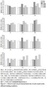
2.1 颈椎C4-5有限元模型的建立 建立C4-5模型M共计37 714个单元和67 013个节点,模型M1 共计97 793个单元和169 184个节点,模型M2共计104 307个单元和180 794个节点。 2.2 各模型不同工况下的椎间盘应力比较 见图4。"

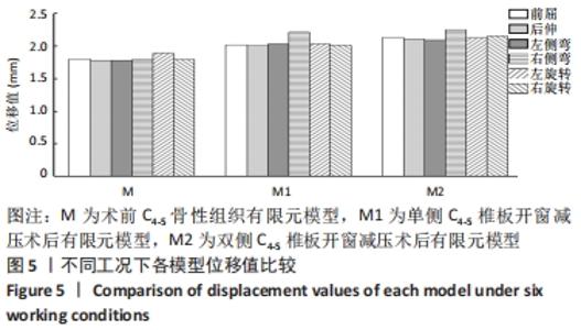
与模型M比较,模型M1右侧弯活动下的椎间盘b、d区域应力增高10%-12%,其他工况下的椎间盘应力无明显差异;模型M2右侧弯活动下的b、d区域应力增高10%-13%,左侧弯活动下的a、c区域增高9%-11%,其他工况下的椎间盘应力无明显差异。 2.3 各模型不同工况下的位移比较 见图5。 "
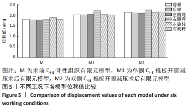
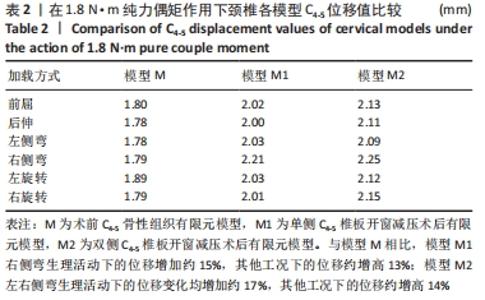
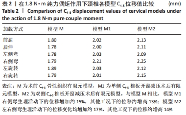
与模型M相比,模型M1右侧弯生理活动下的位移增加约15%,其他工况下的位移约增高13%;模型M2左右侧弯生理活动下的位移变化均增加约17%,其他工况下的位移约增高14%,见表2。 "
| [1] 杨宝林,张绍东,王小虎,等.颈椎后路改良单开门椎管扩大成形术治疗多节段脊髓型颈椎病的疗效分析[J]. 中国脊柱脊髓杂志, 2018,28(4):289-296. [2] NASTO LA, MUQUIT S, PEREZ-ROMERA AB, et al. Clinical outcome and safety study of a newly developed instrumented French-door cervical laminoplasty technique. J Orthop Traumatol. 2017;18(2):135-143. [3] LI Z, LIU H, YANG M, et al. A biomechanical analysis of four anterior cervical techniques to treating multilevel cervical spondylotic myelopathy: a finite element study. BMC Musculoskelet Disord. 2021; 22(1):278. [4] FENG S, ZHENG B, ZHANG L, et al. A systematic review and meta-analysis compare surgical treatment and conservative treatment in patients with cervical spondylotic myelopathy. Ann Palliat Med. 2021;10(7):7671-7680. [5] 刘金玉,崔洪鹏,丁宇,等.椎间孔镜术后腰椎间盘突出复发与后纵韧带完整性关系的有限元生物力学研究[J].中国骨与关节杂志, 2021,10(7):508-514.
[6] OUYANG P, LI J, HE X, et al. Biomechanical Comparison of 1-Level Corpectomy and 2-Level Discectomy for Cervical Spondylotic Myelopathy: A Finite Element Analysis. Med Sci Monit. 2020;26: e919270. [9] YANEZ TOUZET A, BHATTI A, DOHLE E, et al. Clinical outcome measures and their evidence base in degenerative cervical myelopathy: a systematic review to inform a core measurement set (AO Spine RECODE-DCM). BMJ Open. 2022;12(1):e57650. [10] 陈钵,秦大平,张晓刚,等.有限元分析法在脊柱生物力学中的研究进展[J].中国疼痛医学杂志,2020,26(3):208-216. [11] 吴超,王振宇,林国中,等.颈椎单侧半椎板及不同程度小关节切除术后生物力学变化的有限元分析[J]. 中华神经外科疾病研究杂志,2018,17(4):352-356. [12] LI Q, HAN X, WANG R, et al. Clinical recovery after 5 level of posterior decompression spine surgeries in patients with cervical spondylotic myelopathy: A retrospective cohort study. Asian J Surg.2020;43(5):613-624. [13] WANG Y, WANG L, DU C, et al. A comparative study on dynamic stiffness in typical finite element model and multi-body model of C6-C7 cervical spine segment. Int J Numer Method Biomed Eng. 2016;32(6):10. [14] DENG Z, WANG K, WANG H, et al. A finite element study of traditional Chinese cervical manipulation. Eur Spine J. 2017;26(9):2308-2317. [15] VOO LM, KUMARESAN S, YOGANANDAN N, et al. Finite element analysis of cervical facetectomy. Spine (Phila Pa 1976). 1997;22(9): 964-969. [16] SEIPEL RC, PINTAR FA, YOGANANDAN N, et al. Biomechanics of calcaneal fractures: A model for the motor vehicle. Clin Orthop Relat Res. 2001;(388):218-224. [17] ZHAO L, CHEN J, LIU J, et al. Biomechanical analysis on of anterior transpedicular screw-fixation after two-level cervical corpectomy using finite element method. Clin Biomech (Bristol, Avon). 2018;60:76-82. [18] 刘金玉,丁宇,蒋强,等.全内镜下腰椎板开窗减压有限元模拟建模及生物力学变化[J].中国组织工程研究,2020,24(27):4291-4296. [19] YUAN H, ZHANG X, ZHANG L, et al. Comparative study of curative effect of spinal endoscopic surgery and anterior cervical decompression for cervical spondylotic myelopathy. J Spine Surg. 2020;6(S1):S186-S196. [20] WANG XZ, LIU H, LI J Q, et al. Comparison of Anterior Cervical Discectomy and Fusion with Cervical Laminectomy and Fusion in the Treatment of 4‐Level Cervical Spondylotic Myelopathy. Orthop Surg. 2022;14(2):229-237. [21] TETREAULT L, KALSI-RYAN S, BENJAMIN D, et al. Degenerative Cervical Myelopathy: A Practical Approach to Diagnosis. Global Spine J. 2022: 1280955569. [22] KIM HC, JEON H, JEONG YH, et al. Factors Affecting Postoperative Complications and Outcomes of Cervical Spondylotic Myelopathy with Cerebral Palsy : A Retrospective Analysis. J Korean Neurosurg Soc. 2021;64(5):808-817. [23] YAO M, LI G, ZHOU L, et al. Shikonin inhibits neuronal apoptosis via regulating endoplasmic reticulum stress in the rat model of double-level chronic cervical cord compression. Cell Biol Toxicol. 2022. doi: 10.1007/s10565-021-09648-3. [24] AKTER F, YU X, QIN X, et al. The Pathophysiology of Degenerative Cervical Myelopathy and the Physiology of Recovery Following Decompression. Front Neurosci. 2020;14:138. [25] ZHANG M, OU-YANG H, LIU J, et al. Predicting postoperative recovery in cervical spondylotic myelopathy: construction and interpretation of T2*-weighted radiomic-based extra trees models. Eur Radiol. 2022. doi: 10.1007/s00330-021-08383-x. [26] ZHONG W, WANG L, HUANG T, et al. Risk factors for rapid progressive neurological deterioration in patients with cervical spondylotic myelopathy. J Orthop Surg Res. 2021;16(1):75-75. [27] 郑耀超,林彩娜,柯松坚,等.多裂肌核心稳定性训练:超声联合表面肌电监测与无监测的比较[J]. 中国组织工程研究,2018,22(36): 5785-5790. [28] KIMURA A, ENDO T, INOUE H, et al. Impact of axial neck pain on quality of life after laminoplasty. Spine (Phila Pa 1976). 2015;40(24): E1292-E1298. [29] PANJABI MM, CHOLEWICKI J, NIBU K, et al. Critical load of the human cervical spine: An in vitro experimental study. Clin Biomech (Bristol, Avon). 1998;13(1):11-17. [30] ZIKA J, ALEXIOU GA, GIANNOPOULOS S, et al. Outcome factors in surgically treated patients for cervical spondylotic myelopathy. J Spinal Cord Med. 2020;43(2):206-210. [31] HIRABAYASHI S, KITAGAWA T, YAMAMOTO I, et al. Development and achievement of cervical laminoplasty and related studies on cervical myelopathy. Spine Surg Relat Res. 2019;4(1):8-17. [32] SCHOMACHER J, FALLA D. Function and structure of the deep cervical extensor muscles in patients with neck pain. Man Ther. 2013;18(5): 360-366. [33] PILATO F, CALANDRELLI R, DISTEFANO M, et al. Multidimensional assessment of cervical spondylotic myelopathy patients. Usefulness of a comprehensive score system. Neurol Sci. 2021;42(4):1507-1514. [34] CHENG CH, CHIEN A, HSU WL, et al. Investigation of the differential contributions of superficial and deep muscles on cervical spinal loads with changing head postures. PLoS One. 2016;11(3):e0150608. [35] DAVIES BM, MOWFORTH O, WOOD H, et al. Improving Awareness Could Transform Outcomes in Degenerative Cervical Myelopathy [AO Spine RECODE-DCM Research Priority Number 1]. Global Spine J. 2022;12(1):28S-38S. [36] XU C, WANG R, LI J, et al. Intervertebral-spreader-assisted anterior cervical discectomy and fusion prevents postoperative axial pain by alleviating facet joint pressure. J Orthop Surg Res. 2022;17(1):91. [37] JIANG Q, DING Y, LU Z, et al. Comparative analysis of non-full and full endoscopic spine technique via Interlaminar approach for the treatment of degenerative lumbar spinal stenosis: A retrospective, single Institute, propensity score-matched study. Global Spine J. 2021; 18(5):360-366. [38] RYU WHA, PLATT A, DEUTSCH H. Hybrid decompression and reconstruction technique for cervical spondylotic myelopathy: case series and review of the literature. J Spine Surg. 2020;6(1):181-195. [39] DUAN Y, WANG HH, JIN AM, et al. Finite element analysis of posterior cervical fixation. Orthop Traumatol Surg Res. 2015;101(1):23-29. [40] 刘金玉,丁逸苇,卢正操,等.有限元分析全可视化内镜下椎板开窗减压治疗脊髓型颈椎病的生物力学特点[J].中国组织工程研究, 2021,25(24):3850-3854. [41] LU X, ZOU F, LU F, et al. How to reconstruct the lordosis of cervical spine in patients with Hirayama disease? A finite element analysis of biomechanical changes focusing on adjacent segments after anterior cervical discectomy and fusion. J Orthop Surg Res. 2022;17(1):101. [42] 樊瑜波,邓小燕.生物力学建模仿真与应用[M].上海:上海交通大学出版社,2017:1-5. [43] 钱伟长,叶开沅.弹性力学[M].北京:科学出版社,1956:69-102. |
| [1] | Zhong Yizheng, Huang Peizhen, Cai Qunbin, Zheng Liqin, He Xingpeng, Dong Hang. Microstructural indexes that determine the trabecular bone maximum stress of micro-finite element models [J]. Chinese Journal of Tissue Engineering Research, 2023, 27(9): 1313-1318. |
| [2] | Wu Taoguang, Nie Shaobo, Chen Hua, Zhu Zhengguo, Qi Lin, Tang Peifu. Biomechanical characteristics of a new multi-dimensional cross locking plate in the treatment of subtrochanteric nonunion [J]. Chinese Journal of Tissue Engineering Research, 2023, 27(9): 1330-1334. |
| [3] | Peng Zhixin, Yan Wengang, Wang Kun, Zhang Zhenjiang. Finite element analysis and structural optimization design of 3D printed forearm braces [J]. Chinese Journal of Tissue Engineering Research, 2023, 27(9): 1340-1345. |
| [4] | Wu Tianliang, Tao Xiuxia, Xu Hongguang. Influence of different bone mineral densities on cage subsidence after stand-alone oblique lateral interbody fusion: three-dimensional finite element analysis [J]. Chinese Journal of Tissue Engineering Research, 2023, 27(9): 1352-1358. |
| [5] | He Yujie, Kang Zhijie, Xue Mingming, Jin Feng, Li Zhijun, Wang Xing, Xu Yangyang, Gao Mingjie, Li Jiawei, Li Xiaohe, Wang Haiyan. Finite element analysis of transarticular screw fixation of adolescent thoracic vertebra [J]. Chinese Journal of Tissue Engineering Research, 2023, 27(9): 1365-1370. |
| [6] | Wen Xinghua, Ding Huanwen, Cheng Kai, Yan Xiaonan, Peng Yuanhao, Wang Yuning, Liu Kang, Zhang Huiwu. Three-dimensional finite element model analysis of intramedullary nailing fixation design for large femoral defects in Beagle dogs [J]. Chinese Journal of Tissue Engineering Research, 2023, 27(9): 1371-1376. |
| [7] | Li Shihao, Li Qi, Li Zhen, Zhang Yuanyuan, Liu Miaomiao, Ouyang Yi, Xu Weiguo. Plantar pressure and gait analysis in patients with anterior cruciate ligament injury and reconstruction [J]. Chinese Journal of Tissue Engineering Research, 2023, 27(4): 626-631. |
| [8] | Liu Qinghua, Cai Yongqiang, Jin Feng, Yu Jinghong, Wang Haiyan, Zhang Yunfeng, Wang Lidong, Li Jiawei, Wang Xing, He Yujie, Dai Lina, Wang Jianzhong, Wu Chao, Tong Ling, Kang Zhijie, Li Zhijun, Li Xiaohe. Finite element model of the 12-year-old child whole cervical spine: establishment and validity verification based on CT data [J]. Chinese Journal of Tissue Engineering Research, 2023, 27(4): 500-504. |
| [9] | Li Yaping, Liu Hong, Gao Zhen, Chen Xiaolin, Huang Wujie, Jiang Zheng. Three-dimensional motion analysis of lower limb biomechanical performance in Tai Chi practitioners accompanied by knee joint pain [J]. Chinese Journal of Tissue Engineering Research, 2023, 27(4): 520-526. |
| [10] | Zhan Yi, Wang Biao, Ma Yuli, He Simin, Sun Honghui, Hao Dingjun. Biomechanical comparison between a novel bone cement screw system and common surgical methods for the treatment of Kummell’s disease [J]. Chinese Journal of Tissue Engineering Research, 2023, 27(3): 385-390. |
| [11] | Lu Hui, Wu Qimei, Liu Rong. Finite element analysis and application of unilateral and bilateral bone-filling mesh container in treatment of osteoporotic vertebral compression fracture [J]. Chinese Journal of Tissue Engineering Research, 2023, 27(3): 391-397. |
| [12] | Wang Jianping, Zhang Xiaohui, Yu Jinwei, Wei Shaoliang, Zhang Xinmin, Xu Xingxin, Qu Haijun. Application of knee joint motion analysis in machanism based on three-dimensional image registration and coordinate transformation [J]. Chinese Journal of Tissue Engineering Research, 2022, 26(在线): 1-5. |
| [13] | Wei Guoqiang, Li Yunfeng, Wang Yi, Niu Xiaofen, Che Lifang, Wang Haiyan, Li Zhijun, Shi Guopeng, Bai Ling, Mo Kai, Zhang Chenchen, Xu Yangyang, Li Xiaohe. Biomechanical analysis of non-uniform material femur under different loads [J]. Chinese Journal of Tissue Engineering Research, 2022, 26(9): 1318-1322. |
| [14] | Zhang Yufang, Lü Meng, Mei Zhao. Construction and verification of a full spine biomechanical model of adolescent scoliosis [J]. Chinese Journal of Tissue Engineering Research, 2022, 26(9): 1351-1356. |
| [15] | Zhang Jichao, Dong Yuefu, Mou Zhifang, Zhang Zhen, Li Bingyan, Xu Xiangjun, Li Jiayi, Ren Meng, Dong Wanpeng. Finite element analysis of biomechanical changes in the osteoarthritis knee joint in different gait flexion angles [J]. Chinese Journal of Tissue Engineering Research, 2022, 26(9): 1357-1361. |
| Viewed | ||||||||||||||||||||||||||||||||||||||||||||||||||
|
Full text 176
|
|
|||||||||||||||||||||||||||||||||||||||||||||||||
|
Abstract 548
|
|
|||||||||||||||||||||||||||||||||||||||||||||||||