Chinese Journal of Tissue Engineering Research ›› 2023, Vol. 27 ›› Issue (26): 4175-4180.doi: 10.12307/2023.407

Previous Articles     Next Articles

miR-23b effects on transforming growth factor beta1 and osteoclast activity in synovial tissue of rats with rheumatoid arthritis

Song Jianhui, Mu Jihong, Li Shiwen   

  1. The Second Naval Hospital of Southern Theater Command, Sanya 572000, Hainan Province, China
  • Received:2021-12-16 Accepted:2022-06-20 Online:2023-09-18 Published:2023-01-20
  • Contact: Mu Jihong, MD, Attending physician, The Second Naval Hospital of Southern Theater Command, Sanya 572000, Hainan Province, China
  • About author:Song Jianhui, Attending physician, The Second Naval Hospital of Southern Theater Command, Sanya 572000, Hainan Province, China
  • Supported by:
    miR-23b; rheumatoid arthritis; synovial tissue; transforming growth factor-β1; osteoclast activity

Abstract: BACKGROUND: Studies have shown that an elevation in miR-23b is closely related to the occurrence and development of rheumatoid arthritis; therefore, inhibiting the expression of miR-23b can be a new target for the treatment of the disease. 
OBJECTIVE: To explore the effect of miR-23b on the regulation of transforming growth factor-β1 in synovial tissue and the inhibition of osteoclast activity in rats with rheumatoid arthritis. 
METHODS: Sixty SPF male Sprague-Dawley rats were randomly divided into normal control group, model group, miR-NC group, and miR-23b inhibitor group (n=15 per group). Except for the normal control group, animal models of rheumatoid arthritis were prepared in the other three groups using Fredrin complete adjuvant method. After successful modeling, the miR-NC group was injected with 20 μL of 1×10⁸/L miR-NC and the miR-23b inhibitor group was injected with 20 μL of 1×10⁸/L miR-23b inhibitor. Normal control group and model group were injected with the same volume of normal saline at the same time. Twenty-one days later, hematoxylin-eosin staining was used to detect the pathological morphology of the articular tissue of rats, real-time PCR was used to detect the relative mRNA expression of miR-23b in rat serum, and immunohistochemical method was used to detect the protein expression of transforming growth factor-β1 in the synovial tissue of rats. The activity of osteoclasts was detected by tartrate-resistant acid phosphatase staining and phalloidin staining.
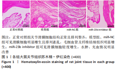
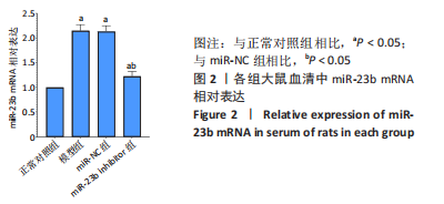
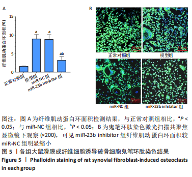
RESULTS AND CONCLUSION: In the normal control group, the structure of synovial cells was normal and neatly arranged. In the model and miR-NC groups, synovial cells proliferated obviously and arranged in disorder. At the same time, capillaries and fibrous connective tissue were obviously proliferated, accompanied by a large number of inflammatory cells infiltrated. In the miR-23b inhibitor group, mild hyperplasia of synovial cells and a small amount of inflammatory cell infiltration appeared, and edema and congestion were significantly improved. The relative expression of miR-23b in the serum was significantly increased in the model group compared with the normal control group (P < 0.05), while there was no significant difference between the model group and the miR-NC group (P > 0.05). The relative expression of miR-23b in the miR-23b inhibitor group was significantly lower than that in the miR-NC group 
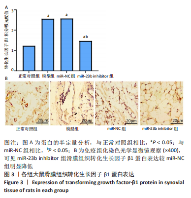
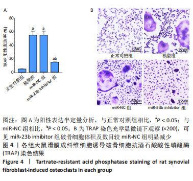
(P < 0.05). Compared with the normal control group, the expression of transforming growth factor-β1 protein in the synovial tissue was significantly increased in the model group (P < 0.05), while there was no significant difference between the model and miR-NC groups (P > 0.05). The expression of transforming growth factor-β1 protein in the synovial tissue was significantly lower in the miR-23b inhibitor group than the miR-NC group (P < 0.05). Compared with the normal control group, a highly positive expression of typical multinucleated osteoclasts was found in the synovial fibroblasts of the model group by the tartrate-resistant acid phosphatase staining (P < 0.05). There was no significant difference between the model and the miR-NC groups (P > 0.05). The volume and number of osteoclasts in the miR-23b inhibitor group were significantly lower than those in the miR-NC group (P < 0.05). Compared with the normal control group, the phalloidin staining of osteoclasts significantly indicated the formation of rings composed of actin filaments in the model group (P < 0.05) and there was no significant difference between the model and miR-NC groups (P > 0.05), while the actin ring area was significantly reduced in the miR-23b inhibitor group compared with the miR-NC group (P < 0.05). The above results indicate that reducing the expression of miR-23b can play a therapeutic effect on rheumatoid arthritis, which can effectively inhibit the expression of transforming growth factor-β1 and reduce the activity of osteoclasts in rat synovial tissue.

Key words: miR-23b, rheumatoid arthritis, synovial tissue, transforming growth factor-β1, osteoclast activity

CLC Number: