[1] LI XR, TONG YH, LI XQ, et al. Total endovascular repair of an intraoperative stent-graft deployed in the false lumen of Stanford type A aortic dissection: A case report. World J Clin Cases. 2020;8(5):954-962.
[2] ZHANG M, TONG YH, LIU C, et al. Treatment of Stanford type A aortic dissection with triple pre-fenestration, reduced diameter, and three-dimensional-printing techniques: A case report. World J Clin Cases. 2021;9(1):183-189.
[3] SASAKI S, WATANABE H, SHIBAYAMA K, et al. Three-dimensional transesophageal echocardiographic evaluation of coronary involvement in patients with acute type A aortic dissection. J Am Soc Echocardiogr. 2013;26(8):837-845.
[4] SUN W, XU H, XIONG J, et al. 3D Morphologic Findings Before and After Thoracic Endovascular Aortic Repair for Type B Aortic Dissection. Ann Vasc Surg. 2021;74:220-228.
[5] WANG CJ, RODRIGUEZ DIAZ CA, TRINH MA. Use of real-time three-dimensional transesophageal echocardiography in type A aortic dissections: advantages of 3D TEE illustrated in three cases. Ann Card Anaesth. 2015;18(1):83-86.
[6] KRÜGER T, OIKONOMOU A, SCHIBILSKY D, et al. Aortic elongation and the risk for dissection: the Tubingen Aortic Pathoanatomy (TAIPAN) project. Eur J Cardiothorac Surg. 2017;51(6):1119-1126.
[7] NIENABER CA, SAKALIHASAN N, CLOUGH RE, et al. Thoracic endovascular aortic repair (TEVAR) in proximal (type A) aortic dissection: Ready for a broader application? J Thorac Cardiovasc Surg. 2017;153(2):S3-S11.
[8] WANG CJ, RODRIGUEZ DIAZ CA, TRINH MA. Use of real-time three-dimensional transesophageal echocardiography in type A aortic dissections: advantages of 3D TEE illustrated in three cases. Ann Card Anaesth. 2015;18(1):83-86.
[9] KRÜGER T, OIKONOMOU A, SCHIBILSKY D, et al. Aortic elongation and the risk for dissection: the Tubingen Aortic Pathoanatomy (TAIPAN) project. Eur J Cardiothorac Surg. 2017;51(6):1119-1126.
[10] PATTERSON BO, VIDAL-DIEZ A, KARTHIKESALINGAM A, et al. Comparison of aortic diameter and area after endovascular treatment of aortic dissection. Ann Thorac Surg. 2015;99(1):95-102.
[11] LONG KO JK, LIU RW, MA D, et al. Pulsatile hemodynamics in patient-specific thoracic aortic dissection models constructed from computed tomography angiography. J Xray Sci Technol. 2017;25(2):233-245.
[12] ULLERY BW, SUH GY, HIROTSU K, et al. Geometric Deformations of the Thoracic Aorta and Supra-Aortic Arch Branch Vessels Following Thoracic Endovascular Aortic Repair..Vasc Endovascular Surg. 2018; 52(3):173-180.
[13] PÉREZ TM, GARCÍA SM, VELASCO ML, et al. Interrupted aortic arch diagnosis by computed tomography angiography and 3-D reconstruction: A case report. Radiol Case Rep. 2017;13(1):35-38.
[14] HIROTSU K, SUH GY, LEE JT, et al. Changes in Geometry and Cardiac Deformation of the Thoracic Aorta after Thoracic Endovascular Aortic Repair. Ann Vasc Surg. 2018;46:83-89.
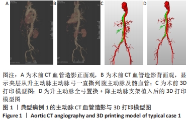
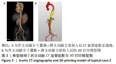
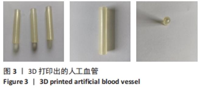
|