Chinese Journal of Tissue Engineering Research ›› 2023, Vol. 27 ›› Issue (5): 726-731.doi: 10.12307/2022.948
Previous Articles Next Articles
Changes in sensory neurons and astrocytes and the expression of interleukin 1beta and glial fibrillary acidic protein in the rat spinal cord after selective dorsal rhizotomy
Chen Guodong1, Zheng Meiyan1, Zhang Peng1, Wang Zhenchao2, Jin Lixin3
- 1Chengwu Hospital, Heze 274000, Shandong Province, China; 2PLA Navy Submarine Academy, Qingdao 266000, Shandong Province, China; 3Department of Anatomy, Medical College of Qingdao University, Qingdao 266000, Shandong Province, China
-
Received:2021-11-27Accepted:2022-01-22Online:2023-02-18Published:2022-07-22 -
Contact:Jin Lixin, Associate professor, Department of Anatomy, Medical College of Qingdao University, Qingdao 266000, Shandong Province, China -
About author:Chen Guodong, Master, Attending physician, Chengwu Hospital, Heze 274000, Shandong Province, China -
Supported by:Shandong Provincial Medical and Health Science and Technology Development Plan, No. 202104070403 (to CGD); the Innovative Experimental Project of National Experimental Teaching Demonstration Center of Qingdao University School of Medicine, No. 201511065055 (to JLX)
CLC Number:
Cite this article
Chen Guodong, Zheng Meiyan, Zhang Peng, Wang Zhenchao, Jin Lixin. Changes in sensory neurons and astrocytes and the expression of interleukin 1beta and glial fibrillary acidic protein in the rat spinal cord after selective dorsal rhizotomy[J]. Chinese Journal of Tissue Engineering Research, 2023, 27(5): 726-731.
share this article
Add to citation manager EndNote|Reference Manager|ProCite|BibTeX|RefWorks
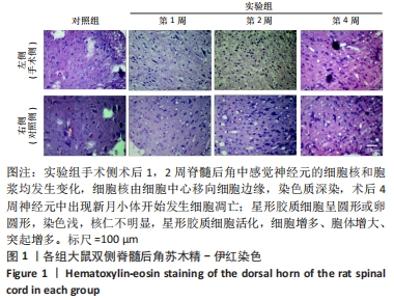
2.1 实验动物数量分析 共纳入33只Wistar大鼠,其中对照组3只、实验组30只,实验组术后1周改良Tarlov评分未达到4级3只,予以剔除,最后共进入结果分析大鼠30只。 2.2 各组脊髓组织苏木精-伊红染色结果 脊神经后根切断后,实验组手术侧术后1,2周对应节段脊髓后角中感觉神经元的细胞核和胞浆均发生变化,细胞核由细胞中心移向细胞边缘,染色质深染,术后4周神经元中出现新月小体开始发生细胞凋亡;星形胶质细胞的核比其他胶质细胞的核大,呈圆形或卵圆形,染色浅,核仁不明显,星形胶质细胞活化,细胞增多、胞体增大、突起增多,见图1。"
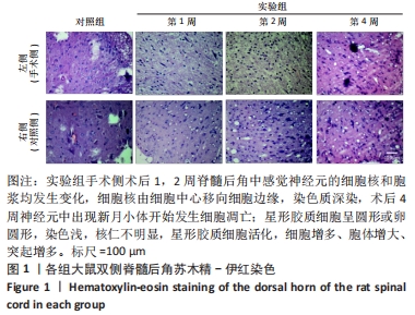

随着术后时间的推移,实验组手术侧脊髓后角感觉神经元计数逐渐减少(P < 0.05);术后1周,实验组手术侧脊髓后角感觉神经元计数与实验组对照侧比较差异无显著性意义(P > 0.05);术后2,4周,实验组手术侧脊髓后角感觉神经元计数少于实验组对照侧(P < 0.05或P < 0.001),见图2A。切断脊神经后根后,实验组手术侧星形胶质细胞数量增多,术后2周的星形胶质细胞数量多于术后1,4周(P < 0.05);实验组手术侧术后1,2,4周的星形胶质细胞数量均多于实验组对照侧与对照组(P < 0.001),见图2B。 "
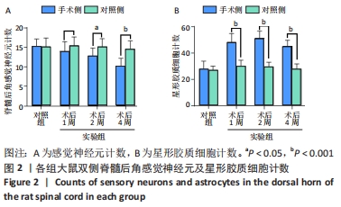
2.3 各组大鼠脊髓组织胶质纤维酸性蛋白免疫组化染色结果 见图3。"
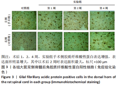
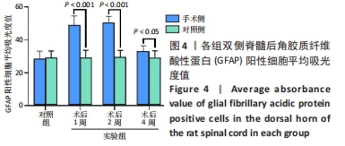
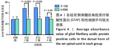
免疫组化下胶质纤维酸性蛋白表达增强表现为棕色或红棕色染色加深,表达面积明显增大。切断脊神经后根后,实验组手术侧胶质纤维酸性蛋白阳性细胞平均吸光度值增大,术后2周的胶质纤维酸性蛋白阳性细胞平均吸光度值大于术后1,4周(P < 0.05);实验组手术侧术后1,2,4周的胶质纤维酸性蛋白阳性细胞平均吸光度值均大于实验组对照侧、对照组(P < 0.05或P < 0.001),见图4。 "
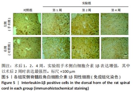
2.4 各组大鼠脊髓组织白细胞介素1β免疫组化染色结果 见图5。"
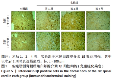
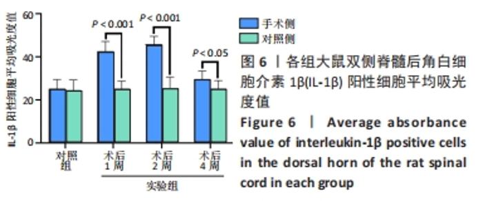
大鼠脊神经后根切断后1,2周,实验组手术侧免疫组化白细胞介素1β表达强烈,可见阳性颗粒,白细胞介素1β阳性颗粒大多分布在手术侧脊髓后角神经胶质细胞和后角浅层神经元上。切断脊神经后根后,实验组手术侧白细胞介素1β阳性细胞平均吸光度值增大,术后2周的白细胞介素1β阳性细胞平均吸光度值大于术后1,4周(P < 0.05);实验组手术侧术后1,2,4周的白细胞介素1β阳性细胞平均吸光度值均大于实验组对照侧、对照组(P < 0.05或P < 0.001),见图6。 "

| [1] JAIN NB, AYERS GD, PETERSON EN, et al. Traumatic spinal cord injury in the United States, 1993-2012. JAMA. 2015;313(22):2236-2243. [2] ZHOU Y, WANG XB, KAN SL, et al. Traumatic spinal cord injury in Tianjin, China: a single-center report of 354 cases. Spinal Cord. 2016;54(9):670-674. [3] KARIMI-ABDOLREZAEE S, BILLAKANTI R. Reactive astrogliosis after spinal cord injury-beneficial and detrimental effects. Mol Neurobiol. 2012;46(2):251-264. [4] BRADBURY EJ, BURNSIDE ER. Moving beyond the glial scar for spinal cord repair. Nat Commun. 2019;10(1):3879. [5] CHENG X, WANG J, SUN X, et al. Morphological and functional alterations of astrocytes responding to traumatic brain injury. J Integr Neurosci. 2019;18(2):203-215. [6] HERGENROEDER GW, REDELL JB, MOORE AN, et al. Biomarkers in the clinical diagnosis and management of traumatic brain injury. Mol Diagn Ther. 2008;12(6):345-358. [7] ANDREIUOLO F, JUNIER MP, HOL EM, et al. GFAPdelta immunostaining improves visualization of normal and pathologic astrocytic heterogeneity. Neuropathology. 2009;29(1):31-39. [8] BLECHINGBERG J, HOLM IE, NIELSEN KB, et al. Identification and characterization of GFAPkappa, a novel glial fibrillary acidic protein isoform. Glia. 2007;55(5):497-507. [9] MEHTA T, FAYYAZ M, GILER GE, et al. Current Trends in Biomarkers for Traumatic Brain Injury. Open Access J Neurol Neurosurg. 2020;12(4):86-94. [10] HALFORD J, SHEN S, ITAMURA K, et al. New astroglial injury-defined biomarkers for neurotrauma assessment. J Cereb Blood Flow Metab. 2017;37(10):3278-3299. [11] SOFRONIEW MV, VINTERS HV. Astrocytes: biology and pathology. Acta Neuropathol. 2010;119(1):7-35. [12] 张亮,黄绍明,潘剑.大鼠脊髓损伤后脑和脊髓胶质纤维酸性蛋白表达变化研究[J].社区医学杂志,2011,9(6):17-19. [13] 潘剑.大鼠脊髓半切损伤后脑和脊髓GFAP表达的变化[D].南宁: 广西医科大学,2010. [14] ALLAN SM, TYRRELL PJ, ROTHWELL NJ. Interleukin-1 and neuronal injury. Nat Rev Immunol. 2005;5(8):629-640. [15] OKADA S, HARA M, KOBAYAKAWA K, et al. Astrocyte reactivity and astrogliosis after spinal cord injury. Neurosci Res. 2018;126:39-43. [16] HU XC, LU YB, YANG YN, et al. Progress in clinical trials of cell transplantation for the treatment of spinal cord injury: how many questions remain unanswered? Neural Regen Res. 2021;16(3):405-413. [17] CHENG H, CAO Y, OLSON L. Spinal cord repair in adult paraplegic rats: partial restoration of hind limb function. Science. 1996;273(5274):510-513. [18] 张冬艳.CNTF对大鼠坐骨神经切断缝合后脊髓前角GFAP表达的影响[D].秦皇岛:华北煤炭医学院,2010. [19] 曹祖懋,时素华,胡煜,等.针刺双向良性调控星形胶质细胞治疗脊髓损伤的研究进展[J].世界科学技术-中医药现代化,2021,23(6): 2100-2104. [20] ISHII T, WARABI E, MANN GE. Circadian control of BDNF-mediated Nrf2 activation in astrocytes protects dopaminergic neurons from ferroptosis. Free Radic Biol Med. 2019;133:169-178. [21] HART AM, TERENGHI G, KELLERTH JO, et al. Sensory neuroprotection, mitochondrial preservation, and therapeutic potential of N-acetyl-cysteine after nerve injury. Neuroscience. 2004;125(1):91-101. [22] SIU AW, ORTIZ GG, BENITEZ-KING G, et al. Effects of melatonin on the nitric oxide treated retina. Br J Ophthalmol. 2004;88(8):1078-1081. [23] 陈宣维,贾连顺,林建华,等.大鼠脊髓损伤后白细胞介素-1β、肿瘤坏死因子-α基因表达的变化[J]. 骨与关节损伤杂志,2003,18(11): 764-766. [24] BUSCH SA, HAMILTON JA, HORN KP, et al. Multipotent adult progenitor cells prevent macrophage-mediated axonal dieback and promote regrowth after spinal cord injury. J Neurosci. 2011;31(3):944-953. [25] MAO L, WANG HD, PAN H, et al. Sulphoraphane enhances aquaporin-4 expression and decreases spinal cord oedema following spinal cord injury. Brain Inj. 2011;25(3):300-306. [26] FAN X, MA W, ZHANG Y, et al. P2X7 Receptor (P2X7R) of Microglia Mediates Neuroinflammation by Regulating (NOD)-Like Receptor Protein 3 (NLRP3) Inflammasome-Dependent Inflammation After Spinal Cord Injury. Med Sci Monit. 2020;26:e925491. [27] LI X, YU Z, ZONG W, et al. Deficiency of the microglial Hv1 proton channel attenuates neuronal pyroptosis and inhibits inflammatory reaction after spinal cord injury. J Neuroinflammation. 2020;17(1):263. [28] 段煜东,张子程,李博,等.低强度脉冲超声在脊髓损伤神经修复中的研究进展[J].第二军医大学学报,2021,42(9):1037-1043. [29] GUTH L. A reassessment of LPS/indomethacin/pregnenolone combination therapy after spinal cord injury in rats. Exp Neurol. 2012; 233(2):686. [30] 武俏丽,李庆国,黄慧玲,等.他克莫司促进神经干细胞移植大鼠脊髓损伤的再生与修复[J].中国组织工程研究与临床康复,2010, 14(23):4235-4238. [31] WU J, ZHU ZY, FAN ZW, et al. Downregulation of EphB2 by RNA interference attenuates glial/fibrotic scar formation and promotes axon growth. Neural Regen Res. 2022;17(2):362-369. [32] LI LM, HAN M, JIANG XC, et al. Peptide-Tethered Hydrogel Scaffold Promotes Recovery from Spinal Cord Transection via Synergism with Mesenchymal Stem Cells. ACS Appl Mater Interfaces. 2017;9(4):3330-3342. [33] NAKAJIMA H, UCHIDA K, GUERRERO AR, et al. Transplantation of mesenchymal stem cells promotes an alternative pathway of macrophage activation and functional recovery after spinal cord injury. J Neurotrauma. 2012;29(8):1614-1625. [34] 张衡,张贤平,戴厚杰,等.天麻素干预脊髓损伤模型兔运动功能及神经生长相关蛋白43的表达[J]. 中国组织工程研究,2021,25(29): 4626-4631. [35] ZHENG G, ZHAN Y, WANG H, et al. Carbon monoxide releasing molecule-3 alleviates neuron death after spinal cord injury via inflammasome regulation. EBioMedicine. 2019;40:643-654. [36] WANG XJ, PENG CH, ZHANG S, et al. Polysialic-Acid-Based Micelles Promote Neural Regeneration in Spinal Cord Injury Therapy. Nano Lett. 2019;19(2):829-838. [37] ROSENZWEIG ES, SALEGIO EA, LIANG JJ, et al. Chondroitinase improves anatomical and functional outcomes after primate spinal cord injury. Nat Neurosci. 2019;22(8):1269-1275. [38] 胡道松,曹福元,徐清贵,等.外周神经损伤后对大鼠脊髓运动神经元及背根节感觉神经元的影响[J]. 同济医科大学学报,1999,28(6): 478-481. [39]. 张光运,段丽,饶志仁,等.大鼠坐骨神经切断后腰脊髓腹角内胶质细胞和神经元的可塑性变化[J]. 中国神经科学杂志,2003,19(6): 345-348. [40] 陈云丰,曾炳芳,张惠箴,等.大鼠腰神经根损伤后脊髓神经胶质细胞激活和细胞因子表达[J].上海医学,2002,25(z1):2-4. [41] LIU SM, XIAO ZF, LI X, et al. Vascular endothelial growth factor activates neural stem cells through epidermal growth factor receptor signal after spinal cord injury. CNS Neurosci Ther. 2019;25(3):3753-85. [42] 谢乐斯,郑林丰,曾志成,等.大鼠脊神经后根切断后脊髓和背根神经节CGRP的表达变化[J].神经解剖学杂志,2007,23(2):209-213. [43] JOHNSON RA, OKRAGLY AJ, HAAK-FRENDSCHO M, et al. Cervical dorsal rhizotomy increases brain-derived neurotrophic factor and neurotrophin-3 expression in the ventral spinal cord. J Neurosci. 2000;20(10):RC77. [44] KINKEAD R, ZHAN WZ, PRAKASH YS, et al. Cervical dorsal rhizotomy enhances serotonergic innervation of phrenic motoneurons and serotonin-dependent long-term facilitation of respiratory motor output in rats. J Neurosci. 1998;18(20):8436-8443. |
| [1] | Zhang Haobo, Zhao Yunan, Yang Xuejun. Role and therapeutic implications of pyroptosis in intervertebral disc degeneration [J]. Chinese Journal of Tissue Engineering Research, 2022, 26(9): 1445-1451. |
| [2] | Wang Shihui, Cheng Yang, Zhu Yunjie, Cheng Shaodan, Mao Jianying. Effect of arc edge needle-scalpel therapy on inflammatory factors and histomorphology of the frozen shoulder in rabbit models [J]. Chinese Journal of Tissue Engineering Research, 2022, 26(5): 706-711. |
| [3] | Zhou Liang, Chen Xingzhen, Li Zhenyu, Zhang Zekun, Duan Guoqing. The mechanism of lncRNA HOTAIR in interleukin-1beta-mediated osteoarthritis [J]. Chinese Journal of Tissue Engineering Research, 2022, 26(35): 5607-5613. |
| [4] | Zhang Lili, Ou Ya, Yuan Xiaodong, Zhang Pingshu. Bax/Bcl-2 protein and apoptosis during adipose-derived stromal cell differentiation into astrocytes [J]. Chinese Journal of Tissue Engineering Research, 2022, 26(31): 4982-4987. |
| [5] | Guan Shihao, Huang Yonghui, Gong Aihua, Cao Xingbing, Sun Haitao, Cai Chuang. Transdifferentiation of rat astrocytes into neurons induced by ectodermal mesenchymal stem cells-derived extracellular vesicles [J]. Chinese Journal of Tissue Engineering Research, 2022, 26(30): 4840-4846. |
| [6] | Tang Fuyu, Zhou Binbin, Wei Weibing, Zhang Hongsheng. Effect of electroacupuncture on glial fibrillary acidic protein expression at the injured site in a rat model of spinal cord injury [J]. Chinese Journal of Tissue Engineering Research, 2022, 26(26): 4113-4117. |
| [7] | Yan Nan, Wu Yanlong, Tang Xiaohui, Zhang Xiaoyan, Wang Hui, Yang Tianze, Zhou Maochun, Wang Zhengdong, Yang Xiaoxia. Bone marrow mesenchymal stem cells may alleviate brain damage caused by the microglial overactivation in the cortex around ischemic site of stroke [J]. Chinese Journal of Tissue Engineering Research, 2022, 26(24): 3790-3795. |
| [8] | Ao Pian, Zhao Xin, Yu Hongrong, Zhang Siqin, Zhang Xinyue, Gu Weili, Wei Li. Laminaria japonica polysaccharide protects radiation-induced brain injury by regulating the blood-brain barrier in mice [J]. Chinese Journal of Tissue Engineering Research, 2022, 26(20): 3158-3163. |
| [9] | Li Jianfeng, Zhang Shulian, Yan Jinyu, Li jiayi, Jin Zhao, Deng Qi. Vimentin silenced by small interfering RNA inhibits glial scar formation in a rat model of acute spinal cord injury [J]. Chinese Journal of Tissue Engineering Research, 2022, 26(20): 3190-3195. |
| [10] | Guan Hong, Zhang Hongbo, Shao Yan, Guo Dong, Zhang Haiyan, Cai Daozhang. PDZ domain containing 1 deficiency promotes chondrocyte senescence in osteoarthritis [J]. Chinese Journal of Tissue Engineering Research, 2022, 26(2): 182-189. |
| [11] | Sun Haitao, Huang Yonghui, Gong Aihua, Cao Xingbing, Li Zhen. Protective effect of lipopolysaccharide-preconditioned astrocyte extracellular vesicles on neurons [J]. Chinese Journal of Tissue Engineering Research, 2022, 26(13): 1985-1992. |
| [12] | Li Shuai, Fan Yiming, Liu Fangyu, Zhang Hongyu, Wang Yansong. Effect and mechanism of astrocytes in spinal cord injury [J]. Chinese Journal of Tissue Engineering Research, 2022, 26(13): 2062-2068. |
| [13] | Shan Wen, Shi Wei. Expression and function of colony-stimulating factor 1 in differentiation of neural stem cells induced by activated astrocyte conditioned medium [J]. Chinese Journal of Tissue Engineering Research, 2022, 26(13): 2069-2074. |
| [14] | Tan Qian, Li Bocun, Li Jing, Li Jia, Xiang Hongchun, Cai Guowei. Acupuncture combined with moxibustion regulates the expression of circadian clock protein in the synovium of rats with osteoarthritis [J]. Chinese Journal of Tissue Engineering Research, 2022, 26(11): 1714-1719. |
| [15] | Xie Xingqi, Hu Wei, Tu Guanjun. Bone marrow mesenchymal stem cells-derived exosomes combined with chondroitinase ABC for treating spinal cord injury in rats [J]. Chinese Journal of Tissue Engineering Research, 2022, 26(1): 20-26. |
| Viewed | ||||||
|
Full text |
|
|||||
|
Abstract |
|
|||||