Chinese Journal of Tissue Engineering Research ›› 2022, Vol. 26 ›› Issue (2): 182-189.doi: 10.12307/2022.031
Previous Articles Next Articles
PDZ domain containing 1 deficiency promotes chondrocyte senescence in osteoarthritis
Guan Hong, Zhang Hongbo, Shao Yan, Guo Dong, Zhang Haiyan, Cai Daozhang
- The Third Affiliated Hospital of Southern Medical University, Guangzhou 510630, Guangdong Province, China
-
Received:2021-01-25Revised:2021-01-27Accepted:2021-02-27Online:2022-01-18Published:2021-10-27 -
Contact:Cai Daozhang, MD, Chief physician, The Third Affiliated Hospital of Southern Medical University, Guangzhou 510630, Guangdong Province, China -
About author:Guan Hong, The Third Affiliated Hospital of Southern Medical University, Guangzhou 510630, Guangdong Province, China -
Supported by:the National Natural Science Foundation of China, No. 81974341 (to CDZ); the Natural Science Foundation of Guangdong Province, No. 2020A1515011062 (to ZHY)
CLC Number:
Cite this article
Guan Hong, Zhang Hongbo, Shao Yan, Guo Dong, Zhang Haiyan, Cai Daozhang. PDZ domain containing 1 deficiency promotes chondrocyte senescence in osteoarthritis[J]. Chinese Journal of Tissue Engineering Research, 2022, 26(2): 182-189.
share this article
Add to citation manager EndNote|Reference Manager|ProCite|BibTeX|RefWorks

2.1 实验动物数量分析 所有小鼠均无脱失,全部进入结果分析。 2.2 PDZK1在骨关节炎关节软骨中的表达情况 为了检测PDZK1在骨关节炎动物模型中的表达情况,对小鼠的关节软骨进行了相关检测。在此次研究中,对内侧半月板不稳定-骨关节炎小鼠模型术后4,8周进行取材。首先,对实验组小鼠术后4周进行取材并染色,关节面出现部分的蛋白聚糖的丢失以及轻微软骨退变;实验组小鼠术后8周取材并染色,可见软骨退变程度加重,出现软骨皲裂、剥脱、丢失,软骨下骨硬化明显,骨赘形成;假手术组关节面完整、无蛋白聚糖丢失。与假手术组相比,实验组小鼠术后4周OARSI病理评分显著升高,差异有显著性意义(P < 0.01)。而且实验组小鼠术后8周OARSI病理评分也显著高于假手术组,差异有显著性意义(P < 0.01)。即证明造模是成功的,见图1。"
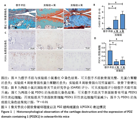

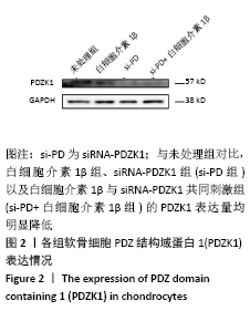
实验组OARSI病理评分标准主要参考Glasson评分细则(标准详见方法部分),接着通过免疫组织化学染色对小鼠的关节软骨的PDZK1表达情况进行检测。结果发现,与假手术组相比,术后4周实验组小鼠关节软骨细胞的PDZK1表达量明显降低(P < 0.01);另外,术后8周实验组小鼠关节软骨细胞的PDZK1表达量也显著低于假手术组(P < 0.01)。通过对结果进行计数分析其阳性率,统计学分析差异有显著性意义,见图1。 2.3 敲低PDZK1后软骨细胞合成代谢减弱,分解代谢增强 分离培养小鼠原代软骨细胞,为在体外骨关节炎模型中查看PDZK1的表达量改变以及验证siRNA-PDZK1的敲低效果,分别利用siRNA-PDZK1与白细胞介素1β刺激软骨细胞48 h,用Western Blot方法检测PDZK1的表达量。作者发现,白细胞介素1β组、si-PD组以及si-PD+白细胞介素1β组软骨细胞的PDZK1表达量均显著低于未处理组,见图2。即体外骨关节炎模型中PDZK1的表达量也是降低的,与动物体内实验结果相符合;而且siRNA-PDZK1有明显敲低效果。"

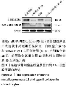
为探究PDZK1与软骨细胞代谢的关系,利用siRNA-PDZK1与白细胞介素1β刺激软骨细胞48 h,建立骨关节炎体外模型,用Western Blot方法检测Ⅱ型胶原a1与基质金属蛋白酶13的表达量。作者发现,si-PD组敲低PDZK1后,与未处理组相比,软骨细胞Ⅱ型胶原a1表达显著减少,即敲低PDZK1后,软骨细胞合成代谢减弱;同时,与白细胞介素1β组相比,si-PD+白细胞介素1β组的基质金属蛋白酶13表达显著增加,即在骨关节炎体外模型中,敲低PDZK1后,软骨细胞分解代谢显著增强,见图3。"

2.4 敲低PDZK1后可使软骨细胞高表达衰老指标 siRNA-PDZK1处理软骨细胞后,软骨细胞衰老指标表达量增加。利用siRNA-PDZK1刺激软骨细胞48 h,通过对软骨细胞衰老指标P16、P21、P53基因表达的相对定量检测,进一步研究PDZK1对软骨细胞衰老的影响,见图4。与对照组相比,siRNA-PDZK1组PDZK1基因相对表达水平明显降低,差异有显著性意义(P < 0.01);与此同时,软骨细胞衰老指标,包括P16、P21、P53,基因相对表达水平均显著升高,差异有显著性意义(P < 0.01)。"

| [1] SAFIRI S, KOLAHI AA, SMITH E, et al. Global, regional and national burden of osteoarthritis 1990-2017: a systematic analysis of the Global Burden of Disease Study 2017. Ann Rheum Dis. 2020;79(6):819-828. [2] HUNTER DJ, SCHOFIELD D, CALLANDER E. The individual and socioeconomic impact of osteoarthritis. Nat Rev Rheumatol. 2014; 10(7):437-441. [3] HULSHOF C, COLOSIO C, DAAMS JG, et al. WHO/ILO work-related burden of disease and injury: Protocol for systematic reviews of exposure to occupational ergonomic risk factors and of the effect of exposure to occupational ergonomic risk factors on osteoarthritis of hip or knee and selected other musculoskeletal diseases. Environ Int. 2019;125:554-566. [4] MCCULLOCH K, LITHERLAND GJ, RAI TS. Cellular senescence in osteoarthritis pathology. Aging Cell. 2017;16(2):210-218. [5] RAHMATI M, NALESSO G, MOBASHERI A, et al. Aging and osteoarthritis: Central role of the extracellular matrix. Ageing Res Rev. 2017;40:20-30. [6] GREENE MA, LOESER RF. Aging-related inflammation in osteoarthritis. Osteoarthr. Cartil. 2015;23(11):1966-1971. [7] XIE J, LIN J, WEI M, et al. Sustained Akt signaling in articular chondrocytes causes osteoarthritis via oxidative stress-induced senescence in mice. Bone Res. 2019;7:23. [8] KAKIUCHI Y, YURUBE T, KAKUTANI K, et al. Pharmacological inhibition of mTORC1 but not mTORC2 protects against human disc cellular apoptosis, senescence, and extracellular matrix catabolism through Akt and autophagy induction. Osteoarthr Cartil. 2019;27(6):965-976. [9] RAPISARDA V, BORGHESAN M, MIGUELA V, et al. Integrin Beta 3 Regulates Cellular Senescence by Activating the TGF-beta Pathway. Cell Rep. 2017;18(10):2480-2493. [10] KANG C, XU Q, MARTIN TD, et al. The DNA damage response induces inflammation and senescence by inhibiting autophagy of GATA4. Sci Adv. 2015;349(6255):aaa5612-aaa5612. [11] KOCHER O, COMELLA N, TOGNAZZI K, et al. Identification and partial characterization of PDZK1: a novel protein containing PDZ interaction domains. Lab Invest. 1998;78(1):117-125. [12] RITTER-MAKINSON SL, PAQUET M, BOGENPOHL JW, et al. Group II Metabotropic Glutamate Receptor Interactions with NHERF Scaffold Proteins: Implications for Receptor Localization in Brain. Neuroscience. 2017;353:58-75. [13] CHA B, YANG J, SINGH V, et al. PDZ domain-dependent regulation of NHE3 protein by both internal Class II and C-terminal Class I PDZ-binding motifs. J. Biol. Chem. 2017;292(20):8279-8290. [14] TAO T, YANG X, ZHENG J, et al. PDZK1 inhibits the development and progression of renal cell carcinoma by suppression of SHP-1 phosphorylation. Oncogene. 2017;36(44):6119-6131. [15] YAN B, XIONG C, HUANG F, et al. Big data-based identification of methylated genes associated with drug resistance and prognosis in ovarian cancer. Medicine (Baltimore). 2020;99(27):e20802. [16] LU X, CHEN M, SHEN J, et al. IL-1beta functionally attenuates ABCG2 and PDZK1 expression in HK-2 cells partially through NF-kB activation. Cell Biol Int. 2019;43(3):279-289. [17] FERNANDEZ-TORRES J, MARTINEZ-NAVA GA, OLIVIERO F, et al. Common gene variants interactions related to uric acid transport are associated with knee osteoarthritis susceptibility. Connect. Tissue Res. 2019;60(3): 219-229. [18] FANG H, BEIER F. Mouse models of osteoarthritis: modelling risk factors and assessing outcomes. Nat Rev Rheumatol. 2014;10(7):413-421. [19] FANG H, HUANG L, WELCH I, et al. Early Changes of Articular Cartilage and Subchondral Bone in The DMM Mouse Model of Osteoarthritis. Sci Rep. 2018;8(1):2855-2859. [20] GLASSON SS, CHAMBERS MG, VAN DEN BERG WB, et al. The OARSI histopathology initiative - recommendations for histological assessments of osteoarthritis in the mouse. Osteoarthr. Cartil. 2010; 18 Suppl 3:S17-23. [21] WANG B, JIANG Y, YAO Z, et al. Aucubin Protects Chondrocytes Against IL-1β-Induced Apoptosis In Vitro And Inhibits Osteoarthritis In Mice Model. Drug Des Devel Ther. 2019;13:3529-3538. [22] GLASSON SS, ASKEW R, SHEPPARD B, et al. Deletion of active ADAMTS5 prevents cartilage degradation in a murine model of osteoarthritis. Nature. 2005;434(7033):644-648. [23] TONG W, ZENG Y, CHOW D, et al. Wnt16 attenuates osteoarthritis progression through a PCP/JNK-mTORC1-PTHrP cascade. Ann Rheum Dis. 2019;78(4):551-561. [24] BLÜHER M. Obesity: global epidemiology and pathogenesis. Nat Rev Endocrinol. 2019;15(5):288-298. [25] HUSSAIN SM, CICUTTINI FM, ALYOUSEF B, et al. Female hormonal factors and osteoarthritis of the knee, hip and hand: a narrative review. Climacteric. 2018;21(2):132-139. [26] LIEBERTHAL J, SAMBAMURTHY N, SCANZELLO CR. Inflammation in joint injury and post-traumatic osteoarthritis. Osteoarthr Cartil. 2015;23(11): 1825-1834. [27] LOESER RF, COLLINS JA, DIEKMAN BO. Ageing and the pathogenesis of osteoarthritis. Nat Rev Rheumatol. 2016;12(7):412-420. [28] KANG D, SHIN J, CHO Y, et al. Stress-activated miR-204 governs senescent phenotypes of chondrocytes to promote osteoarthritis development. Sci Transl Med. 2019;11(486):eaar6659. [29] GAO SG, ZENG C, LI LJ, et al. Correlation between senescence-associated beta-galactosidase expression in articular cartilage and disease severity of patients with knee osteoarthritis. Int J Rheum Dis. 2016;19(3):226-232. [30] MATSUZAKI T, MATSUSHITA T, TAKAYAMA K, et al. Disruption of Sirt1 in chondrocytes causes accelerated progression of osteoarthritis under mechanical stress and during ageing in mice. Ann Rheum Dis. 2014;73(7):1397-1404. [31] KIM JH, LEE G, WON Y, et al. Matrix cross-linking-mediated mechanotransduction promotes posttraumatic osteoarthritis. Proc Natl Acad Sci USA. 2015;112(30):9424-9429. [32] XU M, BRADLEY EW, WEIVODA MM, et al. Transplanted Senescent Cells Induce an Osteoarthritis-Like Condition in Mice. J Gerontol A Biol Sci Med Sci. 2017;72(6):780-785. [33] 谢锦伟, 鲁凌云, 余希杰. 细胞衰老在骨关节炎发病机制中的研究进展[J]. 中国修复重建外科杂志,2021,35(4):519-526. |
| [1] | Zhang Haobo, Zhao Yunan, Yang Xuejun. Role and therapeutic implications of pyroptosis in intervertebral disc degeneration [J]. Chinese Journal of Tissue Engineering Research, 2022, 26(9): 1445-1451. |
| [2] | Jin Tao, Liu Lin, Zhu Xiaoyan, Shi Yucong, Niu Jianxiong, Zhang Tongtong, Wu Shujin, Yang Qingshan. Osteoarthritis and mitochondrial abnormalities [J]. Chinese Journal of Tissue Engineering Research, 2022, 26(9): 1452-1458. |
| [3] | Zhang Jichao, Dong Yuefu, Mou Zhifang, Zhang Zhen, Li Bingyan, Xu Xiangjun, Li Jiayi, Ren Meng, Dong Wanpeng. Finite element analysis of biomechanical changes in the osteoarthritis knee joint in different gait flexion angles [J]. Chinese Journal of Tissue Engineering Research, 2022, 26(9): 1357-1361. |
| [4] | Wang Baojuan, Zheng Shuguang, Zhang Qi, Li Tianyang. Miao medicine fumigation can delay extracellular matrix destruction in a rabbit model of knee osteoarthritis [J]. Chinese Journal of Tissue Engineering Research, 2022, 26(8): 1180-1186. |
| [5] | Liu Dongcheng, Zhao Jijun, Zhou Zihong, Wu Zhaofeng, Yu Yinghao, Chen Yuhao, Feng Dehong. Comparison of different reference methods for force line correction in open wedge high tibial osteotomy [J]. Chinese Journal of Tissue Engineering Research, 2022, 26(6): 827-831. |
| [6] | Zhou Jianguo, Liu Shiwei, Yuan Changhong, Bi Shengrong, Yang Guoping, Hu Weiquan, Liu Hui, Qian Rui. Total knee arthroplasty with posterior cruciate ligament retaining prosthesis in the treatment of knee osteoarthritis with knee valgus deformity [J]. Chinese Journal of Tissue Engineering Research, 2022, 26(6): 892-897. |
| [7] | He Junjun, Huang Zeling, Hong Zhenqiang. Interventional effect of Yanghe Decoction on synovial inflammation in a rabbit model of early knee osteoarthritis [J]. Chinese Journal of Tissue Engineering Research, 2022, 26(5): 694-699. |
| [8] | Lin Xuchen, Zhu Hainian, Wang Zengshun, Qi Tengmin, Liu Limin, Suonan Angxiu. Effect of xanthohumol on inflammatory factors and articular cartilage in a mouse mode of osteoarthritis [J]. Chinese Journal of Tissue Engineering Research, 2022, 26(5): 676-681. |
| [9] | Wang Shihui, Cheng Yang, Zhu Yunjie, Cheng Shaodan, Mao Jianying. Effect of arc edge needle-scalpel therapy on inflammatory factors and histomorphology of the frozen shoulder in rabbit models [J]. Chinese Journal of Tissue Engineering Research, 2022, 26(5): 706-711. |
| [10] | Xu Lei, Han Xiaoqiang, Zhang Jintao, Sun Haibiao. Hyaluronic acid around articular chondrocytes: production, transformation and function characteristics [J]. Chinese Journal of Tissue Engineering Research, 2022, 26(5): 768-773. |
| [11] | Zhang Tong, Cai Jinchi, Yuan Zhifa, Zhao Haiyan, Han Xingwen, Wang Wenji. Hyaluronic acid-based composite hydrogel in cartilage injury caused by osteoarthritis: application and mechanism [J]. Chinese Journal of Tissue Engineering Research, 2022, 26(4): 617-625. |
| [12] | Wang Chong, Zhang Meiying, Zhou Jian, Lao Kecheng. Early gait changes after total hip arthroplasty through direct anterior approach and posterolateral approach [J]. Chinese Journal of Tissue Engineering Research, 2022, 26(3): 359-364. |
| [13] | Zhang Jian, Lin Jianping, Zhou Gang, Fang Yehan, Wang Benchao, Wu Yongchang. Semi-quantitative MRI evaluation of cartilage degeneration in early knee osteoarthritis [J]. Chinese Journal of Tissue Engineering Research, 2022, 26(3): 425-429. |
| [14] | Liu Shaohua, Zhou Guanming, Chen Xicong, Xiao Keming, Cai Jian, Liu Xiaofang. Changes in kinematic parameters after unicompartmental knee arthroplasty and high tibial osteotomy [J]. Chinese Journal of Tissue Engineering Research, 2022, 26(3): 390-396. |
| [15] | Li Anan, Jiang Tao, Zhan Min, Cai Yuning, Song Min, Li Congcong, Lin Wenzheng, Zhang Jiayuan, Liu Wengang. Pharmacological mechanism of Shenling Baizhu San in the treatment of knee osteoarthritis based on network pharmacology and molecular docking [J]. Chinese Journal of Tissue Engineering Research, 2022, 26(2): 197-204. |
| Viewed | ||||||
|
Full text |
|
|||||
|
Abstract |
|
|||||