Chinese Journal of Tissue Engineering Research ›› 2022, Vol. 26 ›› Issue (32): 5148-5154.doi: 10.12307/2022.925
Previous Articles Next Articles
Functional differentiation and visualization of nerve fibers in the human sciatic nerve
Li Xiaojun, Li Jia, Gao Feng, Li Lang, Li Qin, Ouyang Xiangyu
- Department of Orthopedics, Chengdu Office Hospital of the People’s Government of Tibet Autonomous Region, Chengdu 610041, Sichuan Province, China
-
Received:2021-11-10Accepted:2021-12-16Online:2022-11-18Published:2022-05-12 -
Contact:Ouyang Xiangyu, Master, Associate chief physician, Department of Orthopedics, Chengdu Office Hospital of the People’s Government of Tibet Autonomous Region, Chengdu 610041, Sichuan Province, China -
About author:Li Xiaojun, Attending physician, Department of Orthopedics, Chengdu Office Hospital of the People’s Government of Tibet Autonomous Region, Chengdu 610041, Sichuan Province, China -
Supported by:the General Project of Tibet Autonomous Region Science and Technology Department, No. XZ 2019ZR G-131 (to OXY)
CLC Number:
Cite this article
Li Xiaojun, Li Jia, Gao Feng, Li Lang, Li Qin, Ouyang Xiangyu. Functional differentiation and visualization of nerve fibers in the human sciatic nerve[J]. Chinese Journal of Tissue Engineering Research, 2022, 26(32): 5148-5154.
share this article
Add to citation manager EndNote|Reference Manager|ProCite|BibTeX|RefWorks
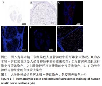
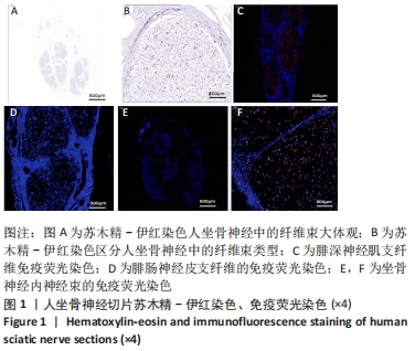
2.1 坐骨神经二维图像横断面的观察结果 二维图像横断面显示,人坐骨神经位于大腿肌间隙,呈小圆点状。将人坐骨神经节段连续切片,切片厚度为5 μm,切片间隔45 μm,间距内的石蜡组织切片的二维横断面图像信息只有极小可忽略不计的改变。电子显微镜下逐个观察切片样本的形态结构,判断坐骨神经内部结构的走向。采用参照物校正位置,保持切片的连续性。将取下的人坐骨神经石蜡包裹固定后连续组织切片,免疫组化染色后使用电子显微镜中的照相功能提取其二维图像信息,观察发现,使用免疫组化的方式染色后,功能束所含有的特异性抗体的不同,能直观明确精准地区分运动神经纤维束及其他纤维束,见图1A,B。人坐骨神经中的运动神经纤维束经过染色后颜色变深,其他神经纤维束未染色。 利用免疫荧光染色,观察了距股骨髁近端20 cm坐骨神经,1 cm各断面神经束的数目和性质。阳性对照选取纯运动神经束(腓深神经肌支),经Anti-HB9/HLXB9 Antibody特异性染色,放大50倍镜下观察显示神经纤维内均有特异性染色,见图1C;阴性对照选取纯感觉神经束(腓肠神经),经Anti-HB9/HLXB9 Antibody特异性染色,观察显示神经纤维内没有特异性染色,见图1D。而实际的人坐骨神经样本,区别于坐骨神经内运动神经束及其他神经束,观察发现,此段坐骨神经内大部分纤维束均有特异性染色,小部分未被染色,此段坐骨神经纤维束由被特异性染色的纯运动神经束、混合神经束及纯感觉神经束组成,见图1E,坐骨神经神经束的数量总共有50束。 标本的坐骨神经平面位于大腿中段,坐骨神经在大腿中部该横切面的神经纤维束数目较多;纤维交叉较频繁,由于重建坐骨神经长度为1 cm,该段坐骨神经纤维束形态、大小变化频率不高;以混合束为主。神经束数目较大腿上部减增多。该横切面总共有50个神经束,其中运动及混合神经束44个,感觉神经束6个,神经束断面形状无规律,见图1F。 "
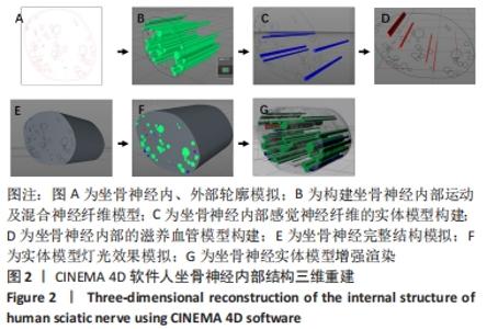
2.2 通过CINEMA 4D软件三维立体重建人坐骨神经可视化 运用CINEMA 4D软件进行三维重组,重建人坐骨神经三维形态。根据假设,神经纤维束可看作管道结构,该管道的表面是由球心沿着中轴线的球包络滚动而成。用管道沿着横断面连续切片,获得该管道的某一段结构的连续200张平行切片,利用显微成像技术将管道横断面保留为可处理图像,记录管道与切片的多组信息,图像按照切片的先后顺序依次命名为0-199.bmp,为方便数据导入和图像编辑,均保留成BMP格式,分辨率宽和高均为512 pixel。每个球的半径固定;切片间距以及图像象素的尺寸均为1[13-14]。取垂直于切片的坐标系为Z轴,平面Z=0代表第1张切片,以此类推,平面Z=199代表第200张切片,那么,坐标所代表的Z=z切片在文件中的先后次序为:(-256,-256,z),(-256,-255,z),…(-256,255,z),(-255,-256,z),(-255,-255,z),…(-255,255,z)。 按照之前的假设,管道是由一组等径的圆球,其圆心按中轴线滚动所形成的包络。每张切面与中轴线有且仅有一个交点,并且这个交点就是切片图像上最大内切圆的圆心,也就是球心的位置。因此,所要寻找的中轴线与半径,就是找图像中最大内切圆的圆心和半径,其解决方案就是求任意一个骨架上的点到所有轮廓点的最小距离,再取所有骨架点对应的最小距离的最大值,此最大值即最大内切圆的半径,对应的点即为最大内切圆的圆心。求出了最大内切圆的半径和球心后,通过拟合的方式来获取中轴线的图像,将最大内切圆的半径取平均值,得到管道半径。在计算得到最大内切圆的球心和半径的基础上,通过拟合的方式获得中轴线的图像,再根据CINEMA 4D软件排列平面的投影图。 按照以上思路,200张切片的最大内切圆圆心坐标及半径,结合体质量的信息,中轴线的z轴从1-200,得到中轴线在空间中的坐标。放样制作得到管道模型。 此次用创建神经管道的模型,主要应用到放样命令的操作。整个神经模型就应用不同切片路径,按照一定到等比间隙进行排列,依次链接,形成某段神经到重建复原。 打开CINEMA 4D导入切面图转化来到路径,将该路径转换为可编辑多边形,按照一定到等比间隙进行排列,依次链接,内部及轮廓做一个组,见图2A。 先选取内部所有轮廓,在快捷菜单栏展开中找到“放样”工具,将轮廓线处于放样的子级中。工作区即可看到一个生成的实体模型,将运动纤维及混合纤维识别出并进行重新建,见图2B。同理生成没有被抗体特异性结合的感觉纤维,见图2C。同理生成此段坐骨神经内部的滋养血管,见图2D。此模型为实体模型无法看到内部结构,对该层赋予半透明材质,点击材质求,对参数进行调节,包含颜色,半透明,在透明图层上加上噪波纹理,使其更加具有真实感,见图2E。将得到的2组模型拼合到一起,配合灯光,见图2F;调节好渲染参数进行渲染,得到效果,见图2G。 "

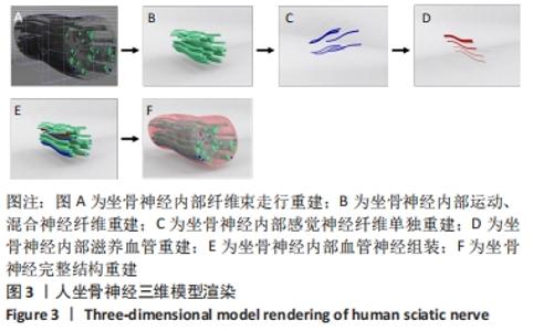
将200张切片的二维图像按照上述方法处理后根据软硬定位重建1 cm坐骨神经内部结构,见图3A;单独重建的坐骨神经内部运动、混合神经纤维,见图3B;单独重建的坐骨神经内部感觉神经纤维,见图3C;单独重建的坐骨神经内部滋养血管,见图3D。 "
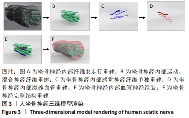

分别将单独重建的运动、混合神经纤维、感觉神经纤维及滋养血管按照原始空间比例组合成坐骨神经内部的三维立体结构,渲染神经比邻软组织逐张扫描、顺序存储输入计算机坐骨神经横断面二维信息,见图3E,F,利用CINEMA 4D软件逐张系统的进行排序、设置原点、比对、定位、匹配和合成;根据被Anti-HB9/HLXB9 Antibody特异性染色的神经束将坐骨神经内部不同类型的神经束及其功能进行区分,清晰准确地呈现各种类型神经束的三维结构;此外,CINEMA 4D软件能实现任意角度的旋转和不同平面的裁剪,较好地重建坐骨神经中各个神经束的三维立体结构,指导临床上对坐骨神经的手术操作以及模拟术后的恢复效果。将神经束简化为一条条中心线,可清楚观察神经束交叉重组的复杂结构。从三维重建的结果可以看到,此段坐骨神经标本出现错综复杂的交叉重组,最短的间隔0.04 mm出现了神经束的交叉,这可能与坐骨神经发出胫神经及坐骨神经支有关。运用CINEMA 4D三维可视化软件建模分析,重建坐骨神经内部神经束,结合免疫荧光染色,区分运动、混合神经束和感觉神经束。 "

| [1] 房娟. 显微高光谱成像技术结合神经分类的应用研究[D]. 上海:华东师范大学,2016. [2] 陈焱, 肖志宏, 邢丹谋. 周围神经损伤再生与修复的研究进展[J]. 中华显微外科杂志,2015,38(4):413-416. [3] SUDERLAND S, RAY LJ. The intraneural topography of the sciatic nerve and its poplitical divisions in man. Brain. 1948;71:242-273. [4] 陈增淦,陈统一,张键,等.臂丛神经显微结构的计算机三维重建[J].中华骨科杂志,2004,24(8):462-466. [5] WATCHMAKER GP, GUMUCIO CA, CRANDALL RE, et al. Fascicular topograpy of the median nerve: A computer based to identify branching patterns. J Hand Surg (Am). 1991;16(1):53-59. [6] 张元智. 臂丛神经影像形态学和虚拟中国人女Ⅰ号臂丛、腰骶丛神经断层解剖学及可视化初步研究[D]. 上海:第一军医大学, 2005. [7] 陈增淦, 张猛, 张健, 等. 人体坐骨神经连续组织切片三维重建研究[J]. 复旦学报(医学版),2008,35(4):510-513. [8] 高宏, 裴国献. 周围神经三维重建与可视化研究进展[J]. 中国修复重建外科杂志,2009,23(2):239-244. [9] 赵文强,温树正,郝增涛,等. 组织工程人工神经修复周围神经损伤的研究进展[J]. 实用手外科杂志,2019,33(4):449-454,457. [10] 孟军.连续大尺度组织切片自动成像及三维重建系统研究[D]. 上海:上海交通大学,2013. [11] 孙廷卿. 人脑薄束核的三维解剖研究[D]. 杭州:杭州师范大学, 2012. [12] 廖礼彬, 陈胜国, 王士平, 等. 人胚胎三叉神经连续切片的三维重建[J]. 解剖学杂志,2016,39(3):308-310. [13] 祝玉杰. 周围神经MicroCT图像中神经束分割的标准化流程研究[D].广州:广东工业大学,2020. [14] 舒茂, 胡立华, 董秋雷,等. 基于学习的鲁棒三维射影重建[J]. 计算机辅助设计与图形学学报,2018,30(2):309-317. [15] Bikis C, Degrugillier L, Thalmann P, et al. Three-dimensional imaging and analysis of entire peripheral nerves after repair and reconstruction. J Neurosci Methods. 2018;295:37-44. [16] 刘小林, 罗鹏, 戚剑, 等. 周围神经可视化虚拟重建关键技术研究[C]. 中华医学会第 10 届全国显微外科学术会议暨世界首例断肢再植成功50周年庆典论文集,2013. [17] SHIMAZU Y, KUDO T, YAGISHITA H, et al. Three-dimensional visualization and quantification for the growth and invasion of oral squamous cell carcinoma. Jap Dent Sci Rev. 2010;46:17-25. [18] GÜLEKON N, PEKER T, TURGUT HB, et al. Qualitative comparison of anatomical microdissection, Sihler’s staining and computerized reconstruction methods for visualizing intramuscular nerve branches. Surg Radiol Anat. 2007;29(5):373-378. [19] 孙廓, 胡平, 张峰,等. 人体正中神经内部显微结构的三维重建与可视化研究[J]. 中国矫形外科杂志,2008,16(18):1385-1388. [20] 罗鹏,张毅,刘小林,等.不同组织染色法在周围神经虚拟三维重建中的应用评估[J]. 实用临床医药杂志,2017,21(7):59-63. [21] 罗鹏, 刘小林, 戚剑, 等. 周围神经三维可视化模型建立中的功能束识别研究进展[J]. 中华创伤骨科杂志,2011,13(2):187-189. [22] BIKIS C, THALMANN P, DEGRUGILLIER L, et al. Three-dimensional and non-destructive characterization of nerves inside conduits using laboratory-based micro computed tomography. J Neurosci Methods. 2018;294:59-66. [23] FARAHANI N, BRAUN A, JUTT D, et al. Three-dimensional imaging and scanning: current and future applications for pathology. J Pathol Inform. 2017;8:36. [24] ZOU ZM, LI J, CAO QY, et al. Clinical value of diffusion tensor imaging parameter value in evaluating the prognosis of spinal cord injury in acute cervical spinal cord injury Zhonghua yi xue za zhi. 2017;97(1): 17-21. [25] POGGENBORG RP, ESHED I, ØSTERGAARD M, et al. Enthesitis in patients with psoriatic arthritis, axial spondyloarthritis and healthy subjects assessed by ‘head-to-toe’whole-body MRI and clinical examination. Ann Rheum Dis. 2015;74(5):823-829. [26] PALADINI D, QUARANTELLI M, SGLAVO G, et al. Accuracy of neurosonography and MRI in clinical management of fetuses referred with central nervous system abnormalities. Ultrasound Obstet Gynecol. 2014;44(2):188-196. [27] 杜永浩,牛刚,杨健. 定量动态增强磁共振技术的影响因素分析[J]. 磁共振成像,2017,8(1):76-80. [28] 于春水,李坤成. 弥散张量成像纤维跟踪技术的研究进展[J]. 中国医学影像技术,2004,20(3):477-479. [29] 熊炜烽.鼻咽癌放疗后颞叶“正常表现脑白质”的磁共振波谱与扩散张量成像初步研究[D]. 广州:南方医科大学,2011 [30] 葛艳明,李耀武,王滨,等. 背景信号抑制扩散加权成像对兔 VX2 肝移植瘤疗效评价的实验研究[J]. 临床放射学杂志,2011,30(9): 1387-1390. [31] ALIZADEH M, FISHER J, SAKSENA S, et al. Reduced field of view diffusion tensor imaging and fiber tractography of the pediatric cervical and thoracic spinal cord injury. J Neurotrauma. 2018;35(3):452-460. [32] LV Z, TEK A, DA SILVA F, et al. Game on, science-how video game technology may help biologists tackle visualization challenges. PloS One. 2013;8(3):e57990. [33] 曹军. MAX 与 C4D 模型制作的实践对比浅谈[J]. 影视制作,2012, 20(5):51-55. [34] QU J, FUNG A, KELLY P, et al. Visualising a rare and complex case of advanced hilar cholangiocarcinoma. J Vis Commun Med. 2017;40(1): 26-31. [35] ALTOUNIAN V. Cover stories: From plot to finish: Visualizing martian atmospheric data from MAVEN in CINEMA 4D with Python. Science. 2015;350(6261):597. [36] TOGAO O, HIWATASHI A, OBARA M, et al. 4D ASL-based MR angiography for visualization of distal arteries and leptomeningeal collateral vessels in moyamoya disease: a comparison of techniques. Eur Radiol. 2018;28(11):4871-4881. [37] 吐尔逊江·达地汗,穆叶沙尔·吾拉木,廖礼彬,等.人胚胎三叉神经连续切片的三维重建[J].解剖学杂志,2016,39(3):308-310+357. [38] 唐仲平,崔权哲,包骥,等.全切片图像扫描技术在病理切片质控中的作用[J].中国医药科学,2020,10(12):33-38. [39] 李征毅. 臂丛超声影像学解剖与可视化研究及其临床意义[D].广州:南方医科大学,2012. [40] 胡智魁. 生物医学图像计算机智能识别关键技术研究[D]. 广州:广东工业大学,2012. [41] 谢华,夏顺仁,张赞超,等. 医学图像识别中多分类器融合方法的研究进展[J]. 国际生物医学工程杂志,2006,29(3):152-157. [42] 亚穆罕默德·阿力克,阿吉木·克热木,伊力扎提·伊力哈木,等. 基于断层扫描的兔坐骨神经显微结构三维可视化研究[J]. 中华实验外科杂志,2017,34(9):1538-1540. [43] 亚穆罕默德·阿力克,伊力扎提·伊力哈木,阿里木江·阿不来提,等. 应用micro-CT实现兔坐骨神经显微三维结构可视化研究[J]. 中国修复重建外科杂志,2017,31(12):1490-1494. [44] 张付龙,刘胜全,闫呈新,等. 3.0T MR扩散张量成像对膝部神经三维重建的应用[J]. 实用放射学杂志,2018,34(12):1953-1955,1969. [45] YAMUHANMODE A, YILIZATI Y, ALIMUJIANG A, et al. Visualization research of three-dimensional microstructure of rabbit sciatic nerve bundles by micro-CT. Zhongguo Xiu Fu Chong Jian Wai Ke Za Zhi. 2017; 31(12):1490-1494. [46] YAO Z, YAN LW, WANG T, et al. A rapid micro-magnetic resonance imaging scanning for three-dimensional reconstruction of peripheral nerve fascicles. Neural Regen Res. 2018;13(11):1953-1960. [47] SINGH A, ASIKAINEN S, TEOTIA AK, et al. Biomimetic Photocurable Three-Dimensional Printed Nerve Guidance Channels with Aligned Cryomatrix Lumen for Peripheral Nerve Regeneration. ACS Appl Mater Interfaces. 2018;10(50):43327-43342. [48] JHA BS, COLELLO RJ, BOWMAN JR, et al. Two pole air gap electrospinning: Fabrication of highly aligned, three-dimensional scaffolds for nerve reconstruction. Acta Biomater. 2011;7(1):203-215. [49] ZAVAN B, ABATANGELO G, MAZZOLENI F, et al. New 3D hyaluronan-based scaffold for in vitro reconstruction of the rat sciatic nerve. Neurol Res. 2008;30(2):190-196. [50] YAN L, LIU S, QI J, et al. Three-dimensional reconstruction of internal fascicles and microvascular structures of human peripheral nerves. Int J Numer Method Biomed Eng. 2019;35(10):e3245. |
| [1] | Cai Zhihao, Xie Zhaoyong. Femoral neck anteversion measurement assessment: how to establish a unified method and standard [J]. Chinese Journal of Tissue Engineering Research, 2023, 27(9): 1448-1454. |
| [2] | Li Daen, Wu Yafei, Qiu Shang, Wang Gang, Gao Xuren, Chen Xiangyang. Factors associated with subscapularis injury and predictive efficacy based on three-dimensional CT reconstruction [J]. Chinese Journal of Tissue Engineering Research, 2023, 27(27): 4351-4356. |
| [3] | Xu Biao, Lu Tan, Yang Junliang, Jiang Yaqiong, Yin Yujiao. Three-dimensional finite element analysis of the stress of the patellofemoral joint after medial patellofemoral ligament reconstruction by different femoral reconstruction insertion points [J]. Chinese Journal of Tissue Engineering Research, 2023, 27(22): 3463-3468. |
| [4] | Jiang Daixiang, Lu Hui, Ma Ling, Zhang Hua, Liu Dingxi, Sheng Zhaoyou, Wu Qimei, Liu Rong. Study on the distribution of calcaneal fracture line based on three-dimensional fracture map technology [J]. Chinese Journal of Tissue Engineering Research, 2023, 27(18): 2842-2847. |
| [5] | Liu Changzhen, Sun Ning, Zhu Kai, Liu Xin, Dou Yongfeng, Wang Jianye, Bi Jingwei, Zhu Tengyue, Sun Zhaozhong. Safety of interbody fusion with one-hole split endoscope for L4/5 spondylolisthesis evaluated by three-dimensional CT [J]. Chinese Journal of Tissue Engineering Research, 2023, 27(18): 2884-2891. |
| [6] | Wei Guoqiang, Li Yunfeng, Wang Yi, Niu Xiaofen, Che Lifang, Wang Haiyan, Li Zhijun, Shi Guopeng, Bai Ling, Mo Kai, Zhang Chenchen, Xu Yangyang, Li Xiaohe. Biomechanical analysis of non-uniform material femur under different loads [J]. Chinese Journal of Tissue Engineering Research, 2022, 26(9): 1318-1322. |
| [7] | Kong Yamin, Yan Juntao, Ma Bingxiang, Li Huawei. Massage vibration intervenes with MyoD expression and proliferation and differentiation of muscle satellite cells in rats with sciatic nerve injury [J]. Chinese Journal of Tissue Engineering Research, 2022, 26(8): 1160-1166. |
| [8] | Zhao Jing, Liu Xiaobo, Zhang Yue, Zhang Jiaming, Zhong Dongling, Li Juan, Jin Rongjiang. Visualization analysis of neuromuscular electrical stimulation therapy based on CiteSpace: therapeutic effects, hot spots, and developmental trends [J]. Chinese Journal of Tissue Engineering Research, 2022, 26(8): 1234-1241. |
| [9] | Yuan Jing, Sun Xiaohu, Chen Hui, Qiao Yongjie, Wang Lixin. Digital measurement and analysis of the distal femur in adults with secondary knee valgus deformity [J]. Chinese Journal of Tissue Engineering Research, 2022, 26(6): 881-885. |
| [10] | Yi Xinrong, Jia Fuquan, He Xin, Zhang Shaojie, Ren Xiaoyan, Li Zhijun. Establishment of cervical bone age equation for male adolescents aged 8-16 years old in Hohhot based on thin-slice CT [J]. Chinese Journal of Tissue Engineering Research, 2022, 26(6): 954-958. |
| [11] | Zhang Qiang, Wu Zongde, Liu Liang, Wei Guohua, Peng Liang. Finite element analysis of medial and lateral locking plates for fixation of externally rotated spiral fractures of the lower tibia [J]. Chinese Journal of Tissue Engineering Research, 2022, 26(36): 5750-5754. |
| [12] | Liu Yang, Zhu Zhiqiang, Zhao Xiaowei, Xiang Yujie, Xiao Jian, Cheng Lifen. Hot topics and international frontiers of electromyography in the field of body movements [J]. Chinese Journal of Tissue Engineering Research, 2022, 26(35): 5707-5715. |
| [13] | She Jian, Zhao Jing, Zhang Jiaming, Xia Haisha, Zhong Dongling, Li Yuxi, Zheng Zhong, Li Juan, Jin Rongjiang. Visualization analysis of literature on eye tracking in cognitive behaviors of autistic patients [J]. Chinese Journal of Tissue Engineering Research, 2022, 26(35): 5724-5732. |
| [14] | Bi Jingwei, Ren Jiabin, Liu Xin, Sun Ning, Li Rui, Li Yuefei, Sun Zhaozhong. 3D-CT-guided unilateral biportal endoscopic localization of L5 and S1 nerve roots and intervertebral space [J]. Chinese Journal of Tissue Engineering Research, 2022, 26(33): 5283-5289. |
| [15] | Deng Zhaoyang, Yang Zhaohui. Three-dimensional model analysis of tibial plateau fracture lines [J]. Chinese Journal of Tissue Engineering Research, 2022, 26(3): 334-339. |
| Viewed | ||||||
|
Full text |
|
|||||
|
Abstract |
|
|||||