Chinese Journal of Tissue Engineering Research ›› 2022, Vol. 26 ›› Issue (24): 3833-3839.doi: 10.12307/2022.562
Previous Articles Next Articles
Co-culture of fibroblasts and vascular endothelial cells affects proliferation and osteogenesis of adipose stem cells
Zhong Ruiying, Wang Fuke, Yang Guiran, Wang Guoliang, Hou Jianfei, Liao Xinyu
- Department of Sports Medicine, First Affiliated Hospital of Kunming Medical University, Kunming 650032, Yunnan Province, China
-
Received:2021-03-19Accepted:2021-04-23Online:2022-08-28Published:2022-01-22 -
Contact:Wang Fuke, PhD, Chief physician, Professor, Department of Sports Medicine, First Affiliated Hospital of Kunming Medical University, Kunming 650032, Yunnan Province, China Yang Guiran, Master, Physician, Department of Sports Medicine, First Affiliated Hospital of Kunming Medical University, Kunming 650032, Yunnan Province, China -
About author:Zhong Ruiying, Master candidate, Department of Sports Medicine, First Affiliated Hospital of Kunming Medical University, Kunming 650032, Yunnan Province, China -
Supported by:Joint Special Fund for Applied Basic Research Department of Science and Technology of Yunnan Province and Kunming Medical University, No. 201701UH00095 (to WFK)
CLC Number:
Cite this article
Zhong Ruiying, Wang Fuke, Yang Guiran, Wang Guoliang, Hou Jianfei, Liao Xinyu. Co-culture of fibroblasts and vascular endothelial cells affects proliferation and osteogenesis of adipose stem cells[J]. Chinese Journal of Tissue Engineering Research, 2022, 26(24): 3833-3839.
share this article
Add to citation manager EndNote|Reference Manager|ProCite|BibTeX|RefWorks

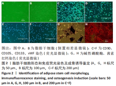
2.1 细胞的培养与鉴定结果 2.1.1 脂肪干细胞形态学观察及鉴定结果 倒置相差显微镜下观察,稳定传至4周3代后可见大量梭形细胞和多角形细胞,胞体大,核居中,呈巢状生长,排列紧凑且规律。表面抗原鉴定显示:第3代脂肪干细胞免疫荧光检测CD90、CD105染色均为阳性表达[8],CD133、vWF均阴性表达,阴性对照未发现阳性细胞;成骨诱导14 d时,碱性磷酸酶染色可见多数细胞呈阳性,细胞融合成团,蓝黑色颗粒状相互融合交错成网状排列;茜素红钙染色可见细胞部分融合,形成片状的钙化,呈大片红色钙结节影。染色结果证明其具有成骨分化能力,故判定所获细胞为脂肪干细胞[9],见图2。"
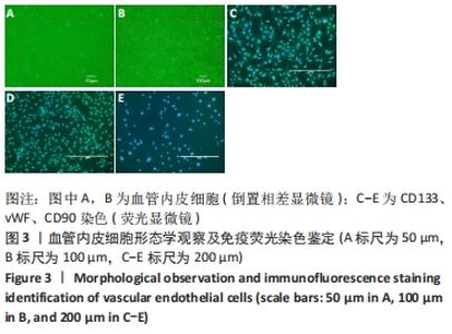
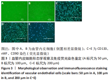
2.1.2 血管内皮细胞形态学观察及鉴定结果 倒置相差显微镜下观察,细胞增长数量多,形态呈多样化,有梭形和多角形混合生长,呈团、束状排列,亦单个散在分布。表面抗原鉴定显示:传代培养至3周时血管内皮细胞免疫荧光检测CD133、vWF染色均为阳性表达[10],CD90染色为阴性表达,阴性对照未发现阳性细胞,见图3。"
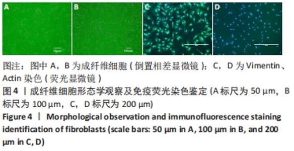
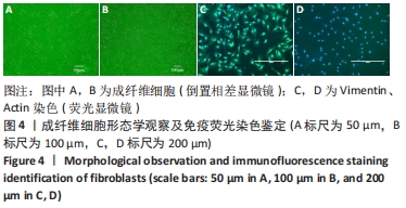
2.1.3 成纤维细胞形态学观察及鉴定结果 倒置相差显微镜下观,细胞数目较多,胞体大,多为梭形、纺锤形的扁平细胞,也有部分形态呈现不规则,细胞间以细长突起相互交联,呈漩涡状密集排列。传代培养至3周时成纤维细胞免疫荧光检测Vimentin(波形蛋白)染色为阳性表达[11],Actin(肌动蛋白)染色为阴性表达,阴性对照未发现阳性细胞,见图4。"
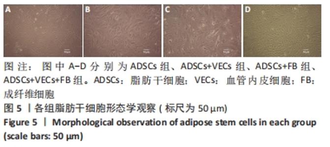
2.2 各组脂肪干细胞形态 各组脂肪干细胞培养至2周时,镜下观察细胞形态,见图5。"
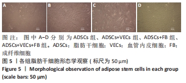
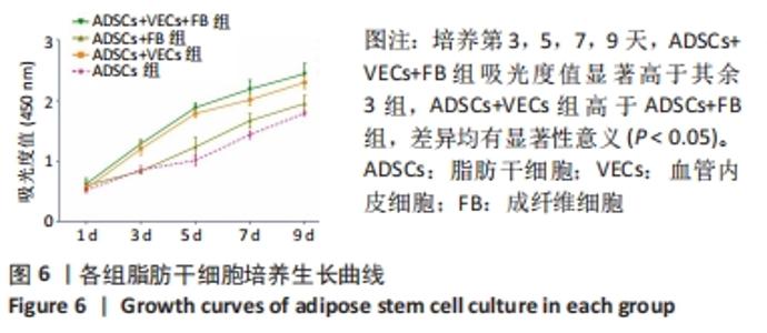
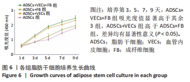
①脂肪干细胞组:形态单一呈梭形,没有细胞重叠现象;②脂肪干细胞+血管内皮细胞共培养组:细胞数量增多,漩涡状生长,形态呈多样化,有梭形和多角形混合生长,梭形多见,少量呈巢样分布;③脂肪干细胞+成纤维细胞共培养组:细胞形态仍较为单一,呈长梭形,排列均匀,胞体均较大,未出现细胞融合团块状;④脂肪干细胞+血管内皮细胞+成纤维细胞共培养组:细胞数量逐渐增多,细胞大多失去梭形,形体变大呈多角形,部分细胞融合成团块状,呈巢样分布。 2.3 各组脂肪干细胞生长曲线 用CCK-8检测各组脂肪干细胞吸光度值,绘制生长曲线。各组细胞生长曲线形态基本相似,细胞吸光度值逐渐升高。采用统计学单因素方差分析进行组间比较,采用q检验进行两两比较各组吸光度值,第1天各组间差异无显著性意义(P > 0.05),第3天脂肪干细胞组与脂肪干细胞+成纤维细胞共培养组比较差异无显著性意义(P > 0.05);第3,5,7,9 天,脂肪干细胞+血管内皮细胞+成纤维细胞共培养组吸光度值显著高于其余3组,脂肪干细胞+血管内皮细胞共培养组高于脂肪干细胞+成纤维细胞共培养组,差异均有显著性意义(P < 0.05),见图6。"
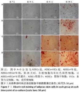
2.4 茜素红及碱性磷酸酶染色结果 2.4.1 各组细胞茜素红染色分析 第7天时各组细胞均呈阴性反应;第14天时,混合培养各组细胞均出现极少量红色阳性细胞;第21天,混合培养各组细胞红色阳性细胞增多,以脂肪干细胞+血管内皮细胞+成纤维细胞共培养组增加最为明显;第28天时,各组细胞均有红色阳性细胞,以脂肪干细胞+血管内皮细胞+成纤维细胞共培养组最多,呈红色灶状,脂肪干细胞+血管内皮细胞共培养组和脂肪干细胞+成纤维细胞共培养组较少,脂肪干细胞组最少,见图7。"

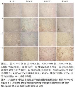
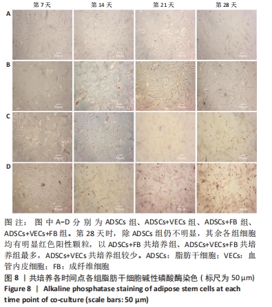
2.4.2 各组细胞碱性磷酸酶染色分析 第7天时除脂肪干细胞组呈阴性反应,其余各组均开始出现阳性颗粒,以脂肪干细胞+血管内皮细胞+成纤维细胞共培养组最多;第14天时,各组细胞均出现红色阳性颗粒,以脂肪干细胞+成纤维细胞共培养组、脂肪干细胞+血管内皮细胞+成纤维细胞共培养组较为明显;第21天,混合培养各组细胞红色阳性颗粒增多,以脂肪干细胞+血管内皮细胞+成纤维细胞共培养组增加最为明显;第28天时,除脂肪干细胞组仍不明显,其余各组细胞均有明显红色阳性颗粒,以脂肪干细胞+成纤维细胞共培养组、脂肪干细胞+血管内皮细胞+成纤维细胞共培养组最多,脂肪干细胞+血管内皮细胞共培养组较少,脂肪干细胞组最少,见图8。"
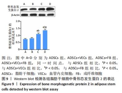
2.5 Western blot检测骨形态发生蛋白2的表达 各组脂肪干细胞均有骨形态发生蛋白2表达,脂肪干细胞+成纤维细胞共培养组、脂肪干细胞+血管内皮细胞+成纤维细胞共培养组较明显;脂肪干细胞组与脂肪干细胞+血管内皮细胞共培养组比较差异无显著性意义(P > 0.05),其余各组比较差异均有显著性意义(P < 0.05),见图9。"

| [1] 王福科,张红,李彦林,等.联合培养血管内皮细胞与脂肪干细胞构建组织工程骨异位成骨[J].中国修复重建外科杂志,2019,33(10): 1310-1319. [2] 陈雄林,曹小明,杨耀防,等.真皮成纤维细胞原代培养及生长曲线检测[J].九江学院学报(自然科学版),2017,32(2):94-95,112. [3] 杨桂然,王福科,李彦林.成纤维细胞的生物学特性及分化潜能[J].中国组织工程研究,2020,24(13):2114-2119. [4] LEE MS, WANG J, YUAN H, et al. Endothelin-1 differentially directs lineage specification of adipose- and bone marrow-derived mesenchymal stem cells. FASEB J. 2019;33(1):996-1007. [5] FUDULU DP, HORN G, HAZELL G, et al. Co-culture of monocytes and zona fasciculata adrenal cells: An in vitro model to study the immune-adrenal cross-talk. Mol Cell Endocrinol. 2021;526:111195. [6] 吕继忠.成纤维细胞来源诱导性多能干细胞定向分化为血管内皮样细胞及与牙髓干细胞的共培养[J].中国组织工程研究,2018, 22(21):3371-3375. [7] 陈娟,叶云金,谢丽华,等.Raji-MG63细胞共培养体系中心肌营养蛋白样细胞因子1(CLCF1)对RANKL/OPG比值及成骨细胞分化的影响[J].中国骨质疏松杂志,2021,27(2):157-161. [8] DAI R, WANG Z, SAMANIPOUR R, et al. Adipose-Derived Stem Cells for Tissue Engineering and Regenerative Medicine Applications. Stem Cells Int. 2016;2016:6737345. [9] YANG H, HONG N, LIU H, et al. Differentiated adipose-derived stem cell cocultures for bone regeneration in RADA16-I in vitro. J Cell Physiol. 2018;233(12):9458-9472. [10] 稂翠玲,孙斌,雷红召,等.血管瘤血管内皮细胞体外分离纯化方法的优化[J].中国美容医学,2018,27(4):61-64. [11] 魏传飞,王伟,刘延明,等.人体皮肤成纤维细胞的快速分离及多向分化能力鉴定[J].中华细胞与干细胞杂志(电子版),2019,9(5): 267-275. [12] WANG Q, DING G, XU X. Immunomodulatory functions of mesenchymal stem cells and possible mechanisms. Histol Histopathol. 2016;31(9):949-959. [13] LI M, ZHANG C, YANG Y. Effects of mechanical forces on osteogenesis and osteoclastogenesis in human periodontal ligament fibroblasts: A systematic review of in vitro studies. Bone Joint Res. 2019;8(1):19-31. [14] 陈晓婷,高艳虹.间充质干细胞成骨分化中多种调控因子的交互作用及其机制[J].上海交通大学学报(医学版),2017,37(5):694-698. [15] HURLEY MM, GRONOWICZ G, ZHU L, et al. Age-Related Changes in FGF-2, Fibroblast Growth Factor Receptors and β-Catenin Expression in Human Mesenchyme-Derived Progenitor Cells. J Cell Biochem. 2016; 117(3):721-729. [16] KATSUYAMA T, OTSUKA F, TERASAKA T, et al. Regulatory effects of fibroblast growth factor-8 and tumor necrosis factor-α on osteoblast marker expression induced by bone morphogenetic protein-2. Peptides. 2015;73:88-94. [17] 韩祥祯,何惠宇,胡杨,等.人骨形态发生蛋白-2重组慢病毒载体转染羊骨髓间充质干细胞及其成骨调节作用[J].中华实用诊断与治疗杂志,2014,28(6):540-542. [18] 张波,韦冰丹,甘坤宁,等.富血小板血浆联合骨髓间充质干细胞对兔股骨头坏死BMP-2/Smads通路的影响[J].中国骨质疏松杂志, 2016,22(2):131-134,227. [19] BETZ VM, REN B, MESSMER C, et al. Bone morphogenetic protein-2 is a stronger inducer of osteogenesis within muscle tissue than heterodimeric bone morphogenetic protein-2/6 and -2/7: Implications for expedited gene-enhanced bone repair. J Gene Med. 2018;20(9): e3042. [20] QIAN C, ZHU C, YU W, et al. Bone morphogenetic protein 2 promotes osteogenesis of bone marrow stromal cells in type 2 diabetic rats via the Wnt signaling pathway. Int J Biochem Cell Biol. 2016;80:143-153. [21] DECAMBRON A, DEVRIENDT N, LAROCHETTE N, et al. Effect of the Bone Morphogenetic Protein-2 Doses on the Osteogenic Potential of Human Multipotent Stromal Cells- Containing Tissue Engineered Constructs. Tissue Eng Part A. 2019;25(7-8):642-651. [22] ZUR NIEDEN NI, TURGMAN CC, LANG X, et al. Fluorescent hydrogels for embryoid body formation and osteogenic differentiation of embryonic stem cells. ACS Appl Mater Interfaces. 2015;7(19):10599-10605. [23] 张磊,付炜,张海波.间充质干细胞与血管内皮细胞的相互作用及其应用[J].中华医学杂志,2016,96(7):585-588. [24] MERFELD-CLAUSS S, LUPOV IP, LU H, et al. Adipose Stromal Cell Contact with Endothelial Cells Results in Loss of Complementary Vasculogenic Activity Mediated by Induction of Activin A. Stem Cells. 2015;33(10):3039-3051. [25] PANINA YA, YAKIMOV AS, KOMLEVA YK, et al. Plasticity of Adipose Tissue-Derived Stem Cells and Regulation of Angiogenesis. Front Physiol. 2018;9:1656. [26] PASCHOS NK, BROWN WE, ESWARAMOORTHY R, et al. Advances in tissue engineering through stem cell-based co-culture. J Tissue Eng Regen Med. 2015;9(5):488-503. [27] LI CJ, MADHU V, BALIAN G, et al. Cross-Talk Between VEGF and BMP-6 Pathways Accelerates Osteogenic Differentiation of Human Adipose-Derived Stem Cells. J Cell Physiol. 2015;230(11):2671-2682. [28] LI N, SANYOUR H, REMUND T, et al. Vascular extracellular matrix and fibroblasts-coculture directed differentiation of human mesenchymal stem cells toward smooth muscle-like cells for vascular tissue engineering. Mater Sci Eng C Mater Biol Appl. 2018;93:61-69. [29] HAMANEH MB, YU YK. Exploring induced pluripotency in human fibroblasts via construction, validation, and application of a gene regulatory network. PLoS One. 2019;14(8):e0220742. [30] QI W, YAN J, SUN H, et al. Multifunctional Nanocomposite Films for Synergistic Delivery of bFGF and BMP-2. ACS Omega. 2017;2(3):899-909. [31] WANG T, GUO S, ZHANG H. Synergistic Effects of Controlled-Released BMP-2 and VEGF from nHAC/PLGAs Scaffold on Osteogenesis. Biomed Res Int. 2018;2018:3516463. |
| [1] | Yao Xiaoling, Peng Jiancheng, Xu Yuerong, Yang Zhidong, Zhang Shuncong. Variable-angle zero-notch anterior interbody fusion system in the treatment of cervical spondylotic myelopathy: 30-month follow-up [J]. Chinese Journal of Tissue Engineering Research, 2022, 26(9): 1377-1382. |
| [2] | Li Qin, Mao Shuangfa, Li Min, Cheng Jiyan. Protective effect and mechanism of dendrobium on fibroblasts damaged by ultraviolet B [J]. Chinese Journal of Tissue Engineering Research, 2022, 26(8): 1228-1233. |
| [3] | Wang Jing, Xiong Shan, Cao Jin, Feng Linwei, Wang Xin. Role and mechanism of interleukin-3 in bone metabolism [J]. Chinese Journal of Tissue Engineering Research, 2022, 26(8): 1260-1265. |
| [4] | Xiao Hao, Liu Jing, Zhou Jun. Research progress of pulsed electromagnetic field in the treatment of postmenopausal osteoporosis [J]. Chinese Journal of Tissue Engineering Research, 2022, 26(8): 1266-1271. |
| [5] | Wen Dandan, Li Qiang, Shen Caiqi, Ji Zhe, Jin Peisheng. Nocardia rubra cell wall skeleton for extemal use improves the viability of adipogenic mesenchymal stem cells and promotes diabetes wound repair [J]. Chinese Journal of Tissue Engineering Research, 2022, 26(7): 1038-1044. |
| [6] | Zhu Bingbing, Deng Jianghua, Chen Jingjing, Mu Xiaoling. Interleukin-8 receptor enhances the migration and adhesion of umbilical cord mesenchymal stem cells to injured endothelium [J]. Chinese Journal of Tissue Engineering Research, 2022, 26(7): 1045-1050. |
| [7] | Luo Xiaoling, Zhang Li, Yang Maohua, Xu Jie, Xu Xiaomei. Effect of naringenin on osteogenic differentiation of human periodontal ligament stem cells [J]. Chinese Journal of Tissue Engineering Research, 2022, 26(7): 1051-1056. |
| [8] | Wang Xinmin, Liu Fei, Xu Jie, Bai Yuxi, Lü Jian. Core decompression combined with dental pulp stem cells in the treatment of steroid-associated femoral head necrosis in rabbits [J]. Chinese Journal of Tissue Engineering Research, 2022, 26(7): 1074-1079. |
| [9] | Zhang Yujie, Yang Jiandong, Cai Jun, Zhu Shoulei, Tian Yuan. Mechanism by which allicin inhibits proliferation and promotes apoptosis of rat vascular endothelial cells [J]. Chinese Journal of Tissue Engineering Research, 2022, 26(7): 1080-1084. |
| [10] | Fang Xiaolei, Leng Jun, Zhang Chen, Liu Huimin, Guo Wen. Systematic evaluation of different therapeutic effects of mesenchymal stem cell transplantation in the treatment of ischemic stroke [J]. Chinese Journal of Tissue Engineering Research, 2022, 26(7): 1085-1092. |
| [11] | Guo Jia, Ding Qionghua, Liu Ze, Lü Siyi, Zhou Quancheng, Gao Yuhua, Bai Chunyu. Biological characteristics and immunoregulation of exosomes derived from mesenchymal stem cells [J]. Chinese Journal of Tissue Engineering Research, 2022, 26(7): 1093-1101. |
| [12] | Huang Chenwei, Fei Yankang, Zhu Mengmei, Li Penghao, Yu Bing. Important role of glutathione in stemness and regulation of stem cells [J]. Chinese Journal of Tissue Engineering Research, 2022, 26(7): 1119-1124. |
| [13] | Hui Xiaoshan, Bai Jing, Zhou Siyuan, Wang Jie, Zhang Jinsheng, He Qingyong, Meng Peipei. Theoretical mechanism of traditional Chinese medicine theory on stem cell induced differentiation [J]. Chinese Journal of Tissue Engineering Research, 2022, 26(7): 1125-1129. |
| [14] | An Weizheng, He Xiao, Ren Shuai, Liu Jianyu. Potential of muscle-derived stem cells in peripheral nerve regeneration [J]. Chinese Journal of Tissue Engineering Research, 2022, 26(7): 1130-1136. |
| [15] | Fan Yiming, Liu Fangyu, Zhang Hongyu, Li Shuai, Wang Yansong. Serial questions about endogenous neural stem cell response in the ependymal zone after spinal cord injury [J]. Chinese Journal of Tissue Engineering Research, 2022, 26(7): 1137-1142. |
| Viewed | ||||||
|
Full text |
|
|||||
|
Abstract |
|
|||||