中国组织工程研究 ›› 2014, Vol. 18 ›› Issue (51): 8297-8300.doi: 10.3969/j.issn.2095-4344.2014.51.018

• 肌肉肌腱韧带组织构建 tissue construction of the muscle, tendon and ligament • 上一篇    下一篇

兔肌腱细胞的分离、培养及鉴定

王玉聪1,张前法2   

  1. 1宁波市第九医院骨科,浙江省宁波市 315020;2上海市第一人民医院宝山分院骨科,上海市 200400
  • 出版日期:2014-12-10 发布日期:2014-12-10
  • 通讯作者: 张前法,主任医师,上海市第一人民医院宝山分院骨科,上海市 200400
  • 作者简介:王玉聪,男,1985年生,安徽省肥东市人,汉族,硕士,主要从事创伤骨科的研究。

Isolation, culture and identification of rabbit tendon cells

Wang Yu-cong1, Zhang Qian-fa2   

  1. 1Department of Orthopedics, Ninth People’s Hospital of Ningbo City, Ningbo 315020, Zhejiang Province, China; 2Department of Orthopedics, Baoshan Branch, First People’s Hospital of Shanghai, Shanghai 200400, China
  • Online:2014-12-10 Published:2014-12-10
  • Contact: Zhang Qian-fa, Chief physician, Department of Orthopedics, Baoshan Branch, First People’s Hospital of Shanghai, Shanghai 200400, China
  • About author:Wang Yu-cong, Master, Department of Orthopedics, Ninth People’s Hospital of Ningbo City, Ningbo 315020, Zhejiang Province, China

摘要:

背景:肌腱细胞是一种高分化的细胞,其增殖相对缓慢,在体外经多次传代后甚至丧失增殖能力,因此有必要建立肌腱细胞良好的体外分离、培养模式。


目的:探讨兔肌腱细胞的分离、培养及鉴定。


方法:无菌条件下切取新西兰乳兔趾屈肌腱,显微镜下剥离腱外膜,采用Henderson分步酶消化法分离肌腱细胞,用含体积分数为20%胎牛血清的F-12培养液进行培养、传代。


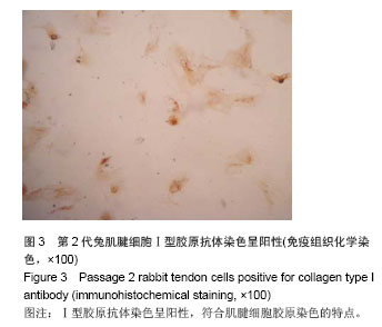
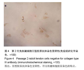
结果与结论:通过不同的酶消化分离可获得较纯肌腱细胞,体外培养细胞表现出良好的细胞增殖能力和传代能力。免疫组织化学染色见分离培养的第2代腱细胞Ⅰ型胶原抗体染色呈阳性,而Ⅲ型胶原抗体染色呈阴性,证明所获细胞为肌腱细胞。提示肌腱细胞能够在体外分离、扩增和传代。


中国组织工程研究
杂志出版内容重点:组织构建;骨细胞;软骨细胞;细胞培养;成纤维细胞;血管内皮细胞;骨质疏松组织工程


全文链接:

关键词: 组织构建, 组织工程, 腱细胞, 兔, 分离, 培养, 鉴定

Abstract:

BACKGROUND: Tendon cells are characterized by high differentiation potentials, slow proliferation rate, and even lost proliferation capacity after passage in vitro. Therefore it is necessary to establish the ideal isolation and culture patterns of tendon cells in vitro.

 
OBJECTIVE: To investigate the isolation, culture and identification of tendon cells.
METHODS: The flexor tendon of New Zealand fetal rabbits were cut under sterile conditions, the peritenon of flexor tendon was removed by microsurgical technique. Tendon cells were isolated with Henderson-step enzymatic digestion method and cultured in a complete medium consisting of DMEM/F12 and 20% fetal bovine serum for primary culture and passage.
RESULTS AND CONCLUSION: The tendon cells with high purity can be successfully isolated by different enzymatic digestion methods, and the cultured cells well proliferated and passaged in vitro. The immunohistochemical staining showed that, passage 2 cells were positive for collagen type I antibody, but negative for collagen type III antibody. These evidences confirmed that the cultured cells are tendon cells. Tendon cells can be isolated, proliferated and passaged in vitro.


中国组织工程研究
杂志出版内容重点:组织构建;骨细胞;软骨细胞;细胞培养;成纤维细胞;血管内皮细胞;骨质疏松组织工程


全文链接:

Key words: cell, cultured, immunohistochemistry, rabbits, tendon

中图分类号: