中国组织工程研究 ›› 2025, Vol. 29 ›› Issue (34): 7369-7375.doi: 10.12307/2025.499
• 生物材料综述 biomaterial review • 上一篇 下一篇
不同年龄阶段膝关节有限元计算模型的材料特性分析
陈 静1,张 楠2,孟庆华1,鲍春雨2
- 天津体育学院,1体育经济管理学院,2社会体育学院,天津市 301617
-
收稿日期:2024-07-01接受日期:2024-08-28出版日期:2025-12-08发布日期:2025-01-17 -
通讯作者:孟庆华,教授,天津体育学院体育经济管理学院,天津市 301617 -
作者简介:陈静,女,1976年生,天津市人,汉族,博士,副教授,主要从事膝关节有限元及智能计算方面的研究。 -
基金资助:国家自然科学基金项目(11372223,11102135),项目负责人:孟庆华;天津市自然科学基金项目(17JCZDJC36000),项目负责人:鲍春雨;天津市自然科学基金项目(18JCZDJC35900),项目负责人:孟庆华;国家体育总局科技创新项目(22KJCX077),项目负责人:鲍春雨;天津市研究生创新项目(2022SKYZ318),项目负责人:张楠
Material characterization of finite element computational models of knee joints at different ages
Chen Jing1, Zhang Nan2, Meng Qinghua1, Bao Chunyu2
- 1School of Economics Management, 2Academy of Social Sports, Tianjin University of Sport, Tianjin 301617, China
-
Received:2024-07-01Accepted:2024-08-28Online:2025-12-08Published:2025-01-17 -
Contact:Meng Qinghua, Professor, School of Economics Management, Tianjin University of Sport, Tianjin 301617, China -
About author:Chen Jing, PhD, Associate professor, School of Economics Management, Tianjin University of Sport, Tianjin 301617, China -
Supported by:National Natural Science Foundation Project, No. 11372223, 11102135 (to MQH); Tianjin Natural Science Foundation Project, No. 17JCZDJC36000 (to BCY); Tianjin Natural Science Foundation Project, No. 18JCZDJC35900 (to MQH); Science and Technology Innovation Project of General Administration of Sport of China, No. 22KJCX077 (to BCY); Tianjin Graduate Innovation Project, No. 2022SKYZ318 (to ZN)
摘要:
文题释义:
弹性模量:是材料的一种力学性质,描述材料受到应力时的弹性变形程度。弹性模量越大,说明材料的弹性越好,即在受到应力后能够迅速恢复原状。在中年到老年阶段,膝关节中股骨、胫骨、腓骨、髌骨的弹性模量会随着年龄的增加而降低,进而回归到儿童时期的弹性模量。泊松比:是材料的另一种力学性质,描述了材料在受到应力时沿着一个方向的收缩程度与沿着垂直方向的膨胀程度之比。
背景:有限元计算模型作为一种重要的工程分析技术已被广泛应用于生物工程研究的各个领域,然而,很少有文献介绍不同年龄阶段膝关节有限元计算模型每个解剖结构的材料性能,以满足不同的研究目的。
目的:在前人膝关节有限元研究的基础上,总结不同年龄阶段膝关节有限元计算模型的材料特性。方法:英文检索词为“Knee,finite element,material selection,ligament injury,osteoarthritis,elderly,children,young people”等,中文检索词为“膝关节,有限元,材料选择,韧带损伤,骨关节炎,老年人,儿童,年轻人”等,分别检索中国知网、PubMed数据库,文献检索时限为1950-2024年,就纳入与排除标准,最终纳入108篇文献进行归纳总结。
结果与结论:儿童膝关节骨密度会随着年龄的增长而增加,在成年后达到峰值。中年到老年阶段,膝关节股骨、胫骨、腓骨、髌骨的弹性模量会随着年龄的增加而降低,进而回归到儿童时期的弹性模量。儿童与成年人软骨的弹性模量基本一致,而老年人的软骨弹性模量增大。随着年龄的增长,膝关节韧带的弹性模量会出现一定程度降低,但年轻人与老年人膝关节韧带的弹性模量无显著差异。随着年龄的增长,膝关节半月板机械完整性的丧失会损害组织的生物力学功能,扰乱组织有效承载和传递的各向异性生物力学响应。膝关节有限元建模可以深入了解膝关节的生物力学特性,开发新的植入材料、预测膝关节疾病、改进手术技术,并指导患者康复锻炼。
https://orcid.org/0009-0007-8015-6783 (陈静)
中国组织工程研究杂志出版内容重点:生物材料;骨生物材料;口腔生物材料;纳米材料;缓释材料;材料相容性;组织工程
中图分类号:
引用本文
陈 静, 张 楠, 孟庆华, 鲍春雨. 不同年龄阶段膝关节有限元计算模型的材料特性分析[J]. 中国组织工程研究, 2025, 29(34): 7369-7375.
Chen Jing, Zhang Nan, Meng Qinghua, Bao Chunyu. Material characterization of finite element computational models of knee joints at different ages[J]. Chinese Journal of Tissue Engineering Research, 2025, 29(34): 7369-7375.
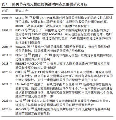
2.1 膝关节有限元模型研究情况 膝关节有限元模型的关键时间点及重要研究介绍,见表1。STEELE等[10]使用0.5 T MRI单元对没有膝关节创伤史的运动女性右膝进行了成像。FUCHS等[11]提出了一种能够通过CT扫描确定膝关节表面坐标的方法。WAWRO等[12]提出一种新的非线性有限元分析软件。DONG等[13]提出了一个3D膝关节有限元模型,以确定软骨缺损对缺损边缘周围应力分布的影响。í?IGUEZ-MACEDO等[14]分析并比较了人造和健康膝关节有限元模型皮质骨和小梁骨的应力分布情况。CHEN等[15]基于CT图像数据建立动态膝关节有限元模型,并验证该模型的有效性。LAHKAR等[16]报道了一种从双平面X射线片和CT数据得到韧带预应力的新方法。LU等[17]通过模拟方法来预测反向运动跳跃期间膝关节内的应力分布,该模拟方法将肌肉骨骼模型与膝关节的有限元模型相结合。ALONSO等[18]施加恒定的负荷提高矫形器在治疗儿童期和青春期早期膝关节侧偏方面的有效性。
2.2 骨(股骨、胫骨、腓骨、髌骨) 膝关节中骨(股骨、胫骨、腓骨、髌骨)的解剖学、生理学、生化、矿化和病理学方面随年龄的变化已有大量文献,而关于膝关节骨材料特性随年龄变化的文献很少。
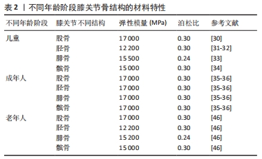
2.2.1 骨密度与年龄之间的关系 皮质骨是一种有机结构,矿物质含量约占人骨骼的80%。影响人类皮质骨质量的病理因素包括成人骨质疏松症[19]、儿童骨质疏松症[20]、克罗恩-S病或骨化症[21-22]。相关研究发现,由于人口老龄化的加剧,预测到2050年骨质疏松性骨折的直接成本将达到767亿欧元[23]。儿童时期的低骨量被认为是晚年骨质疏松的高危因素[24],相关研究认为评估儿童骨密度非常重要[25-26]。骨密度是评估矿物质状况的黄金标准参数之一,首先需要使用双能X射线吸收仪进来检测。相关研究使用霍洛奇双能X射线吸收仪(霍洛奇公司,Waltham,12 MA,USA)对7-11岁儿童进行骨密度扫描[27]。骨密度与骨微结构无关。多项研究表明,超声波测量能够评估探索骨区域(弹性和结构)的数量和质量[28-29]。KIM等[30]将儿童膝关节有限元计算模型中股骨材料的弹性模量与泊松比分别设置为17 000 MPa、0.3;LIMBERT等[31]与LANCIANESE等[32]将儿童胫骨材料的弹性模量与泊松比分别设置为12 200 MPa、0.3;BERTEAU等[33]将儿童腓骨材料的弹性模量与泊松比分别设置为15 500 MPa、0.24;KIAPOUR等[34]将儿童髌骨材料的弹性模量与泊松比分别设置为15 000 MPa、0.3。而有关成年人膝关节有限元计算模型中股骨、胫骨、腓骨、髌骨的弹性模量和泊松比设置为17 000 MPa、0.3[35-36]。这是由于骨密度随着年龄的增长而增加,在成年后达到峰值[37]。
2.2.2 骨密度与弹性模量之间的关系 王倞等[38]将10例成人新鲜尸体作为样本源,选取胫骨近端、大转子、股 骨颈、肱骨头和椎体5个部位的松质骨,采用CT扫描试样骨密度,通过力学测试分析分析不同部位松质骨的弹性模量,发现骨密度与弹性模量之间存在较高的线性相关性(0.850 > R2 > 0.785)和幂次相关性(0.871> R2 > 0.825)。袁嘉尧等[39]通过Pearson相关性分析发现,中老年人整体骨密度与身高、体质量、骨矿含量(整体、头部、主干、上肢、下肢)、肌肉质量(整体、头部、主干、下肢)、头部脂肪质量呈显著正相关,而与年龄呈显著负相关。从中年到老年阶段,膝关节中股骨、胫骨、腓骨、髌骨的弹性模量会随着年龄的增长而降低,进而回归到儿童时期的骨弹性模量。
2.2.3 年龄与弹性模量之间的关系 有关年轻骨骼强度和弹性模量的数据非常缺乏,多篇论文报道了超声轴向透射数据的年龄依赖性[40-42],只有2项体外机械研究对儿童皮质骨弹性模量进行了定量研究[43-44],这2项研究都是对干燥样本进行破坏性测试,儿童骨骼实验值都支持理论优化假设[45],即骨骼弹性模量会从新生儿值上升到成人值,这一假设目前被用于儿科计算方法。儿童骨骼体外超声波速度和弹性模量值与现有文献中的老年成人值接近[46-47]。
评估儿童皮质骨弹性特性是生物力学工程界及更广泛医疗保健专业人员面临的重大挑战[48]。即使采用X射线断层扫描、MRI成像和超声波成像等经典临床模式,由于缺乏儿童骨骼的参考值,也可能导致诊断不当。相关研究利用超声波和机械测量方法提供了儿童(4-16岁,平均年龄10岁)皮质骨的弹性特性值,并与老年人(75 岁以上)进行了比较,首先通过超声波方法评估动态弹性模量和泊松比,其次通过三点微弯曲测试估算静态弹性模量,结果显示:儿童样本在 10 MHz 频率下测得的纵向和横向波速平均值分别为(3.2±0.5) mm/μs 和(1.8±0.1) mm/μs,老年人样本的波速分别为(3.5±0.2) mm/μs 和(1.9±0.1) mm/μs,儿童样本的平均动态弹性模量和平均静态弹性模量分别为 (15.5±3.4) GPa 和(9.1±3.5) GPa;老年人样本的平均动态弹性模量和平均静态弹性模量分别为(16.7±1.9) GPa和(5.8±2.1) GPa,表明儿童和老年人骨骼的动态弹性模量、泊松比和静态弹性模量在相同范围内,没有统计差异[49]。
UEHIRA[50]发现成年人皮质骨的抗压和抗拉强度都随着年龄的增长而下降。抗压强度的下降归因于骨密度的下降,抗拉强度的下降归因于哈弗管横截面积的增加。根据SUGIYAMA[51]的说法,皮质骨的洛氏硬度随着年龄的增长而降低,这种降低是由骨质疏松症引起的,而不是由于骨骼中钙含量的变化。BLACK等[52]对8例胫骨中段的成活皮质骨动态力学性能进行了研究,发现35.4 Hz处的拉伸弹性模数随年龄的增加而下降,而353.6 Hz处的拉伸弹性模数则没有这种变化。WILSON[53]研究了35-89岁个体24个骨骼标本的压缩强度和单位体积骨矿物质的变化,35岁以后骨骼标本的最大抗压强度每10年下降约7%,单位体积骨矿含量每10年下降约3.3%。
不同年龄阶段骨结构的材料特性,见表2。
2.3 软骨 在膝关节有限元计算模型中,由于软骨结构光滑、柔软(刚性较小)、厚度较薄,因此在膝关节有限元建模过程中较为困难[54],也很难追踪软骨形态随年龄的演变。SI等[55]利用3-T超导扫描仪采集膝关节MRI图像,通过深度学习自动、精确地分割软骨,计算出14个解剖区域的软骨厚度,发现股骨软骨厚度始终大于胫骨软骨相应部位,除胫骨软骨内侧和外侧外,所有区域的软骨都随着年龄的增长而变薄。BLAZEK等[56]发现软骨厚度与体质量×身高呈正相关。相关研究发现,兔膝关节软骨弹性模量与关节软骨厚度呈负相关[57]。
由上可知,随着年龄的增加股骨软骨厚度在减少,而随着股骨软骨厚度的减少其弹性模量在增加。但是DéMARTEAU等[58]通过比较老年人股骨软骨与成年牛肱骨软骨发现,老年人股骨软骨具有更大的弹性模量。作者认为这是由于研究不同类型软骨得出的结论,不影响前文的结论。该文对后续学者研究软骨厚度与弹性模量直接的线性或非线性关系具有一定的指导意义。
2.4 韧带 膝关节韧带是由软组织组成的弹性带,具有复杂的微观结构和生物力学特性,对膝关节的运动学特性和应力承受能力至关重要[59-60]。从弹性一维单元到具有解剖逼真形状的复杂超弹性三维结构,已被广泛应用于膝关节韧带研究[61]。在膝关节3D模型中包含韧带的最直观方法是使用实体元素[62]。MRI扫描与3D重建软件相结合,提供了一种方便的方法来创建关节的详细模型,包括特定患者的韧带几何形状及其插入位置[63]。MRI扫描也可以很容易与CT相结合,CT提供了骨骼结构的高分辨率成像,这种方法还便于通过使用面对面接触来模拟韧带包裹,这在有限元程序中都可以实现[64]。韧带的材料特性(弹性模量、泊松比)选择对于计算精度非常重要,相关研究发现,青壮年人体标本中韧带的弹性模量、极限拉伸应力和破坏应变大约是60岁及以上人体标本的两三
倍[65],青壮年人体标本的主要破坏模式是韧带断裂,而老年人标本的主要破坏模式是韧带插入部位下方的骨骼撕裂,产生的差异和老年人标本韧带连接部位骨量减少有关。实验结果表明,随着年龄的增长,韧带的强度和刚度特性会显著降低,其程度超出预期。将年龄较大人体韧带标本实验结果应用于年轻或运动型个体的韧带破坏机制时,应谨慎解释,人们认为老年人的较差韧带生物力学特性减弱,同时关节固定也会导致韧带出现类似的功能性损伤。KASPERCZYK等[66]通过对6名年轻(平均年龄30岁)和6名年长(平均年龄64.7岁)活动水平相似供者的前交叉韧带和后交叉韧带进行了生物力学测试,发现年轻/年长前交叉韧带的最大应力为24/21 N/mm2)、年轻/年长前交叉韧带的弹性模量为144/129 MPa,年轻/年长前交叉韧带应变为0.25/0.28 mm。
综上所述,随着年龄的增长韧带弹性模量会出现一定程度的降低(出现减低的原因是由于随着年龄的增长韧带成纤维细胞的力学和生物学特性出现一定程度的退化),但是年轻人与老年人韧带之间的弹性模量无统计学差异,表明老年人并不一定会出现交叉韧带功能性损伤。
2.5 半月板
2.5.1 半月板的纳米力学特征随年龄的变化情况 半月板为人类膝关节提供基本支持并包含一系列功能,例如调节膝关节的负荷传递[67-69]。半月板含有沿圆周排列的胶原纤维,能够分布胫骨平台上的载荷,从而强化关节软骨并预防骨关节炎[70-73]。半月板通过称为“半月板附着物”的韧带结构与胫骨相连,其基质主要由蛋白多糖、弹性蛋白、糖脂、成纤维细胞以及60%-70%的水分组成[74-75]。这些强化的胶原纤维帮助半月板将载荷扩散到附着点,逐渐将软组织过渡到骨骼中,减少应力集中[76-78]。
随着年龄的增长,半月板的细胞外基质发生分子和结构变化,导致基质退化[79-80],这一退化过程的特征是合成代谢和分解代谢失衡,细胞减少了重要生长因子的产生,增加了基质降解酶和炎症因子的产生[81]。研究表明,衰老会导致半月板的胶原纤维排列紊乱、细胞减少等生化变化[77-78,82-84]。尽管衰老和骨关节炎引起的半月板退化已通过组织学和免疫组织化学分析研究,但尚未在微观结构层面详细研究其生物力学变化[85-87]。基于现有知识,衰老会改变半月板细胞外基质的生物力学特性,进而影响关节负荷时的反应,导致骨关节炎的发生[88]。研究还显示,随着年龄和疾病的进展,半月板的区域性机械性能逐渐模糊,机械完整性下降,这会破坏组织的生物力学功能,增加骨关节炎的风险。随着年龄增长,纳米力学性能的异质性逐渐增大[89-91]。
2.5.2 半月板的纳米力学特征与弹性模量之间的关系 正常、老年及骨关节炎患者的半月板组织在纳米力学特性上表现出显著差异[92]。年轻健康组织的弹性模量呈单峰分布,各区域表现出明确的峰值:外侧130-150 kPa、内侧70-90 kPa。老化组织同样呈现单峰分布,但其范围更宽,通常呈双峰,并且偏向较高的硬度值。整体上,双峰分布由一个较窄的第一峰和一个较宽的第二峰构成,表明退变相关的硬化区域有所增加。
骨关节炎不仅作为一种独立的疾病表现出特异的临床和生化特征,还随着组织的硬化而导致压缩刚度的增加,最终导致抗拉强度的下降,引发生物力学失效[93-95]。基质硬化源于细胞外基质中与年龄相关的分子变化,例如细胞外基质分子的异常沉积及交联,已知这些变化能够影响基质的机械特性,进而导致细胞功能异常[96-100]。半月板细胞对机械应力高度敏感,尤其是在关节受力时。细胞外基质中这些变化会促发或加剧与骨关节炎相关的细胞表型改变[101-103]。然而,基质力学与骨关节炎发展的确切关联仍有待进一步研究。
不同年龄阶段软骨、韧带与半月板结构的材料特性,见表3-5。
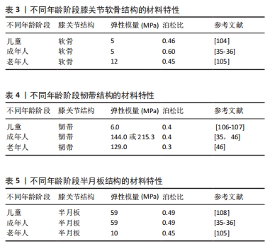
综上所述,人类半月板组织的局部纳米力学特性会随着年龄的增长而发生变化。文献表明,儿童与成人半月板的弹性模量大致相同,但在从成人到老年阶段,弹性模量逐渐降低[35-36,105,108],这一变化与半月板退行性变和骨关节炎的发生密切相关。因此,未来研究应深入探讨半月板的生物力学和生化特性之间的关联,尤其是在退变和骨关节炎的发展过程中。了解这些年龄相关的退变机制,将为预防和治疗骨关节炎提供重要的理论依据和策略支持。

| [1] TAPASVI S, SHEKHAR A, PATIL S, et al. Limb position influences component orientation in Oxford mobile bearing unicompartmental knee arthroplasty: an experimental cadaveric study. Bone Joint Res. 2020;9(6):272-278. [2] ARAUZ P, PENG Y, CASTILLO T, et al. In vitro kinematic analysis of single axis radius posterior-substituting total knee arthroplasty. J Knee Surg. 2021;34(11):1253-1259. [3] KONO K, TOMITA T, YAMAZAKI T, et al. Patellar resurfacing has minimal impact on in vitro tibiofemoral kinematics during deep knee flexion in total knee arthroplasty. Knee. 2021;30:163-169. [4] 潘正晔,马勇,郑伟涛.有限元分析立定跳远状态时不同摆臂动作和触地姿势的膝关节损伤[J].中国组织工程研究,2023,27(36): 5778-5783. [5] NAGHIBI H, JANSSEN D, VAN DEN BOOGAARD T, et al. The implications of non-anatomical positioning of a meniscus prosthesis on predicted human knee joint biomechanics. Med Biol Eng Comput. 2020;58(6): 1341-1355. [6] 韩广弢,李皓桓,高冯.创伤后膝骨关节炎发展中前交叉韧带损伤的作用与意义[J].中国组织工程研究,2020,24(15):2440-2446. [7] 王珂,张泽毅,张力文,等.不同年龄膝骨关节炎患者坐起生物力学特征的系统综述和Meta分析[J].中国组织工程研究,2024,28(18):2939-2946. [8] 张楠,孟庆华,鲍春雨.基于XCM模型预测排球运动员前交叉韧带应力[J].医用生物力学,2024,39(6):1146-1153. [9] KNOWLES NK, WHITTIER DE, BESLER BA, et al. Proximal tibia bone stiffness and strength in HR-pQCT-and QCT-based finite element models. Ann Biomed Eng. 2021;49(9):2389-2398. [10] STEELE JR, BASU A, JOB A. A three-dimensional representation of an athletic female knee joint using magnetic resonance imaging. Med Eng Phys. 1994;16(5): 363-369. [11] FUCHS S, KULLMER G, RICHARD H. Darstellung der Konstruktion eines FE-Modells am Beispiel des Kniegelenkes - Designing an FE Model as Exemplified by the Knee Joint. Biomedical Engineering. 1997;42(12): 347-351. [12] WAWRO M, FATHI-TORBAGHAN M. A parallel framework for the FE-based simulation of knee joint motion. IEEE Trans Biomed Eng. 2004;51(8):1490-1494. [13] DONG YF, HU GH, ZHANG LL, et al. Accurate 3D reconstruction of subject-specific knee finite element model to simulate the articular cartilage defects. J Shanghai Jiaotong Univ Sci. 2011;16:620-627. [14] ÍÑIGUEZ-MACEDO S, SOMOVILLA-GÓMEZ F, LOSTADO-LORZA R, et al. The process of designing a rotating platform artificial knee prosthesis with posterior stabilizers by finite element analysis. Int J Interact Des Manuf. 2018;12:853-864. [15] CHEN YF, LU C, ZHAO Y, et al. [Construction and simulation mechanical analysis of dynamic knee joint finite element model based on CT image]. Zhongguo Gu Shang. 2020;33(5):479-484. [16] LAHKAR BK, ROHAN PY, PILLET H, et al. Development and evaluation of a new procedure for subject-specific tensioning of finite element knee ligaments. Comput Methods Biomech Biomed Engin. 2021;24(11):1195-1205. [17] LU Z, SUN D, KOVÁCS B, et al. Case study: The influence of Achilles tendon rupture on knee joint stress during counter-movement jump - Combining musculoskeletal modeling and finite element analysis. Heliyon. 2023;9(8):e18410. [18] ALONSO MG, YAWNY A, BERTOLINO G. A numerical study towards shape memory alloys application in orthotic management of pediatric knee lateral deviations. Sci Rep. 2023;13(1):2134. [19] MCDONNELL P, MCHUGH PE, O’MAHONEY D. Vertebral osteoporosis and trabecular bone quality. Ann Biomed Eng. 2007;35(2):170-189. [20] MEHLMAN CT, SHEPHERD MA, NORRIS CS, et al. Diagnosis and Treatment of Osteopenic Fractures in Children. Curr Osteoporos Rep. 2012;10(4):317-321. [21] WALTERS TD. IBD: Is measuring bone age in children with Crohn’s disease useful. Nat Rev Gastroenterol Hepatol. 2012;9(11):620-622. [22] ENGIZ O, KARA S, BAGRUL D, et al. Infantile malignant osteopetrosis: a rare cause of neonatal hypocalcemia. J Pediatr Endocrinol Metab. 2012;25(11-12):1205-1207. [23] KANIS JA, COOPER C, HILIGSMANN M, et al. Partial adherence: a new perspective on health economic assessment in osteoporosis. Osteoporos Int. 2011;22(10):2565-2573. [24] JAVAID MK, COOPER C. Prenatal and childhood influences on osteoporosis. Best Pract Res Clin Endocrinol Metab. 2002;16(2):349-367. [25] BACHRACH LK. Osteoporosis in children: still a diagnostic challenge. J Clin Endocrinol Metab. 2007;92(6):2030-2032. [26] ZHANG C, LIU Z, KLEIN GL. Overview of pediatric bone problems and related osteoporosis. J Musculoskelet Neuronal Interact. 2012;12(3):174-182. [27] KALKWARF HJ, ZEMEL BS, GILSANZ V, et al. The bone mineral density in childhood study: bone mineral content and density according to age, sex, and race. J Clin Endocrinol Metab. 2007;92(6):2087-2099. [28] LAUGIER P. Instrumentation for in vivo ultrasonic characterization of bone strength. IEEE Trans Ultrason Ferroelectr Freq Control. 2008; 55(6):1179-1196. [29] CRETIN D, SUETOSHI R, UODOME A, et al. Assessment of cortical bone density and anisotropy in human femur using ultrasound and x-ray. J Acoust Soc Am. 2008;123(5):3785. [30] KIM JE, LI Z, ITO Y,e t al. Finite element model development of a child pelvis with optimization-based material identification. J Biomech. 2009;42:2191-2195. [31] LIMBERT G, ESTIVALÈZES E, HOBATHO MC, et al. In vivo determination of homogenized mechanical characteristics of human tibia: application to the study of tibial torsion in vivo. Clin Biomech. 1998;13:473-479. [32] LANCIANESE SL, GUSHUE DL, YAO J, et al. Risks of trabecular bone fatigue damage in children who are overweight.//52nd annual meeting of the Orthopaedic Research Society. 2005:0321. [33] BERTEAU JP, BARON C, PITHIOUX M, et al. Children cortical bone characterisation: the ultrasonic issue. Proceedings of acoustics 2012 nantes conference. 2012:217-221. [34] KIAPOUR A, KIAPOUR AM, KAUL V, et al. Finite element model of the knee for investi-gation of injury mechanisms: development and validation. J Biomech Eng. 2014;136:011002. [35] 鲍春雨,郭宝川,孟庆华.人体膝关节有限元模型建立及其有效性验证[J].应用力学学报,2017,34(3):559-563+615. [36] 杨骏良,路坦,徐彪,等.前交叉韧带部分断裂对膝关节应力影响的三维有限元分析[J].中国组织工程研究,2024,28(9):1347-1353. [37] 刘蒙飞,马鹏程,尹灿,等.不同骨密度对膝关节单髁置换后关节内各结构影响的三维有限元分析[J].中国组织工程研究,2024,28(24):3801-3806. [38] 王倞,李元超,汪方,等.人体松质骨矿质密度与弹性模量关系[J].医用生物力学,2014,29(5):465-470. [39] 袁嘉尧,林燕平,林贤灿,等.中老年人体成分与骨密度的关系[J].中国组织工程研究,2022,26(15):2394-2399. [40] WEISS M, BEN-SHLOMO AB, HAGAG P, et al. Reference database for bone speed of sound measurement by a novel quantitative multi-site ultrasound device. Osteoporos Int. 2000;11(8):688-696. [41] TALMANT M, KOLTA S, ROUX C, et al. In vivo performance evaluation of bi-directional ultrasonic axial transmission for cortical bone assessment. Ultrasound Med Biol. 2009;35(6):912-919. [42] KILAPPA V, MOILANEN P, XU L, et al. Low-frequency axial ultrasound velocity correlates with bone mineral density and cortical thickness in the radius and tibia in pre- and postmenopausal women. Osteoporos Int. 2011;22(4):1103-1113. [43] CURREY JD. Changes in the impact energy absorption of bone with age. J Biomech. 1979;12(6):459-469. [44] ÖHMAN C, BALEANI M, PANI C, et al.Compressive behaviour of child and adult cortical bone. Bone. 2011;49(4):769-776. [45] Child Occupant Protection Symposium, Society of Automotive Engineers, Association for the Advancement of Automotive Medicine, International Research Council on the Biomechanics of Impact, Stapp Car Crash Conference. Child Occupant Protection 2nd Symposium proceedings. Society of Automobile Engineers. 1997. [46] EVANS FG. Mechanical properties and histology of cortical bone from younger and older men. Anat Rec. 1976;185(1):1-11. [47] LOTZ JC, GERHART TN, HAYES WC. Mechanical properties of metaphyseal bone in the proximal femur. J Biomech. 1991;24(5):317-329. [48] ALBERT CI, JAMESON J, HARRIS G. Design and validation of bending test method for characterization of miniature pediatric cortical bone specimens. Proc Inst Mech Eng H. 2013;227(2):105-113. [49] BERTEAU JP, BARON C, PITHIOUX M, et al. In vitro ultrasonic and mechanic characterization of the modulus of elasticity of children cortical bone. Ultrasonics. 2014;54(5):1270-1276. [50] UEHIRA T. On the relation between the chemical components and the strength of the compact bone. J Kyoto Prefect Led. Univ. 1960;68:923-940. [51] SUGIYAMA Y. A study on the hardness of huinan bones. J Kyoto Prefect Med Univ. 1960;68:557-569. [52] BLACK J, KOROSTOFF E. Dynamic mechanical properties of viable human cortical bone. J Biomech. 1973;6(5):435-438. [53] WILSON CR. Age related changes in the compressive strength and bone mineral per unit volume of compact bone. Phys Med Biol. 1973;18:587. [54] SHAH RF, MARTINEZ AM, PEDOIA V, et al. Variation in the thickness ofknee cartilage. The use ofa novel machine learning algorithm for cartilage segmentation of magnetic resonance images. J Arthropl. 2019;34:2210-2215. [55] SI L, XUAN K, ZHONG J, et al. Knee Cartilage Thickness Differs Alongside Ages: A 3-T Magnetic Resonance Research Upon 2,481 Subjects via Deep Learning. Front Med (Lausanne). 2021;7:600049. [56] BLAZEK K, FAVRE J, ASAY J, et al. Age and obesity alter the relationship between femoral articular cartilage thickness and ambulatory loads in individuals without osteoarthritis. J Orthop Res. 2014;32:394-402. [57] RÄSÄNEN T. MESSNER K. Regional variations of indentation stiffness and thickness of normal rabbit knee articular cartilage. J Biomed Mater Res. 1996;31:519-524. [58] DÉMARTEAU O, PILLET L, INAEBNIT A, et al. Biomechanical characterization and in vitro mechanical injury of elderly human femoral head cartilage: comparison to adult bovine humeral head cartilage. Osteoarthritis Cartilage. 2006;14(6):589-596. [59] TRENT PS, WALKER PS, WOLF B. Ligament length patterns, strength, and rotational axes of the knee joint. Clin Orthop Relat Res. 1976;(117): 263-270. [60] VERONDA DR, WESTMANN RA. Mechanical characterization of skin-finite deformations. J Biomech. 1970;3(1):111-124. [61] VICECONTI M, OLSEN S, NOLTE LP, et al. Extracting clinically relevant data from finite element simulations. Clin Biomech (Bristol, Avon). 2005;20(5):451-454. [62] WANG Y, FAN Y, ZHANG M. Comparison of stress on knee cartilage during kneeling and standing using finite element models. Med Eng Phys. 2014;36(4):439-447. [63] PIERRAT B, MURPHY JG, MACMANUS DB, et al. Finite element implementation of a new model of slight compressibility for transversely isotropic materials. Comput Methods Biomech Biomed Engin. 2016;19(7):745-758. [64] WEISS JA, GARDINER JC. Computational modeling of ligament mechanics. Crit Rev Biomed Eng. 2001;29(3):303-371. [65] NOYES FR, GROOD ES. The strength of the anterior cruciate ligament in humans and Rhesus monkeys. J Bone Joint Surg Am. 1976;58(8):1074-1082. [66] KASPERCZYK WJ, ROSOCHA S, BOSCH U, et al. Alter, Aktivität und die Belastbarkeit von Kniebändern [Age, activity and strength of knee ligaments]. Unfallchirurg. 1991;94(7):372-375. [67] ALHALKI MM, HOWELL SM. How three methods for fixing a medial meniscal autograft affect tibial contact mechanics. Am J Sports Med. 1999;27(3):320-358. [68] BENJAMIN M, EVANS EJ. Quantitative differences in the histology of the attachment zones of the meniscal horns in the knee joint of man. J Anat. 1991; 177:127-134. [69] BENJAMIN M, RALPHS JR. Fibrocartilage in tendons and ligaments--an adaptation to compressive load. J Anat. 1998;193(Pt 4):481-494. [70] AHN JH, LEE YS. Arthroscopic all inside repair of the lateral meniscus root tear. Knee. 2009;16(1):77-80. [71] GAO J, OQVIST G. The attachments of the rabbit medial meniscus. A morphological investigation using image analysis and immunohistochemistry. J Anat. 1994;185(Pt 3):6639-6667. [72] GARDINER JC, WEISS JA. Subject-specific finite element analysis of the human medial collateral ligament during valgus knee loading. J Orthop Res. 2003; 21(6):1098-1106. [73] GOERTZEN D, GILLQUIST J. Tensile strength of the tibial meniscal attachments in the rabbit. J Biomed Mater Res. 1996;30(1):125-128. [74] HAUCH KN, VILLEGAS DF. Geometry time-dependent and failure properties of human meniscal attachments. J Biomech. 2010;43(3):463-468. [75] HAUT DONAHUE TL, HULL ML. How the stiffness of meniscal attachments and meniscal material properties affect tibio-femoral contact pressure computed using a validated finite element model of the human knee joint. J Biomech. 2003;36(1):19-34. [76] ADAMS ME, BILLINGHAM ME, MUIR H. The glycosaminoglycans in menisci in experimental and natural osteoarthritis. Arthritis Rheum. 1983;26:69-76. [77] HELLIO L, GRAVERAND MP, VIGNON E, et al. Early changes in lapine menisci during osteoarthritis development: Part I: cellular and matrix alterations. Osteoarthritis Cartilage. 2001;9:56-64. [78] HELLIO LE GRAVERAND MP, VIGNON E, et al. Early changes in lapine menisci during osteoarthritis development Part II: Molecular alterations. Osteoarthritis Cartilage. 2001;9:65-72. [79] HERWIG J, EGNER E, BUDDECKE E. Chemical changes of human knee joint menisci in various stages of degeneration. Ann Rheum Dis. 1984;43:635-640. [80] ISHIHARA G, KOJIMA T, SAITO Y,et al. Roles of metalloproteinase-3 and aggrecanase 1 and 2 in aggrecan cleavage during human meniscus degeneration. Orthop Rev (Pavia). 2009;1(2):e14. [81] PAULI C, GROGAN S, PATIL S, et al. Macroscopic and histopathologic analysis of human knee menisci in aging and osteoarthritis. Osteoarthritis Cartilage. 2011;19:1132-1141. [82] BURMAN MS, SUTRO CJ. A study of the degenerative changes of the menisci of the knee joint, and the clinical significance thereof. Bone Joint Surg. 1933;15: 835-861. [83] ENGLUND M. The role of the meniscus in osteoarthritis genesis. Med Clin North Am.2009; 93:37-43. [84] LOESER RF. Age-related changes in the musculoskeletal system and the development of osteoarthritis. Clin Geriatr Med. 2010;26:371. [85] BHATTACHARYYA T, GALE D, DEWIRE P, et al. The clinical importance of meniscal tears demonstrated by magnetic resonance imaging in osteoarthritis of the knee. J Bone Joint Surg Am. 2003;85(1):4-9. [86] ENGLUND M, GUERMAZI A, GALE D,et al. Incidental meniscal findings on knee MRI in middle-aged and elderly persons. N Engl J Med. 2008;359(11):1108-1115. [87] MAKRIS EA, HADIDI P, ATHANASIOU KA. The knee meniscus: structure–function, pathophysiology,current repair techniques, and prospects for regeneration. Biomaterials. 2011;32:7411-7431. [88] FELSON DT, LAWRENCE RC, DIEPPE PA, et al. Osteoarthritis: new insights. Part 1: the disease and its risk factors. Ann Intern Med. 2000;133:635-646. [89] WANG X, SHEN X, LI X, et al. Age-related changes in the collagen network and toughness of bone. Bone. 2002;31:1-7. [90] VERZIJL N, DEGROOT J, OLDEHINKEL E, et al. Age-related accumulation of Maillard reaction products in human articular cartilage collagen. Biochem J. 2000;350: 381-387. [91] VERZIJL N, DEGROOT J, ZAKEN CB, et al. Crosslinking by advanced glycation end products increases the stiffness of the collagen network in human articular cartilage: a possible mechanism through which age is a risk factor for osteoarthritis. Arthritis Rheum. 2002;46:114-123. [92] KWOK J, GROGAN S, MECKES B, et al. Atomic force microscopy reveals age-dependent changes in nanomechanical properties of the extracellular matrix of native human menisci: implications for joint degeneration and osteoarthritis. Nanomedicine. 2014;10(8):1777-1785. [93] FELSON DT, LAWRENCE RC, DIEPPE PA, et al. Osteoarthritis: new insights. Part 1: the disease and its risk factors. Ann Intern Med. 2000;133:635-646. [94] WANG X, SHEN X, LI X, et al. Age-related changes in the collagen network and toughness of bone. Bone. 2002;31:1-7. [95] INGBER DE. Tensegrity: the architectural basis of cellular mechanotransduction. Annu Rev Physiol. 1997;59:575-599. [96] INGBER DE. Mechanobiology and diseases of mechanotransduction. Annu Rev Physiol. 2003;35:564-577. [97] WANG N, BUTLER JP, INGBER DE. Mechanotransduction across the cell surface and through the cytoskeleton. Science. 1993;260:1124-1127. [98] WANG N, TYTELL JD, INGBER DE. Mechanotransduction at a distance: mechanically coupling the extracellular matrix with the nucleus. Nat Rev Mol Cell Biol. 2009;10:75-82. [99] DISCHER DE, JANMEY P, WANG YL. Tissue cells feel and respond to the stiffness of their substrate.Science. 2005;310:1139-1143. [100] FINK C, FERMOR B, WEINBERG J, et al. The effect of dynamic mechanical compression on nitric oxide production in the meniscus. Osteoarthritis Cartilage. 2001;9:481-487. [101] FERMOR B, JEFFCOAT D, HENNERBICHLER A, et al. The effects of cyclic mechanical strain and tumor necrosis factor alpha on the response of cells of the meniscus. Osteoarthritis cartilage. 2004;12:956-962. [102] UPTON ML, CHEN J, GUILAK F, et al. Differential effects of static and dynamic compression on meniscal cell gene expression. J Orthop Res. 2003;21:963-969. [103] UPTON ML, GUILAK F, LAURSEN TA, et al. Finite element modeling predictions of region-specific cell-matrix mechanics in the meniscus. Biomech Model Mechanobiol. 2006;5:140-149. [104] LI G, LOPEZ O, RUBASH H. Variability of a three-dimensional finite element model constructed using magnetic resonance images of a knee for joint contact stress analysis. J Biomech Eng. 2001;123(4):341-346. [105] MESFAR W, SHIRAZI-ADL A. Biomechanics of the knee joint in flexion under various quadriceps forces. Knee. 2005;12(6):424-434. [106] SIEGLER S, BLOCK J, SCHNECK CD. The mechanical characteristics of the collateral ligaments of the human ankle joint. Foot Ankle. 1998;8(5):234-242. [107] PEÑA E, CALVO B, MARTÍNEZ MA, et al. Finite element analysis of the effects of meniscal tears and meniscetomies on human knee biomechanics. Clin Biomech. 2005;20:498-507. [108] LEROUX MA, SETTON LA. Experimental and biphasic FEM determinations of the material properties and hydraulic permeability of meniscus in tension. J Biomech Eng. 2002;124(3):315-321. |
| [1] | 钟彩红, 肖晓歌, 李 明, 林剑虹, 洪 靖. 运动相关髌腱炎发病的生物力学机制[J]. 中国组织工程研究, 2026, 30(6): 1417-1423. |
| [2] | 周 坚, 张 涛, 周威力, 赵星丞, 王 军, 沈 杰, 钱 丽, 陆 明. 抗阻训练对骨质疏松并肌少症患者股四头肌质量及膝关节功能的影响[J]. 中国组织工程研究, 2026, 30(5): 1081-1088. |
| [3] | 周 峰, 符鹏飞, 钱宇帆, 许平成, 郭炯炯, 张 磊. 全身骨骼三维建模成像系统检测脊柱矢状位失衡与膝关节参数的相关性[J]. 中国组织工程研究, 2026, 30(3): 596-603. |
| [4] | 章安琪, 化昊天, 蔡恬媛, 王子成, 孟 卓, 占效谦, 陈国茜. 全膝关节置换后疼痛:研究现状及趋势分析[J]. 中国组织工程研究, 2026, 30(3): 795-804. |
| [5] | 姜 侃, 阿力木江·阿不都肉苏力, 沙拉依丁·艾尔西丁, 艾克拜尔江·艾赛提, 库提鲁克·守克尔, 艾克热木江·木合热木. 生物材料与骨再生:研究热点及有影响力的500篇文献分析[J]. 中国组织工程研究, 2026, 30(2): 528-536. |
| [6] | 郭敬文, 王庆伟, 何子俊, 胡梓航, 陈 志, 朱 荣, 王煜明, 刘文菲, 罗庆禄. 不同浓度硅基生物陶瓷关节腔内注射治疗大鼠膝骨关节炎[J]. 中国组织工程研究, 2026, 30(2): 288-295. |
| [7] | 张 茜, 王福霞, 王 文, 张 坤. 纳米水凝胶复合系统特性分析及在可视化影像诊疗中的应用策略[J]. 中国组织工程研究, 2026, 30(2): 480-488. |
| [8] | 董 超, 赵漠涵, 刘宇楠, 杨泽丽, 陈乐琴, 王兰芳. 磁性纳米药物载体对大鼠运动性肌肉损伤及炎性反应的影响[J]. 中国组织工程研究, 2026, 30(2): 345-353. |
| [9] | 李丛丛, 吾凡别克·巴合提, 赵 莉, 陈晓涛, 孔垂范, 俞 敏. 羟基磷灰石/氧化石墨烯/白细胞介素4涂层材料的理化性质及生物相容性[J]. 中国组织工程研究, 2026, 30(2): 404-413. |
| [10] | 周金海, 李江伟, 王序全, 庄 颖, 赵 瑛, 杨渝勇, 王嘉嘉, 杨 阳, 周仕炼. 不同骨强度下全膝置换过程中发生股骨前皮质切迹的三维有限元分析[J]. 中国组织工程研究, 2025, 29(9): 1775-1782. |
| [11] | 王佩光, 张小文, 麦美斯, 黎璐茜, 黄 浩. 广义估计方程评估浮针法联合穴位埋线治疗不同分期膝骨关节炎的疗效[J]. 中国组织工程研究, 2025, 29(8): 1565-1571. |
| [12] | 王 娟, 王广兰, 左会武. 运动疗法对前交叉韧带重建后康复疗效影响的网状Meta分析[J]. 中国组织工程研究, 2025, 29(8): 1714-1726. |
| [13] | 张 斐, 左 俊. 载β-谷甾醇介孔硅纳米颗粒抑制大鼠增生性瘢痕[J]. 中国组织工程研究, 2025, 29(34): 7301-7309. |
| [14] | 潘 春, 范臻成, 洪润洋, 时语婕, 陈 昊. 聚苯乙烯微塑料对雄性小鼠前列腺的影响及机制[J]. 中国组织工程研究, 2025, 29(34): 7353-7361. |
| [15] | 王冠元, 李文莉, 刘婧琳, 张 洁. 全自动智能静脉用药调配机器人ML300在静脉用药调配中心的开发与应用[J]. 中国组织工程研究, 2025, 29(34): 7362-7368. |
该文在总结前人膝关节有限元分析研究成果的基础上,对不同年龄阶段膝关节有限元计算模型中骨、软骨、韧带、半月板材料特性(弹性模量、泊松比)进行了全面综述,为膝关节模型的构建和研究设计提供参考。 中国组织工程研究杂志出版内容重点:生物材料;骨生物材料;口腔生物材料;纳米材料;缓释材料;材料相容性;组织工程
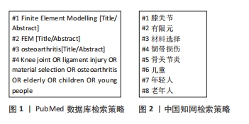
1.1.1 检索人及检索时间 由第一作者在2024年1-6月进行检索。
1.1.2 文献检索时限 1950年1月至2024年6月。
1.1.3 检索数据库 中国知网、PubMed数据库。
1.1.4 检索词 英文检索词为“Knee,finite element,material selection,ligament injury,osteoarthritis,elderly,children,young people”等;中文检索词为“膝关节,有限元,材料选择,韧带损伤,骨关节炎,老年人,儿童,年轻人”等。
1.1.5 检索文献类型 研究原著、综述、述评等。
1.1.6 检索策略 PubMed数据库和中国知网检索策略,见图1,2。
1.2 入选标准
1.2.1 纳入标准 有关膝关节三维有限元计算模型的文献;研究主题是有限元分析中解剖结构的材料特性。
1.2.2 排除标准 全文不可用;与该文主题不相关;全文质量差。
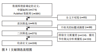
1.3 文献质量评估 数据库共检索到文献558篇,严格按照纳入和排除标准进行筛选,最终纳入文献108篇文献进行综述。文献筛选流程图见图3。
膝关节中骨密度会随着儿童年龄的增长而增加,在成年后骨密度会逐渐增加并达到峰值[37]。王倞等[38]发现骨密度与弹性模量之间存在较高的线性相关性(0.850 > R2 > 0.785)和幂次相关性(0.871 > R2 > 0.825),进而可推断出儿童到成人阶段膝关节骨的弹性模量会随着年龄的增长而增加。袁嘉尧等[39]的研究表明,中老年人的骨密度与身高、体质量、骨矿含量(包括整体、头部、主干、上肢、下肢)以及肌肉质量(整体、头部、主干、下肢)呈显著正相关,而与年龄呈显著负相关。这意味着在中年到老年阶段,随着年龄的增加,膝关节内的股骨、胫骨、腓骨和髌骨的弹性模量逐渐下降,接近儿童时期的水平。
此外,膝关节软骨厚度随着年龄的增加而逐渐减少,然而随着软骨厚度的降低,软骨的弹性模量则呈现出上升趋势。在儿童至成年阶段,软骨厚度的变化微乎其微,成年人的软骨弹性模量基本保持一致[35-36,105]。
膝关节韧带的弹性模量也会随着年龄增长而略有下降,主要原因是韧带成纤维细胞的力学和生物学特性出现退化。然而统计数据显示,年轻人和老年人之间的韧带弹性模量并无显著差异。
随着年龄增长,半月板机械完整性的丧失会损害组织的生物力学功能,扰乱组织有效承载和传递的各向异性生物力学响应[90]。半月板组织的局部纳米力学特性会随着年龄的增长而发生变化。文献表明,儿童与成人的半月板弹性模量大致相同,但在从成人到老年阶段,半月板的弹性模量逐渐降低[35-36,105,108],这一变化与半月板退行性变和骨关节炎的发生密切相关。
膝关节结构复杂,准确的解剖结构和材料是建立仿真模型的基础。该文为不同年龄阶段组织材料的选择提供参考。有限元分析可以有效地模拟膝关节力学变化情况,以较低的成本和风险了解膝关节力学变化情况。未来相关学者可以研究膝关节软骨厚度与弹性模量的线性或非线性关系,以及研究骨密度与弹性模量的线性或非线性关系。
该文在前人膝关节有限元研究的基础上总结了不同年龄阶段膝关节有限元计算模型的材料特性,为膝关节模型的构建和研究设计提供参考。儿童至成人阶段的骨弹性模量会随着年龄的增长而增加,成人至老年人阶段的骨弹性模量会随着年龄增长而减小。膝关节中软骨厚度会随着年龄的增加而减少,而随着软骨厚度的减少其弹性模量在增加。儿童与成人弹性模量基本一致,而老年人的软骨弹性模量较大。韧带与半月板的弹性模量会随着年龄的增长出现一定程度的降低。膝关节有限元建模可以深入了解膝关节的生物力学特性,开发新的植入材料,预测膝关节疾病,改进手术技术,并指导康复锻炼。 中国组织工程研究杂志出版内容重点:生物材料;骨生物材料;口腔生物材料;纳米材料;缓释材料;材料相容性;组织工程
#br#
文题释义:
弹性模量:是材料的一种力学性质,描述材料受到应力时的弹性变形程度。弹性模量越大,说明材料的弹性越好,即在受到应力后能够迅速恢复原状。在中年到老年阶段,膝关节中股骨、胫骨、腓骨、髌骨的弹性模量会随着年龄的增加而降低,进而回归到儿童时期的弹性模量。泊松比:是材料的另一种力学性质,描述了材料在受到应力时沿着一个方向的收缩程度与沿着垂直方向的膨胀程度之比。
该文说明了不同年龄段的膝关节有限元计算模型中不同结构(骨、软骨、半月板、韧带)材料特性(弹性模量、泊松比)的变化情况,为膝关节模型的构建和研究设计提供参考。膝关节的有限元建模可以深入了解膝关节的生物力学特性,开发新的植入材料,预测膝关节疾病,改进手术技术,并指导康复锻炼。未来相关学者可以研究膝关节软骨厚度与弹性模量的线性或非线性关系,以及研究骨密度与弹性模量的线性或非线性关系。
| 阅读次数 | ||||||
|
全文 |
|
|||||
|
摘要 |
|
|||||