[1] BUCK DW, DUMANIAN GA. Bone biology and physiology: Part I. The fundamentals. Plast Reconstr Surg. 2012;129(6):1314-1320.
[2] FLORENCIO-SILVA R, SASSO GR DA S, SASSO-CERRI E, et al. Biology of Bone Tissue: Structure, Function, and Factors That Influence Bone Cells. Biomed Res Int. 2015;2015:421746.
[3] CHAVASSIEUX P, CHAPURLAT R. Interest of Bone Histomorphometry in Bone Pathophysiology Investigation: Foundation, Present, and Future. Front Endocrinol (Lausanne). 2022;13:907914.
[4] AY S, ME D, EA G, et al. Recent progress in tissue optical clearing for spectroscopic application. Spectrochim Acta A Mol Biomol Spectrosc. 2018; 197:216-229.
[5] ZHANG D, CLEVELAND AH, KRIMITZA E, et al. Spatial analysis of tissue immunity and vascularity by light sheet fluorescence microscopy. Nat Protoc. 2024;19(4):1053-1082.
[6] TAINAKA K, KUNO A, KUBOTA SI, et al. Chemical Principles in Tissue Clearing and Staining Protocols for Whole-Body Cell Profiling. Annu Rev Cell Dev Biol. 2016;32:713-741.
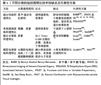
[7] 李茜, 吴科锋. 疏水性组织透明化技术的研究进展[J]. 临床医学研究与实践,2022,7(14):191-195.
[8] SMITH AC, WATAMANIUK L, ROGERS TL. Use of laser-scanning confocal microscopy in the detection of diagenesis in bone. J Forensic Sci. 2022; 67(1):92-101.
[9] FENG F, MAO H, WANG A, et al. Two-Photon Fluorescence Imaging. Adv Exp Med Biol. 2021;3233:45-61.
[10] RICHARDSON DS, LICHTMAN JW. Clarifying Tissue Clearing. Cell. 2015; 162(2):246-257.
[11] 吴昊妍, 詹琰静, 张士文. 水性组织透明化技术的研究进展与应用[J]. 北京生物医学工程,2021,40(3):318-323+329.
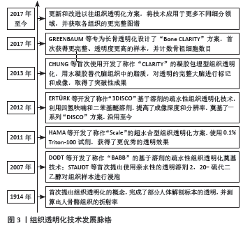
[12] SPALTEHOLZ W. Über das Durchsichtigmachen von menschlichen und tierischen Präparaten und seine theoretischen Bedingungen: nebst Anhang: Über Knochenfärbung. Verlag Von S Hirzel. 1914.
[13] STAUDT T, LANG MC, MEDDA R, et al. 2,2’-thiodiethanol: a new water soluble mounting medium for high resolution optical microscopy. Microsc Res Tech. 2007;70(1):1-9.
[14] DODT HU, LEISCHNER U, SCHIERLOH A, et al. Ultramicroscopy: three-dimensional visualization of neuronal networks in the whole mouse brain. Nat Methods. 2007;4(4):331-336.
[15] HAMA H, KUROKAWA H, KAWANO H, et al. Scale: a chemical approach for fluorescence imaging and reconstruction of transparent mouse brain. Nat Neurosci. 2011;14(11):1481-1488.
[16] ERTÜRK A, BECKER K, JÄHRLING N, et al. Three-dimensional imaging of solvent-cleared organs using 3DISCO. Nat Protoc. 2012;7(11):1983-1995.
[17] CHUNG K, WALLACE J, KIM SY, et al. Structural and molecular interrogation of intact biological systems. Nature. 2013;497(7449):332-337.
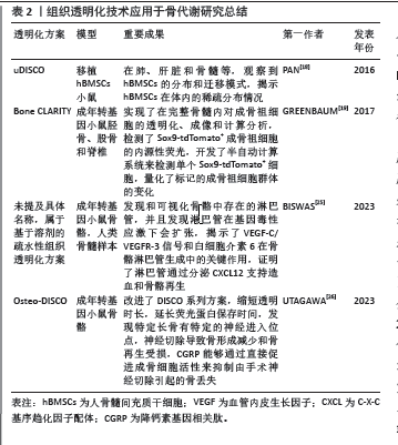
[18] PAN C, CAI R, QUACQUARELLI FP, et al. Shrinkage-mediated imaging of entire organs and organisms using uDISCO. Nat Methods. 2016;13(10):859-867.
[19] GREENBAUM A, CHAN KY, DOBREVA T, et al. Bone CLARITY: Clearing, imaging, and computational analysis of osteoprogenitors within intact bone marrow. Sci Transl Med. 2017;9(387):eaah6518.
[20] JING D, ZHANG S, LUO W, et al. Tissue clearing of both hard and soft tissue organs with the PEGASOS method. Cell Res. 2018;28(8):803-818.
[21] QI Y, YU T, XU J, et al. FDISCO: Advanced solvent-based clearing method for imaging whole organs. Sci Adv. 2019;5(1):eaau8355.
[22] TREWEEK JB, BERES A, JOHNSON N, et al. Phenotyping Intact Mouse Bones Using Bone CLARITY. Exp Mol Med. 2021;2230:217-230.
[23] XU C. Optical clearing of the mouse skull. Light Sci Appl. 2022;11(1):284.
[24] KARTHIKEYAN S, ASAKURA Y, VERMA M, et al. Tissue Clearing and Confocal Microscopic Imaging for Skeletal Muscle. Methods Mol Biol. 2023;2640: 453-462.
[25] BISWAS L, CHEN J, DE ANGELIS J, et al. Lymphatic vessels in bone support regeneration after injury. Cell. 2023;186(2):382-397.e24.
[26] UTAGAWA K, SHIN T, YAMADA H, et al. Three-dimensional visualization of neural networks inside bone by Osteo-DISCO protocol and alteration of bone remodeling by surgical nerve ablation. Sci Rep. 2023;13(1):4674.
[27] BRÉS EF, BARRY JC, HUTCHISON JL. High-resolution electron microscope and computed images of human tooth enamel crystals. J Ultrastruct Res. 1985;90(3):261-274.
[28] JING D, YI Y, LUO W, et al. Tissue Clearing and Its Application to Bone and Dental Tissues. J Dent Res. 2019;98(6):621-631.
[29] KE M-T, FUJIMOTO S, IMAI T. SeeDB: a simple and morphology-preserving optical clearing agent for neuronal circuit reconstruction. Nat Neurosci. 2013;16(8):1154-1161.
[30] WOO J, LEE M, SEO JM, et al. Optimization of the optical transparency of rodent tissues by modified PACT-based passive clearing. Exp Mol Med. 2016;48(12):e274.
[31] AFFATICATI P, SIMION M, DE JOB E, et al. zPACT: Tissue Clearing and Immunohistochemistry on Juvenile Zebrafish Brain. Bio Protoc. 2017;7(23):e2636.
[32] MURAKAMI TC, MANO T, SAIKAWA S, et al. A three-dimensional single-cell-resolution whole-brain atlas using CUBIC-X expansion microscopy and tissue clearing. Nat Neurosci. 2018;21(4):625-637.
[33] 李娜, 马静, 程妍. Passive-CLARITY法透明化小鼠脑及鼠脑组织的荧光检测[J]. 华西药学杂志,2021,36(6):625-627.
[34] RENIER N, WU Z, SIMON DJ, et al. iDISCO: a simple, rapid method to immunolabel large tissue samples for volume imaging. Cell. 2014;159(4): 896-910.
[35] PERIN P, VOIGT FF, BETHGE P, et al. iDISCO+ for the Study of Neuroimmune Architecture of the Rat Auditory Brainstem. Front Neuroanat. 2019;13:15.
[36] ACAR M, KOCHERLAKOTA KS, MURPHY MM, et al. Deep imaging of bone marrow shows non-dividing stem cells are mainly perisinusoidal. Nature. 2015;526(7571):126-130.
[37] JACOB L, BOISSERAND LSB, GERALDO LHM, et al. Anatomy and function of the vertebral column lymphatic network in mice. Nat Commun. 2019; 10(1):4594.
[38] CAI R, KOLABAS ZI, PAN C, et al. Whole-mouse clearing and imaging at the cellular level with vDISCO. Nat Protoc. 2023;18(4):1197-1242.
[39] TSAI PS, KAUFHOLD JP, BLINDER P, et al. Correlations of neuronal and microvascular densities in murine cortex revealed by direct counting and colocalization of nuclei and vessels. J Neurosci. 2009;29(46):14553-14570.
[40] HOU B, ZHANG D, ZHAO S, et al. Scalable and DiI-compatible optical clearance of the mammalian brain. Front Neuroanat. 2015;9:19.
[41] SUSAKI EA, TAINAKA K, PERRIN D, et al. Whole-brain imaging with single-cell resolution using chemical cocktails and computational analysis. Cell. 2014;157(3):726-739.
[42] HAMA H, HIOKI H, NAMIKI K, et al. ScaleS: an optical clearing palette for biological imaging. Nat Neurosci. 2015;18(10):1518-1529.
[43] CHUNG K, DEISSEROTH K. CLARITY for mapping the nervous system. Nat Methods. 2013;10(6):508-513.
[44] TREWEEK JB, CHAN KY, FLYTZANIS NC, et al. Whole-body tissue stabilization and selective extractions via tissue-hydrogel hybrids for high-resolution intact circuit mapping and phenotyping. Nat Protoc. 2015;10(11):1860-1896.
[45] 殷俊辉, 周隽, 陈逸炜, 等. 新型硬组织切片技术在临床骨与骨髓病理诊断中的应用价值[J]. 中华医学杂志,2022,102(45):3617-3623.
[46] LI W, GERMAIN RN, GERNER MY. Multiplex, quantitative cellular analysis in large tissue volumes with clearing-enhanced 3D microscopy (Ce3D). Proc Natl Acad Sci U S A. 2017;114(35):E7321-E7330.
[47] CAI R, PAN C, GHASEMIGHARAGOZ A, et al. Panoptic imaging of transparent mice reveals whole-body neuronal projections and skull-meninges connections. Nat Neurosci. 2019;22(2):317-327.
[48] MOATTI A, CAI Y, LI C, et al. Tissue clearing and three-dimensional imaging of the whole cochlea and vestibular system from multiple large-animal models. STAR Protoc. 2023;4(2):102220.
[49] THAI J, FULLER-JACKSON J-P, IVANUSIC JJ. Using tissue clearing and light sheet fluorescence microscopy for the three-dimensional analysis of sensory and sympathetic nerve endings that innervate bone and dental tissue of mice. J Comp Neurol. 2024;532(1):e25582.
[50] YI Y, LI Y, ZHANG S, et al. Mapping of individual sensory nerve axons from digits to spinal cord with the transparent embedding solvent system. Cell Res. 2024;34(2):124-139.
[51] GORELASHVILI MG, HEINZE KG, STEGNER D. Optical Clearing of Murine Bones to Study Megakaryocytes in Intact Bone Marrow Using Light-Sheet Fluorescence Microscopy. Methods Mol Biol. 2018;1812:233-253.
[52] 中华医学会骨质疏松和骨矿盐疾病分会. 原发性骨质疏松症诊疗指南(2022)[J]. 中国全科医学,2023,26(14):1671-1691.
[53] 陈光华, 黄贵芝, 林颢, 等. 骨髓间充质干细胞移植对去卵巢骨质疏松大鼠骨密度的影响[J]. 中国组织工程研究,2017,21(1):49-53.
[54] 冯皓, 张斌, 王建平. 骨髓间充质干细胞移植可提高骨质疏松大鼠骨代谢水平[J]. 中国组织工程研究,2023,27(1):72-75.
[55] CHOI JH, RO JY. The 2020 WHO Classification of Tumors of Bone: An Updated Review. Adv Anat Pathol. 2021;28(3):119-138.
[56] FERGUSON JL, TURNER SP. Bone Cancer: Diagnosis and Treatment Principles. Am Fam Physician. 2018;98(4):205-213.
[57] PAN C, SCHOPPE O, PARRA-DAMAS A, et al. Deep Learning Reveals Cancer Metastasis and Therapeutic Antibody Targeting in the Entire Body. Cell. 2019;179(7):1661-1676.e19.
[58] LLOYD-LEWIS B, DAVIS FM, HARRIS OB, et al. Imaging the mammary gland and mammary tumours in 3D: optical tissue clearing and immunofluorescence methods. Breast Cancer Res. 2016;18(1):127.
[59] CAMAL RUGGIERI IN, CÍCERO AM, ISSA JPM, et al. Bone fracture healing: perspectives according to molecular basis. J Bone Miner Metab. 2021;39(3):311-331.
[60] 来钰栋, 蒋振松. 骨质疏松性骨折的预防与临床治疗进展[J]. 山东第一医科大学(山东省医学科学院)学报,2023,44(1):67-73.
[61] 黄韬. 淫羊藿苷在骨折愈合中的疗效及机制研究[D]. 汕头:汕头大学, 2022.
[62] 王伟, 李文波, 张亚强, 等. 感染性骨缺损的诊断与临床治疗进展[J]. 中国骨与关节损伤杂志,2022,37(3):331-334.
[63] MORIARTY TF, METSEMAKERS WJ, MORGENSTERN M, et al. Fracture-related infection. Nat Rev Dis Primers. 2022;8(1):67.
[64] MATSUMOTO K, MITANI TT, HORIGUCHI SA, et al. Advanced CUBIC tissue clearing for whole-organ cell profiling. Nat Protoc. 2019;14(12):3506-3537.
[65] ZHAN ZY, HUANG YR, ZHAO LW, et al. Use of a tissue clearing technique combined with retrograde trans-synaptic viral tracing to evaluate changes in mouse retinorecipient brain regions following optic nerve crush. Neural Regen Res. 2023;18(4):913-921.
[66] HOFMANN J, KEPPLER SJ. Tissue clearing and 3D imaging - putting immune cells into context. J Cell Sci. 2021;134(15):jcs258494.
[67] 宋会平, 王志强. 骨移植的过去、现在和未来[J]. 中国修复重建外科杂志,2009,23(5):513-516.
[68] 袁冰, 韦卓. 骨缺损修复的研究进展[J]. 生物骨科材料与临床研究, 2014,11(3):38-41.
[69] 宁钰, 赵红斌. 骨缺损修复方法的研究进展[J]. 世界最新医学信息文摘, 2019,19(50):115+117.
[70] YI Y, MEN Y, JING D, et al. 3-dimensional visualization of implant-tissue interface with the polyethylene glycol associated solvent system tissue clearing method. Cell Prolif. 2019;52(3):e12578.
[71] 赵延洁. 适用于活体皮层成像的光透明颅窗研究[D]. 武汉:华中科技大学, 2019.
[72] 李东宇, 俞婷婷, 朱京谭, 等. 活体颅骨光透明方法及应用(特邀)[J]. 光子学报,2022,51(8):252-268.
[73] 田彬, 刘长松, 程为. 骨质疏松兔颅骨骨缺损模型的建立[J]. 口腔颌面修复学杂志,2023,24(1):20-24, 37.
[74] 张雅雯, 朱光旭, 李雅喆, 等. 一种改良大鼠颅骨缺损动物模型的构建及应用[J]. 实验动物与比较医学,2020,40(3):227-231.
[75] WANG J, ZHANG Y, XU TH, et al. An innovative transparent cranial window based on skull optical clearing. Laser Phys Lett. 2012;9(6):469.
[76] ZHANG C, FENG W, ZHAO Y, et al. A large, switchable optical clearing skull window for cerebrovascular imaging. Theranostics. 2018;8(10):2696-2708.
[77] CHEN Y, LIU S, LIU H, et al. Coherent Raman Scattering Unravelling Mechanisms Underlying Skull Optical Clearing for Through-Skull Brain Imaging. Anal Chem. 2019;91(15):9371-9375.
[78] LI DY, ZHENG Z, YU TT, et al. Visible-near infrared-II skull optical clearing window for in vivo cortical vasculature imaging and targeted manipulation. J Biophotonics. 2020;13(10):e202000142.
[79] 宣昂. 组织光透明结合光学成像研究小鼠卒中后神经肌肉接头三维结构变化[D]. 武汉:华中科技大学,2022.
[80] 田婷, 杨朝阳, 李晓光. 组织透明化技术的研究与应用[J]. 中国组织工程研究,2020,24(21):3363-3371.
[81] 陈小玉, 罗连响, 潘韵琪, 等. 组织透明化技术在神经退行性疾病中的应用研究进展[J]. 解放军医学杂志,2022,47(3):305-313.
[82] 陈影, 黄金智, 吴科锋, 等. 组织透明化三维成像技术在卵巢组织中的应用进展[J]. 国际妇产科学杂志,2022,49(5):492-496. |