[1] CAI X, YUAN S, ZENG Y, et al. New Trends in Pharmacological Treatments for Osteoarthritis. Front Pharmacol. 2021;12:645842.
[2] LONG H, LIU Q, YIN H, et al. Prevalence Trends of Site-Specific Osteoarthritis From 1990 to 2019: Findings From the Global Burden of Disease Study 2019. Arthritis Rheumatol. 2022;74(7):1172-1183.
[3] HE Y, LI Z, ALEXANDER PG, et al. Pathogenesis of Osteoarthritis: Risk Factors, Regulatory Pathways in Chondrocytes, and Experimental Models. Biology (Basel). 2020;9(8):194.
[4] FUGGLE N, CURTIS E, SHAW S, et al. Safety of Opioids in Osteoarthritis: Outcomes of a Systematic Review and Meta-Analysis. Drugs Aging. 2019;36(Suppl 1):129-143.
[5] YAO Q, WU X, TAO C, et al. Osteoarthritis: pathogenic signaling pathways and therapeutic targets. Signal Transduct Target Ther. 2023; 8(1):56.
[6] BLASIOLI DJ, KAPLAN DL. The Roles of Catabolic Factors in the Development of Osteoarthritis. Tissue Eng Part B Rev. 2014;20(4): 355-363.
[7] PARK C, JEONG JW, LEE DS, et al. Sargassum serratifolium Extract Attenuates Interleukin-1β-Induced Oxidative Stress and Inflammatory Response in Chondrocytes by Suppressing the Activation of NF-κB, p38 MAPK, and PI3K/Akt. Int J Mol Sci. 2018;19(8):2308.
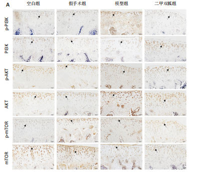
[8] SUN K, LUO J, GUO J, et al. The PI3K/AKT/mTOR signaling pathway in osteoarthritis: a narrative review. Osteoarthritis Cartilage. 2020;28(4): 400-409.
[9] ALA M, ALA M. Metformin for Cardiovascular Protection, Inflammatory Bowel Disease, Osteoporosis, Periodontitis, Polycystic Ovarian Syndrome, Neurodegeneration, Cancer, Inflammation and Senescence: What Is Next? ACS Pharmacol Transl Sci. 2021;4(6):1747-1770.
[10] PARK MJ, MOON SJ, BAEK JA, et al. Metformin Augments Anti-Inflammatory and Chondroprotective Properties of Mesenchymal Stem Cells in Experimental Osteoarthritis. J Immunol. 2019;203(1):127-136.
[11] LI J, ZHANG B, LIU WX, et al. Metformin limits osteoarthritis development and progression through activation of AMPK signalling. Ann Rheum Dis. 2020;79(5):635-645.
[12] YAN J, FENG G, MA L, et al. Metformin alleviates osteoarthritis in mice by inhibiting chondrocyte ferroptosis and improving subchondral osteosclerosis and angiogenesis. J Orthop Surg Res. 2022;17(1):333.
[13] YAN S, DONG W, LI Z, et al. Metformin regulates chondrocyte senescence and proliferation through microRNA-34a/SIRT1 pathway in osteoarthritis. J Orthop Surg Res. 2023;18(1):198.
[14] WANG J, WANG X, CAO Y, et al. Therapeutic potential of hyaluronic acid/chitosan nanoparticles for the delivery of curcuminoid in knee osteoarthritis and an in vitro evaluation in chondrocytes. Int J Mol Med. 2018;42(5):2604-2614.
[15] 冯晓峰,张荣凯,祁伟仲,等.二甲双胍干预骨关节炎模型小鼠早期骨关节炎软骨及软骨下骨变化[J]. 中国组织工程研究,2019, 23(19):3031-3036.
[16] YAN J, DING D, FENG G, et al. Metformin reduces chondrocyte pyroptosis in an osteoarthritis mouse model by inhibiting NLRP3 inflammasome activation. Exp Ther Med. 2022;23(3):222.
[17] 罗臻,李宏栩,卢启贵,等.红姜提取物保护早期膝骨关节炎模型大鼠的关节软骨[J].中国组织工程研究,2021,25(32):5155-5161.
[18] HWANG HS, KIM HA. Chondrocyte Apoptosis in the Pathogenesis of Osteoarthritis. Int J Mol Sci. 2015;16(11):26035-26054.
[19] FANELLI A, GHISI D, APRILE PL, et al. Cardiovascular and cerebrovascular risk with nonsteroidal anti-inflammatory drugs and cyclooxygenase 2 inhibitors: latest evidence and clinical implications. Ther Adv Drug Saf. 2017;8(6):173-182.
[20] LIN H, AO H, GUO G, et al. The Role and Mechanism of Metformin in Inflammatory Diseases. J Inflamm Res. 2023;16:5545-5564.
[21] JIN P, JIANG J, ZHOU L, et al. Disrupting metformin adaptation of liver cancer cells by targeting the TOMM34/ATP5B axis. EMBO Mol Med. 2022;14(12):e16082.
[22] MARTIN-MONTALVO A, MERCKEN EM, MITCHELL SJ, et al. Metformin improves healthspan and lifespan in mice. Nat Commun. 2013;4:2192.
[23] SANCHEZ-LOPEZ E, CORAS R, TORRES A, et al. Synovial inflammation in osteoarthritis progression. Nat Rev Rheumatol. 2022;18(5): 258-275.
[24] KRISHNAN Y, GRODZINSKY AJ. Cartilage Diseases. Matrix Biol. 2018;71-72:51-69.
[25] MARTÍNEZ-MORENO D, JIMÉNEZ G, GÁLVEZ-MARTÍN P, et al. Cartilage biomechanics: A key factor for osteoarthritis regenerative medicine. Biochim Biophys Acta Mol Basis Dis. 2019;1865(6):1067-1075.
[26] HUSSAIN S, SUN M, GUO Y, et al. SFMBT2 positively regulates SOX9 and chondrocyte proliferation. Int J Mol Med. 2018;42(6):3503-3512.
[27] XU R, WU J, ZHENG L, et al. Undenatured type II collagen and its role in improving osteoarthritis. Ageing Res Rev. 2023;91:102080.
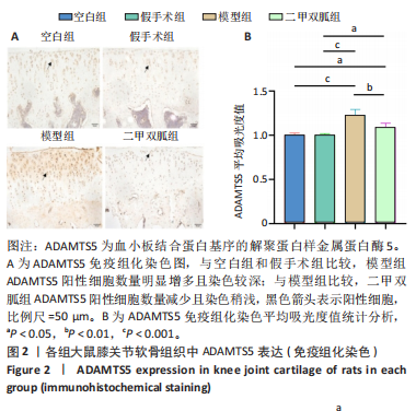
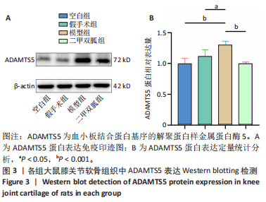
[28] LI T, PENG J, LI Q, et al. The Mechanism and Role of ADAMTS Protein Family in Osteoarthritis. Biomolecules. 2022;12(7):959.
[29] YAO M, ZHANG C, NI L, et al. Cepharanthine Ameliorates Chondrocytic Inflammation and Osteoarthritis via Regulating the MAPK/NF-κB-Autophagy Pathway. Front Pharmacol. 2022;13:854239.
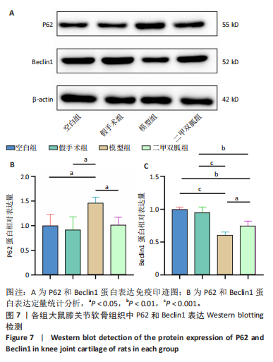
[30] SUN K, JING X, GUO J, et al. Mitophagy in degenerative joint diseases. Autophagy. 2021;17(9):2082-2092.
[31] 谢一潋,杨乃彬,王丽萍,等.姜黄素通过激活肝细胞自噬减轻脂多糖/D-氨基半乳糖诱导的大鼠急性肝损伤[J].中国病理生理杂志, 2020,36(5):860-864.
[32] 管津智,姜泉,夏聪敏,等.清热活血方对CIA大鼠自噬相关蛋白p62表达的影响[J].中国中医基础医学杂志,2023,29(4):588-592.
[33] BOUDERLIQUE T, VUPPALAPATI KK, NEWTON PT, et al. Targeted deletion of Atg5 in chondrocytes promotes age-related osteoarthritis. Ann Rheum. 2016;75(3):627-631.
[34] LV X, ZHAO T, DAI Y, et al. New insights into the interplay between autophagy and cartilage degeneration in osteoarthritis. Front Cell Dev Biol. 2022;10:1089668.
[35] HAYER S, PUNDT N, PETERS MA, et al. PI3Kgamma regulates cartilage damage in chronic inflammatory arthritis. FASEB J. 2009;23(12): 4288-4298.
[36] AZIZ AUR, FARID S, QIN K, et al. PIM Kinases and Their Relevance to the PI3K/AKT/mTOR Pathway in the Regulation of Ovarian Cancer. Biomolecules. 2018;8(1):E7.
[37] ZHOU J, JIANG Y, CHEN H, et al. Tanshinone I attenuates the malignant biological properties of ovarian cancer by inducing apoptosis and autophagy via the inactivation of PI3K/AKT/mTOR pathway. Cell Prolif. 2019;53(2): e12739.
[38] WANG K, CHU M, WANG F, et al. Putative functional variants of PI3K/AKT/mTOR pathway are associated with knee osteoarthritis susceptibility. J Clin Lab Anal. 2020;34(6):e23240.
[39] LI J, JIANG M, YU Z, et al. Artemisinin relieves osteoarthritis by activating mitochondrial autophagy through reducing TNFSF11 expression and inhibiting PI3K/AKT/mTOR signaling in cartilage. Cell Mol Biol Lett. 2022;27(1):62. |