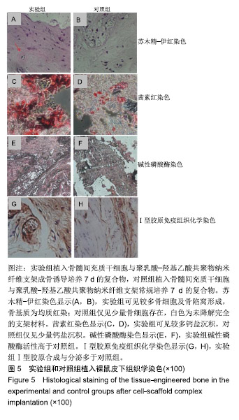
| [1]韦应明,雷利红,陈莉丽.骨组织工程支架与生长因子结合方法研究进展[J].中国实用口腔科杂志,2018,11(6):374-378.[2]Turnbull G,Clarke J,Picard F,et al.3D bioactive composite scaffolds for bone tissue engineering.Bioact Mater. 2017;3(3):278-314.[3]Wu T,Yu S,Chen D,et al.Bionic Design, Materials and Performance of Bone Tissue Scaffolds. Materials. 2017;10(10):1187.[4]Henkel J,Woodruff MA,Epari DR,et al.Bone Regeneration Based on Tissue Engineering Conceptions -A 21st Century Perspective.Bone Resh.2013;1(3):216-248.[5]Gu M,Liu Y,Chen T,et al.Is Graphene a Promising Nano-Material for Promoting Surface Modification of Implants or Scaffold Materials in Bone Tissue Engineering.Tissue Eng Part B Rev.2014;20(5): 477-491.[6]Yu H, Adesida AB,Jomha NM.Meniscus repair using mesenchymal stem cells – a comprehensive review. Stem Cell Res Ther.2015; 6(1):86.[7]Potier E,Noailly J,Ito K.Directing bone marrow-derived stromal cell function with mechanics. J Biomech. 2010;43(5):807-817.[8]Ge M,Ge K,Gao F,et al.Biomimetic mineralized strontium-doped hydroxyapatite on porous poly(L-lactic acid) scaffolds for bone defect repair.Int J Nanomedicine. 2018;13:1707-1721.[9]杨文峰,任远飞,梁武,等.PLGA电纺丝对大鼠骨髓间充质干细胞成骨分化的影响[J].现代医学,2017,45(5):623-627.[10]Yang X,Li Y,Liu X,et al. Incorporation of silica nanoparticles to PLGA electrospun fibers for osteogenic differentiation of human osteoblast-like cells.Regen Biomater. 2018;5(4):229-238.[11]Guimarães P,Oliveira S,Castro Rodrigues G,et al. Development of Sulfadiazine-Decorated PLGA Nanoparticles Loaded with 5-Fluorouracil and Cell Viability.Molecules.2015;20(1):879-899.[12]Jun I, Han HS, Edwards JR,et al.Electrospun Fibrous Scaffolds for Tissue Engineering: Viewpoints on Architecture and Fabrication.Int J Mol Sci.2018;19(3):745.[13]Sankar S,Sharma CS,Rath SN.Enhanced osteodifferentiation of MSC spheroids on patterned electrospun fiber mats - An advanced 3D double strategy for bone tissue regeneration.Mater Sci Eng C Mater Biol Appl. 2019;94:703-712.[14]Yang X,Li Y,He W,et al. Hydroxyapatite/collagen coating on PLGA electrospun fibers for osteogenic differentiation of bone marrow mesenchymal stem cells. J Biomed Mater Res A. 2018;106(11): 2863-2870.[15]Perez JR,Dimitrios K,Li DJ,et al.Tissue Engineering and Cell-Based Therapies for Fractures and Bone Defects.Front Bioeng Biotechnol. 2018;6:105.[16]O'Brien FJ.Biomaterials & scaffolds for tissue engineering. Mater Today. 2011;14(3):88-95.[17]Roseti L,Parisi V,Petretta M,et al.Scaffolds for Bone Tissue Engineering: State of the art and new perspectives.Mater Sci Eng C Mater Biol Appl. 2017;78:1246.[18]Valencia C,Valencia C,Zuluaga F,et al.Synthesis and Application of Scaffolds of Chitosan-Graphene Oxide by the Freeze-Drying Method for Tissue Regeneration. Molecules. 2018;23(10):2651.[19]Shichao Z,Malcolm X,Bingyun L.Biomimetic Layer-by-Layer Self-Assembly of Nanofilms, Nanocoatings and 3D Scaffolds for Tissue Engineering.Int J Mol Sci. 2018;19(6):1641.[20]Du MH,Ding Y,Shi X,et al.The Periosteal Autografts Transplantation for Cartilage Defects of the Hip in Older Children With Developmental Dysplasia as an Adjunctive Procedure.Medicine. 2016;95(17):e3432.[21]Hoffman MD,Xie C,Zhang X,et al.The effect of mesenchymal stem cells delivered via hydrogel-based tissue engineered periosteum on bone allograft healing. Biomaterials. 2013; 34(35):8887-8898.[22]Deval B,Ludwig A,Chan P,et al.A Review on Properties of Natural and Synthetic Based Electrospun Fibrous Materials for Bone Tissue Engineering.Membranes.2018;8(3):62.[23]Gentile P,Chiono V,Carmagnola L,et al.An Overview of Poly(lactic-co-glycolic) Acid (PLGA)-Based Biomaterials for Bone Tissue Engineering.Int J Mol Sci. 2014;15(3):3640-3659.[24]Fu C,Bai H,Zhu J,et al.Enhanced cell proliferation and osteogenic differentiation in electrospun PLGA/hydroxyapatite nanofibre scaffolds incorporated with graphene oxide.PLoS One. 2017; 12(11):e0188352.[25]Kwak S,Haider A,Gupta KC,et al.Micro/Nano Multilayered Scaffolds of PLGA and Collagen by Alternately Electrospinning for Bone Tissue Engineering.Nanoscale Res Lett. 2016;11(1):323. [26]Park BH,Zhou L,Jang KY, et al.Enhancement of tibial regeneration in a rat model by adipose-derived stromal cells in a PLGA scaffold. Bone. 2012;51(3):313-323.[27]Chiesa E,Pisani S,Colzani B,et al.Intra-Articular Formulation of GE11-PLGA Conjugate-Based NPs for Dexamethasone Selective Targeting—In Vitro Evaluation.Int J Mol Sci. 2018;19(8):2304.[28]Mir M,Ali MN,Barakullah A,et al.Synthetic polymeric biomaterials for wound healing: a review. Prog Biomater. 2018;7(1):1-21.[29]Wu J,Hong Y.Enhancing cell infiltration of electrospun fibrous scaffolds in tissue regeneration.Bioact Mater. 2016;1(1):56-64.[30]Wang W,Nie W,Liu D,et al.Macroporous nanofibrous vascular scaffold with improved biodegradability and smooth muscle cells infiltration prepared by dual phase separation technique. Int J Nanomedicine.2018;13:7003-7018.[31]Sascha Z,Toby B,Johannes R,et al.Poly(ε-caprolactone) Scaffolds Fabricated by Melt Electrospinning for Bone Tissue Engineering. Materials.2016;9(4):232.[32]Tibbitt MW,Anseth KS.Hydrogels as extracellular matrix mimics for 3D cell culture. Biotechnol Bioeng. 2010;103(4):655-663.[33]Ma MX,Liu Q,Ye C,et al.Preparation of P3HB4HB/(Gelatin + PVA) Composite Scaffolds by Coaxial Electrospinning and Its Biocompatibility Evaluation.Biomed Res Int. 2017;2017:9251806. [34]Julia S, Moritz P, Alexandra D, et al. Journey into Bone Models: A Review. Genes.2018;9(5):247.[35]Shao W,He J, Sang F,et al.Coaxial electrospun aligned tussah silk fibroin nanostructured fiber scaffolds embedded with hydroxyapatite–tussah silk fibroin nanoparticles for bone tissue engineering. Mater Sci Eng C Mater Biol Appl. 2016;58:342-351. |
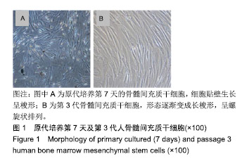
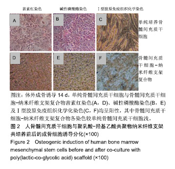
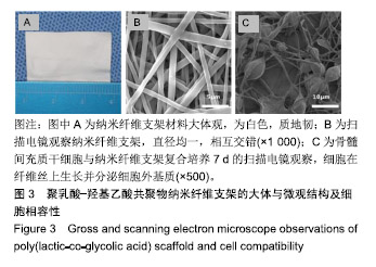
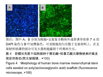
.jpg)
.jpg)
.jpg)