| [1] Rubin C, Bolander M, Ryaby JP, et al. The use of low-intensity ultrasound to accelerate the healing of fractures. J Bone Joint Surg Am.2001;83-A(2): 259-270.[2] Moed BR, Kim EC,van Holsbeeck M,et al.Ultrasound for the early diagnosis of tibial fracture healing after static interlocked nailing without reaming: histologic correlation using a canine model. J Orthop Trauma.1998;12(3): 200-205.[3] Moed BR, Subramanian S,van Holsbeeck M, et al. Ultrasound for the early diagnosis of tibial fracture healing after static interlocked nailing without reaming: clinical results. J Orthop Trauma.1998;12(3): 206-213.[4] Buchtala V. Present state of ultrasound therapy. Dia Med. 1950;22(70): 2944-2950.[5] Maintz G.Animal experiments in the study of the effect ofultrasonic waves on bone regeneration. Strahlentherapie.1950; 82(4): 631-638.[6] Parvizi J, Parpura V, Greenleaf JF, et al. Calcium signaling is required for ultrasound-stimulated aggrecan synthesis by rat chondrocytes. J Orthop Res.2002; 20(1): 51-57.[7] Alvarenga EC,Rodrigues R,Caricati-Neto A,et al.Low-intensity pulsed ultrasound-dependent osteoblast proliferation occurs by via activation of the P2Y receptor: role of the P2Y1 receptor. Bone.2010;46(2): 355-362.[8] Doroudi M,Plaisance MC,Boyan BD,et al.Membrane actions of 1alpha,25(OH)2D3 are mediated by Ca(2+)/calmodulin- dependent protein kinase II in bone and cartilage cells. J Steroid Biochem Mol Biol.2015;145: 65-74.[9] Sun S,Liu Y,Lipsky S,et al.Physical manipulation of calcium oscillations facilitates osteodifferentiation of human mesenchymal stem cells. FASEB J.2007;21(7):1472-1480.[10] Kim TJ, Seong J, Ouyang M, et al. Substrate rigidity regulates Ca2+ oscillation via RhoA pathway in stem cells. J Cell Physiol. 2009;218(2): 285-293.[11] Tang CH, Yang RS, Huang TH, et al. Ultrasound stimulates cyclooxygenase-2 expression and increases bone formation through integrin, focal adhesion kinase, phosphatidylinositol 3-kinase, and Akt pathway in osteoblasts. Mol Pharmacol. 2006; 69(6): 2047-2057.[12] Watabe H,Furuhama T,Tani-Ishii N,et al.Mechanotransduction activates alpha(5)beta(1) integrin and PI3K/Akt signaling pathways in mandibular osteoblasts. Exp Cell Res.2011; 317(18): 2642-2649.[13] Zhou S, Schmelz A, Seufferlein T, et al. Molecular mechanisms of low intensity pulsed ultrasound in human skin fibroblasts.J Biol Chem.2004;279(52): 54463-54469.[14] Sato M, Nagata K, Kuroda S, et al. Low-intensity pulsed ultrasound activates integrin-mediated mechanotransduction pathway in synovial cells. Ann Biomed Eng.2014;42(10): 2156-2163.[15] Angle SR,Sena K,Sumner DR, et al. Osteogenic differentiation of rat bone marrow stromal cells by various intensities of low-intensity pulsed ultrasound. Ultrasonics. 2011; 51(3): 281-288.[16] Cheng K, Xia P, Lin Q, et al. Effects of low-intensity pulsed ultrasound on integrin-FAK-PI3K/Akt mechanochemical transduction in rabbit osteoarthritis chondrocytes. Ultrasound Med Biol.2014;40(7): 1609-1618.[17] Whitney NP,Lamb AC,Louw TM,et al.Integrin-mediated mechanotransduction pathway of low-intensity continuous ultrasound in human chondrocytes. Ultrasound Med Biol. 2012;38(10): 1734-1743.[18] Xia P, Shen S, Lin Q, et al. Low-Intensity Pulsed Ultrasound Treatment at an Early Osteoarthritis Stage Protects Rabbit Cartilage From Damage via the Integrin/Focal Adhesion Kinase/Mitogen-Activated Protein Kinase Signaling Pathway. J Ultrasound Med.2015; 34(11):1991-1999.[19] Saura M, Tarin C, Zaragoza C. Recent insights into the implication of nitric oxide in osteoblast differentiation and proliferation during bone development. Scientific World J. 2010;10: 624-632.[20] Wang FS,Kuo YR,Wang CJ,et al.Nitric oxide mediates ultrasound-induced hypoxia-inducible factor-1alpha activation and vascular endothelial growth factor-A expression in human osteoblasts. Bone.2004;35(1):114-123.[21] Tang CH, Lu DY, Tan TW, et al. Ultrasound induces hypoxia-inducible factor-1 activation and inducible nitric-oxide synthase expression through the integrin/integrin-linked kinase/Akt/mammalian target of rapamycin pathway in osteoblasts. J Biol Chem.2007;282(35): 25406-25415.[22] Hou CH, Lin J, Huang SC, et al. Ultrasound stimulates NF-kappaB activation and iNOS expression via the Ras/Raf/MEK/ERK signaling pathway in cultured preosteoblasts. J Cell Physiol. 2009;220(1): 196-203.[23] Herrera BS, Martins-Porto R, Maia-Dantas A, et al. iNOS-derived nitric oxide stimulates osteoclast activity and alveolar bone loss in ligature-induced periodontitis in rats. J Periodontol.2011;82(11): 1608-1615.[24] Jung DH, Kim KH, Byeon HE, et al. Involvement of ATF3 in the negative regulation of iNOS expression and NO production in activated macrophages. Immunol Res.2015; 62(1): 35-45.[25] Wang G, Yan Q, Woods A, et al. Inducible nitric oxide synthase-nitric oxide signaling mediates the mitogenic activity of Rac1 during endochondral bone growth. J Cell Sci.2011; 124(Pt 20): 3405-3413.[26] Kawaguchi H,Pilbeam CC,Harrison JR,et al.The role of prostaglandins in the regulation of bone metabolism. Clin Orthop Relat Res.1995;(313): 36-46.[27] Kokubu T, Matsui N, Fujioka H, et al. Low intensity pulsed ultrasound exposure increases prostaglandin E2 production via the induction of cyclooxygenase-2 mRNA in mouse osteoblasts. Biochem Biophys Res Commun.1999;256(2): 284-287.[28] Reher P, Harris M, Whiteman M, et al. Ultrasound stimulates nitric oxide and prostaglandin E2 production by human osteoblasts. Bone.2002;31(1): 236-241.[29] Rego EB, Inubushi T, Kawazoe A, et al. Ultrasound stimulation induces PGE(2) synthesis promoting cementoblastic differentiation through EP2/EP4 receptor pathway. Ultrasound Med Biol.2010;36(6): 907-915.[30] Naruse K, Sekiya H, Harada Y, et al. Prolonged endochondral bone healing in senescence is shortened by low-intensity pulsed ultrasound in a manner dependent on COX-2. Ultrasound Med Biol.2010;36(7):1098-1108.[31] Hidaka K, Miyamoto C, Kawata A, et al.13. Effect of Low-Intensity Pulsed Ultrasound (LIPUS) on Remote Bone Marrow in Rats With Healing Socket. J Orthop Trauma, 2016; 30(8): S5-6.[32] Saini V, Yadav S, McCormick S. Low-intensity pulsed ultrasound modulates shear stress induced PGHS-2 expression and PGE2 synthesis in MLO-Y4 osteocyte-like cells. Ann Biomed Eng.2011;39(1): 378-393.[33] Murray SS, Brochmann Murray EJ, Wang JC, et al. The history and histology of bone morphogenetic protein. Histol Histopathol.2016;31(7): 721-732.[34] Ruschke K, Hiepen C, Becker J, et al. BMPs are mediators in tissue crosstalk of the regenerating musculoskeletal system. Cell Tissue Res.2012;347(3):521-544.[35] Suzuki A, Takayama T, Suzuki N, et al. Daily low-intensity pulsed ultrasound stimulates production of bone morphogenetic protein in ROS 17/2.8 cells. J Oral Sci.2009; 51(1): 29-36.[36] Shen B, Wei A, Whittaker S, et al. The role of BMP-7 in chondrogenic and osteogenic differentiation of human bone marrow multipotent mesenchymal stromal cells in vitro. J Cell Biochem.2010;109(2): 406-416.[37] Hernandez-Hurtado AA, Borrego-Soto G, Marino-Martinez IA, et al. Implant Composed of Demineralized Bone and Mesenchymal Stem Cells Genetically Modified with AdBMP2/AdBMP7 for the Regeneration of Bone Fractures in Ovis aries. Stem Cells Int. 2016;2016: 7403890.[38] Huang W, Hasegawa T, Imai Y, et al. Low-intensity pulsed ultrasound enhances bone morphogenetic protein expression of human mandibular fracture haematoma-derived cells. Int J Oral Maxillofac Surg.2015;44(7): 929-935.[39] Lai CH, Chen SC, Chiu LH, et al. Effects of low-intensity pulsed ultrasound, dexamethasone/TGF-beta1 and/or BMP-2 on the transcriptional expression of genes in human mesenchymal stem cells: chondrogenic vs. osteogenic differentiation. Ultrasound Med Biol.2010;36(6): 1022-1033.[40] Lee SY,Koh A,Niikura T,et al.Low-intensity pulsed ultrasound enhances BMP-7-induced osteogenic differentiation of human fracture hematoma-derived progenitor cells in vitro. J Orthop Trauma.2013;27(1): 29-33.[41] Naruse K, Miyauchi A, Itoman M, et al. Distinct anabolic response of osteoblast to low-intensity pulsed ultrasound. J Bone Miner Res.2003;18(2): 360-369.[42] Celil AB, Campbell PG. BMP-2 and insulin-like growth factor-I mediate Osterix (Osx) expression in human mesenchymal stem cells via the MAPK and protein kinase D signaling pathways. J Biol Chem.2005; 280(36): 31353-31359.[43] Sena K, Leven RM, Mazhar K, et al. Early gene response to low-intensity pulsed ultrasound in rat osteoblastic cells. Ultrasound Med Biol.2005;31(5):703-708.[44] Bozec A, Bakiri L, Jimenez M, et al. Fra-2/AP-1 controls bone formation by regulating osteoblast differentiation and collagen production. J Cell Biol.2010;190(6):1093-1106.[45] Gleizal A, Li S, Pialat JB, et al. Transcriptional expression of calvarial bone after treatment with low-intensity ultrasound: an in vitro study. Ultrasound Med Biol.2006;32(10): 1569-1574.[46] Yang RS, Lin WL, Chen YZ, et al. Regulation by ultrasound treatment on the integrin expression and differentiation of osteoblasts. Bone.2005;36(2):276-283.[47] Ting SY, Montagne K, Nishimura Y,et al. Modulation of the Effect of Transforming Growth Factor-beta3 by Low-Intensity Pulsed Ultrasound on Scaffold-Free Dedifferentiated Articular Bovine Chondrocyte Tissues. Tissue Eng Part C Methods. 2015; 21(10):1005-1014.[48] Guha Thakurta S,Budhiraja G,Subramanian A.Growth factor and ultrasound-assisted bioreactor synergism for human mesenchymal stem cell chondrogenesis. J Tissue Eng.2015;6: 2041731414566529.[49] Shafaei H, Esfandiari E, Esmaeili A, et al. Optimizing a novel method for low intensity ultrasound in chondrogenesis induction. Adv Biomed Res.2013;2:79.[50] Kobayashi Y,Sakai D,Iwashina T,et al.Low-intensity pulsed ultrasound stimulates cell proliferation, proteoglycan synthesis and expression of growth factor-related genes in human nucleus pulposus cell line. Eur Cell Mater.2009;17: 15-22.[51] Subramanian A, Turner JA, Budhiraja G, et al. Ultrasonic bioreactor as a platform for studying cellular response. Tissue Eng Part C Methods.2013;19(3): 244-255.[52] Jia L, Chen J, Wang Y, et al. Focused Low-intensity Pulsed Ultrasound Affects Extracellular Matrix Degradation via Decreasing Chondrocyte Apoptosis and Inflammatory Mediators in a Surgically Induced Osteoarthritic Rabbit Model. Ultrasound Med Biol.2016;42(1): 208-219.[53] 杜登悝,易刚,唐赢. 低强度脉冲超声对人骨性关节炎软骨细胞的影响[J]. 第三军医大学学报, 2016,38(21): 2320-2325.[54] 徐守宇,张丽梅,姚新苗. 低强度脉冲超声对兔膝骨关节炎软骨细胞外基质的影响及机制[J]. 中国骨伤, 2014,27(9): 766-771.[55] 刘洋,刘宁,刘昭铭. 低强度脉冲式超声对关节软骨的修复[J].中国组织工程研究, 2016,20(29): 4284-4289.[56] Uchida K, Urabe K, Naruse K, et al. 5. Accelerated Fracture Healing Targeting Periosteal Cells: Possibility of Combined Therapy of Low-Intensity Pulsed Ultrasound (LIPUS), Bone Graft, and Growth Factor (bFGF). J Orthop Trauma.2016; 30(8): S3.[57] Bernardi M, Albiero E, Alghisi A, et al. Production of human platelet lysate by use of ultrasound for ex vivo expansion of human bone marrow-derived mesenchymal stromal cells. Cytotherapy.2013;15(8): 920-929.[58] Nolte P, Anderson R, Strauss E, et al. Heal rate of metatarsal fractures: A propensity-matching study of patients treated with low-intensity pulsed ultrasound (LIPUS) vs. surgical and other treatments. Injury.2016;47(11): 2584-2590.[59] Kinami Y, Noda T, Ozaki T. Efficacy of low-intensity pulsed ultrasound treatment for surgically managed fresh diaphyseal fractures of the lower extremity: multi-center retrospective cohort study. J Orthop Sci.2013;18(3): 410-418.[60] Hannemann PF, Mommers EH, Schots JP, et al. The effects of low-intensity pulsed ultrasound and pulsed electromagnetic fields bone growth stimulation in acute fractures: a systematic review and meta-analysis of randomized controlled trials. Arch Orthop Trauma Surg.2014;134(8): 1093-1106.[61] Watanabe Y, Arai Y, Takenaka N, et al.Three key factors affecting treatment results of low-intensity pulsed ultrasound for delayed unions and nonunions: instability, gap size, and atrophic nonunion. J Orthop Sci.2013;18(5): 803-810.[62] Salem KH, Schmelz A. Low-intensity pulsed ultrasound shortens the treatment time in tibial distraction osteogenesis. Int Orthop.2014;38(7):1477-1482.[63] Dudda M, Hauser J, Muhr G, et al. Low-intensity pulsed ultrasound as a useful adjuvant during distraction osteogenesis: a prospective, randomized controlled trial. J Trauma.2011;71(5):1376-1380.[64] Tsumaki N,Kakiuchi M,Sasaki J,et al.Low-intensity pulsed ultrasound accelerates maturation of callus in patients treated with opening-wedge high tibial osteotomy by hemicallotasis. J Bone Joint Surg Am.2004; 86-A(11): 2399-2405.[65] Urita A, Iwasaki N, Kondo M, et al. Effect of low-intensity pulsed ultrasound on bone healing at osteotomy sites after forearm bone shortening. J Hand Surg Am. 2013;38(3): 498-503.[66] Jia L,Wang Y, Chen J, et al. Efficacy of focused low-intensity pulsed ultrasound therapy for the management of knee osteoarthritis: a randomized, double blind, placebo-controlled trial. Sci Rep.2016;6: 35453.[67] Loyola-Sanchez A, Richardson J, Beattie KA, et al. Effect of low-intensity pulsed ultrasound on the cartilage repair in people with mild to moderate knee osteoarthritis: a double-blinded, randomized, placebo-controlled pilot study. Arch Phys Med Rehabil.2012;93(1): 35-42.[68] Zhou X, Castro NJ, Zhu W, et al. Improved Human Bone Marrow Mesenchymal Stem Cell Osteogenesis in 3D Bioprinted Tissue Scaffolds with Low Intensity Pulsed Ultrasound Stimulation. Sci Rep.2016; 6: 32876.[69] Nagasaki R, Mukudai Y, Yoshizawa Y, et al. A Combination of Low-Intensity Pulsed Ultrasound and Nanohydroxyapatite Concordantly Enhances Osteogenesis of Adipose-Derived Stem Cells From Buccal Fat Pad. Cell Med.2015;7(3): 123-131.[70] 郭杨,马勇,董睿.低强度脉冲超声对同种异体软骨细胞-藻酸钙凝胶复合物修复兔膝关节软骨缺损的影响[J].中国修复重建外科杂志, 2013,27(8): 928-934.[71] 刘青,梁建飞.低强度的脉冲超声对钛种植体骨结合的影响[J].中国超声医学杂志, 2015,31(2):164-166.[72] 秦丽梅,吴琳. 低强度脉冲超声波作用下羟基磷灰石/磷酸三钙三维支架上成骨细胞的生物学行为的研究[J]. 生物医学工程与临床, 2014,18(6): 517-522.[73] Poolman RW, Agoritsas T, Siemieniuk RA, et al. Low intensity pulsed ultrasound (LIPUS) for bone healing: a clinical practice guideline. BMJ.2017;356:j576. |
.jpg) 文题释义:
低强度脉冲超声:是一种频率超过人耳听阈(20 kHz)以上,强度为1-100 mW/cm2的声波。由于其对骨及软骨修复有显著促进作用,并且具有安全、廉价及便捷的特点,美国食品药品监督总局于1994年批准其作为加速骨折愈合的治疗手段。经过数十年的努力,目前临床上已逐渐探索出一条简单、合理、有效利用低强度脉冲超声的治疗方法,在促进患者机体功能康复的同时,最大程度上节约医疗资源,提高社会生产力。
细胞力学:是生物力学的前沿领域,也是组织工程学的一个重要分支。它涉及细胞在力学载荷作用下细胞膜、细胞骨架的变形,弹性常数、黏弹性、黏附力等力学性能的研究,以及力学因素对细胞生长、发育、成熟、增殖、衰老和死亡等的影响及其机制研究。细胞力学关注人体各类细胞,尤其是与骨骼运动系统、血液循环系统、消化系统等有关的细胞。
文题释义:
低强度脉冲超声:是一种频率超过人耳听阈(20 kHz)以上,强度为1-100 mW/cm2的声波。由于其对骨及软骨修复有显著促进作用,并且具有安全、廉价及便捷的特点,美国食品药品监督总局于1994年批准其作为加速骨折愈合的治疗手段。经过数十年的努力,目前临床上已逐渐探索出一条简单、合理、有效利用低强度脉冲超声的治疗方法,在促进患者机体功能康复的同时,最大程度上节约医疗资源,提高社会生产力。
细胞力学:是生物力学的前沿领域,也是组织工程学的一个重要分支。它涉及细胞在力学载荷作用下细胞膜、细胞骨架的变形,弹性常数、黏弹性、黏附力等力学性能的研究,以及力学因素对细胞生长、发育、成熟、增殖、衰老和死亡等的影响及其机制研究。细胞力学关注人体各类细胞,尤其是与骨骼运动系统、血液循环系统、消化系统等有关的细胞。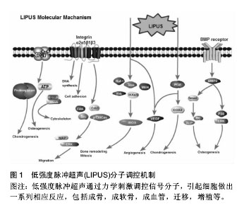
.jpg)
.jpg) 文题释义:
低强度脉冲超声:是一种频率超过人耳听阈(20 kHz)以上,强度为1-100 mW/cm2的声波。由于其对骨及软骨修复有显著促进作用,并且具有安全、廉价及便捷的特点,美国食品药品监督总局于1994年批准其作为加速骨折愈合的治疗手段。经过数十年的努力,目前临床上已逐渐探索出一条简单、合理、有效利用低强度脉冲超声的治疗方法,在促进患者机体功能康复的同时,最大程度上节约医疗资源,提高社会生产力。
细胞力学:是生物力学的前沿领域,也是组织工程学的一个重要分支。它涉及细胞在力学载荷作用下细胞膜、细胞骨架的变形,弹性常数、黏弹性、黏附力等力学性能的研究,以及力学因素对细胞生长、发育、成熟、增殖、衰老和死亡等的影响及其机制研究。细胞力学关注人体各类细胞,尤其是与骨骼运动系统、血液循环系统、消化系统等有关的细胞。
文题释义:
低强度脉冲超声:是一种频率超过人耳听阈(20 kHz)以上,强度为1-100 mW/cm2的声波。由于其对骨及软骨修复有显著促进作用,并且具有安全、廉价及便捷的特点,美国食品药品监督总局于1994年批准其作为加速骨折愈合的治疗手段。经过数十年的努力,目前临床上已逐渐探索出一条简单、合理、有效利用低强度脉冲超声的治疗方法,在促进患者机体功能康复的同时,最大程度上节约医疗资源,提高社会生产力。
细胞力学:是生物力学的前沿领域,也是组织工程学的一个重要分支。它涉及细胞在力学载荷作用下细胞膜、细胞骨架的变形,弹性常数、黏弹性、黏附力等力学性能的研究,以及力学因素对细胞生长、发育、成熟、增殖、衰老和死亡等的影响及其机制研究。细胞力学关注人体各类细胞,尤其是与骨骼运动系统、血液循环系统、消化系统等有关的细胞。