中国组织工程研究 ›› 2017, Vol. 21 ›› Issue (31): 4975-4982.doi: 10.3969/j.issn.2095-4344.2017.31.010
• 脊柱植入物 spinal implant • 上一篇 下一篇
Lenke3型成人特发性脊柱侧凸有限元模型的参数修正及有效性验证
辛大奇1,胡侦明2,汉 迪3,杨学军1,肖宇龙1,邢文华1,赵 岩1,付 裕1,祝 勇1
- 1内蒙古医科大学第二附属医院,内蒙古自治区呼和浩特市 010030;2重庆医科大学附属第一医院骨科,重庆市 400016;3内蒙古医科大学附属医院,内蒙古自治区呼和浩特市 010030
Modification and validation of Lenke3 type adult idiopathic scoliosis finite element model
Xin Da-qi1, Hu Zhen-ming2, Han Di3, Yang Xue-jun1, Xiao Yu-long1, Xing Wen-hua1, Zhao Yan1, Fu Yu1, Zhu Yong1
- 1the Second Affiliated Hospital of Inner Mongolia Medical University, Hohhot 010030, Inner Mongolia Autonomous Region, China; 2Department of Orthopedics, the First Affiliated Hospital of Chongqing Medical University, Chongqing 400016, China; 3the Affiliated Hospital of Inner Mongolia Medical University, Hohhot 010030, Inner Mongolia Autonomous Region, China
摘要:
文章快速阅读:
.jpg) 文题释义:
文题释义:脊柱有限元参数修正:利用有限元分析软件构建的模型,与脊柱的真实结构存在差异,需要利用统计学试验进行精细化修正,使其最大程度的与真实结构接近。
脊柱有限元有效性验证:利用有限元软件构建的模型,经过修正后,为了解其与真实结构的差异,需要利用模拟试验进行验证,观察修正后的模型与真实的吻合度。
摘要
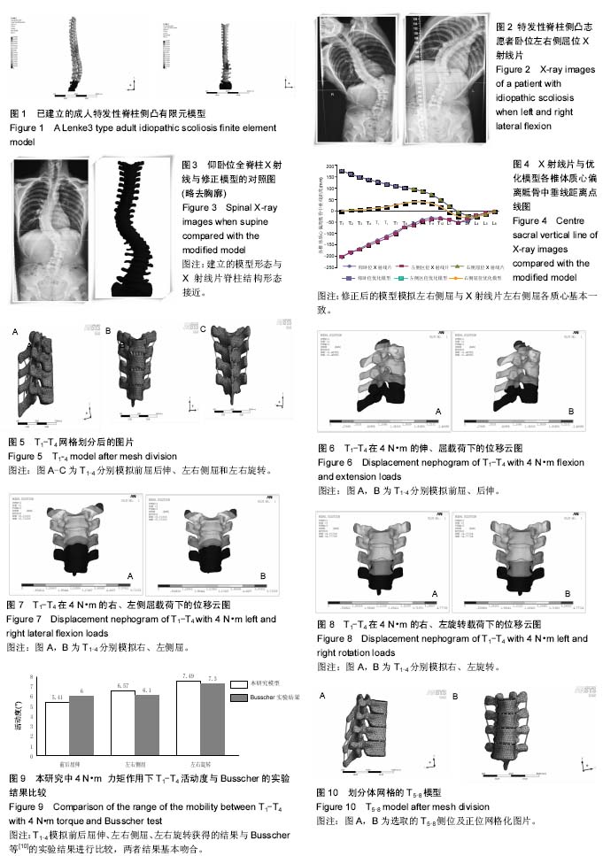
背景:作者利用Mimics等有限元软件成功建立了Lenke3型成人特发性脊柱有限元模型,但模型是否最大程度的符合个体化患者的真实情况,需要进一步进行模型修正及有效性验证。
目的:利用有限元分析软件对Lenke3型成人特发性脊柱有限元模型进行修正及有效性验证。
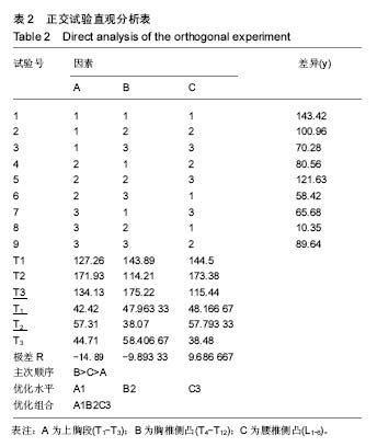
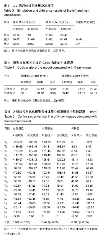
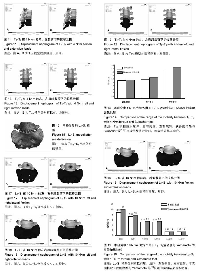
方法:根据Lenke3型成人特发性脊柱侧凸模型的特点,利用三因素三水平正交试验优化有限元模型,使模型特点最大程度的接近真实,通过模拟左右侧屈实验,分段加载选取T1-T4、T5-T8、L6-S1(骶椎腰化)节段分别模拟左右侧屈、前屈后伸,左右旋转活动度与体外Busscher、Yamamoto实验进行对比研究,多方位验证模型有效性。
结果与结论:①根据正交实验计算各因素各水平的平均差异和极差R,最后计算出A1B2C3的最佳组合可使模拟实验结果最符合个体的真实情况,使得有限元模拟实验结果与患者临床真实情况的差异最小。临床侧屈试验和参数修正前模型模拟的Cobb角度的变化差异值为 54.44°,经过参数修正后模型的差异值减小为2.11°。修正后模型各侧凸Cobb角的最大差异为4.29°;②修正后的模型与仰卧左右侧屈位X射线片对比,2组配对数据均服从正态分布,故利用配对t检验进行计算,左侧屈时,P=0.082,P > 0.05;右侧屈时,P=0.421,P > 0.05;仰卧位P=0.160,P > 0.05;③修正后的模型T1-T4节段各个方向的ROM:左屈3.25°,右屈3.32°,前屈2.52°,后伸2.89°,左侧旋转3.73°,右侧旋转3.76°,T5-T8节段各个方向的ROM:左屈1.39°,右屈1.43°,前屈1.35°,后伸1.34°,左旋2.09°,右旋2.11°;L6/S1节段各个方向的ROM:左屈5.17°,右屈5.19°,前屈8.92°,后伸7.35°,左旋1.41°,右旋1.42° ,获得的结果与Busscher及Yamamoto等的实验结果进行比较,结果基本吻合;④结果提示,通过对初始模型进行参数修正处理,使得模型与患者真实的材料属性基本符合。修正后的模型具有较好的可靠性和有效性,为下一步模拟临床手术操作提供了有效的数据平台。
中国组织工程研究杂志出版内容重点:人工关节;骨植入物;脊柱;骨折;内固定;数字化骨科;组织工程
ORCID: 0000-0003-2202-5308(辛大奇)
中图分类号:
.jpg)
.jpg) 文题释义:
文题释义: