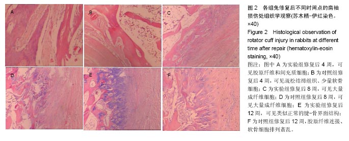
| [1]Bishop J,Klepps S,Lo IK,et al.Cuff integrity after arthroscopic versus open rotator cuff repair: a prospective study.J Shoulder Elbow Surg.2006;15(3):290-299.[2]Lorbach O, Baums MH, Kostuj T, et al. Advances in biology and mechanics of rotator cuff repair.Knee Surg Sports Traumatol Arthrosc.2015;23(2):530-541.[3]Al-Hakim W,Noorani A,Lambert S.Assessment and treatment strategies for rotator cuff tears.Shoulder Elbow.2015;7(2): 76-84.[4]李小溪,赵金忠.细胞工程技术在肩袖损伤修复中的应用[J].中华骨科杂志, 2015,35(6):681-685.[5]Lannotti JP,Codsi MJ,Kwon YW,et al.Porcine small intestine submucosa augmentation of surgical repair of chronic two-tendon rotator cuff tears. A randomized ,controlled trial.J Bone Joint Surg Am.2006;88(6):1238-1244.[6]张颉鸿,吴海山.肩袖修复后促进腱骨界面软骨再生的研究进展[J].中国矫形外科杂志, 2016, 24(14):1299-1303.[7]Thangarajah T,Pendegrass CJ,Shahbazi S,et al. Augmentation of rotator cuff repair with soft tissue scaffolds. Orthop J Sports Med.2015;3(6):1-8.[8]Carvalho AL,Martinelli F,Tramujas L,et al.Rotator cuff injuries and factors associated with reoperation.Rev Bras Ortop. 2016;51(3):298-302.[9]Sevivas N,Serra SC,Portugal R,et al.Animal model for chronic massive rotator cuff tear: behavioural and histologic analysis. Knee Surg Sports Traumatol Arthrosc.2015; 23(2):608-618.[10]Lee HJ,Kim YS,Ok JH,et al.Effect of a single subacromial prednisolone injection in acute rotator cuff tears in a rat model.Knee Surg Sports Traumatol Arthrosc.2015; 23(2):555-561.[11]Lorbach O,Baums MH,Kostuj T,et al.Advances in biology and mechanics of rotator cuff repair.Knee Surg Sports Traumatol Arthrosc.2015;23(2):530-541.[12]Mori D,Funakoshi N,Yamashita F.Arthroscopic surgery of irreparable large or massive rotator cuff tears with low-grade fatty degeneration of the infraspinatus: patch autograft procedure versus partial repair procedure.Arthroscopy. 2013;29(12): 1911-1921.[13]Ciampi P,Scotti C,Nonis A,et al.The benefit of synthetic versus biological patch augmentation in the repair of posterosuperior massive rotator cuff tears a 3-year follow-up study.Am J Sports Med.2014;42:1169-1175.[14]Wong I,Burns J,Snyder S.Arthroscopic graftJacket repair of rotator cuff tear.J Shoulder Elbow Surg.2010;19(2 Suppl): 104-109.[15]Milgrom C,Schaffler M,Gilbert S,et al.Rotator-cuff changes in asymptomatic adults. The effect of age, hand dominance and gender.J Bone Joint Surg Br.1995;77(2):296-298.[16]Murray TF Jr,Lajtai G,Mileski RM,et al.Arthroscopic repair of medium to large full-thickness rotaor cuff tears:outcome at 2-to 6-year follow—up.J Shoulder Elbow Surg.2002;11(1):19-24.[17]Wlk MV,Abdelkafy A,Hexel M,et al.Biomechanical evaluation of suture–tendon interface and tissue holding of three suture configurations in torn and degenerated versus intact human rotator cuffs.Knee Surg Sports Traumatol Arthrosc. 2015; 23(2):386-392.[18]Breidenbach AP, Gilday SD, Lalley AL,et al.Functional tissue engineering of tendon: establishing biological success criteria for improving tendon repair.J Biomech.2014;47(9):1941-1948.[19]Ross D,Maerz T,Kurdziel M,et al.The effect of granulocyte-colony stimulating factor on rotator cuff healing after injury and repair. Clin orthop Relat Res.2015;473(5):1655-1664.[20]Jo CH,Shin JS,Shin WH,et al.Platelet-Rich Plasma for Arthroscopic Repair of Medium to Large Rotator Cuff Tears. Am J Sports Med.2015;43(9):2102-2110.[21]Kabiri A,Esfandiari E,Esmaeili A,et al.Platelet-rich plasma application in chondrogenesis.Adv Biomed Res. 2014;25(3): 138.[22]Antuna S,Barco R,Martinez Diez JM,et al.Platelet rich fibrin in arthroscopic repair of massive rotator cuff tears:A prospective randomized pilot clinical trial.Acta Orthop Belg. 2013;79(1):25-30.[23]Ciampi P,Scotti C,Nonis A,et al.The benefit of synthetic versus biological patch augmentation in the repair of posterosuperior massive rotator cuff tears: A 3-year follow-up study.Am J Sports Med.2014;42(5):1169-1175.[24]Levy O,Relwani J,Zaman T,et al.Measurement of blood flow in the rotator cuff using laser Doppler flowmetry.J Bone Joint Surg Br.2008;90(7):893-898.[25]Kuzel BR,Grindel S,Papandrea R,et a1.Fatty infiltration and rotator cuff atrophy.J Am Acad Orthop Surg.2013;10:613-623.[26]Adams JE,Zobitz ME,Reach JS Jr,et a1.Rotator cuff repak using an acellular dermal matrix graft: an in vivostudy in a canine model.Arthroscopy.2006;22(7):700-709.[27]Hutmacher DW,Sittinger M.Periosteal cells in bone tissue engineering.Tissue Eng. 2003;9(1):S45-S64.[28]张俊玮,陆海涛,杨宇明,等.低温培养对骨膜细胞成软骨分化的影响[J].中华实验外科杂志, 2016,33(5):1281-1283.[29]Deponti D,Di Giancamillo A,Gervaso F,et al.Collagen scaffold for cartilage tissue engineering: the benefit of fibrin glue and the proper culture time in an infant cartilage mode.Tissue Eng Part A.2014;20:1113-1126.[30]Elnikety S,Pendegrass CJ,De Godoy RF,et al.Augmentation and repair of tendons using demineralised cortical bone.BMC Musculoskelet Disord.2016;17(1):483-49.[31]Valencia Mora M,Ruiz lban MA,Diaz Heredia J,et al.Stem cell therapy in the management of shoulder rotator cuff disorders. World J Stem Cells.2015;7(4):691-699.[32]Hernigou P, Flouzat Lachaniette CH, Delambre J,et al. Biologic augmentation of rotator cuff repair with mesenchymal stem cells during arthroscopy improves healing and prevents further tears: a case-controlled study.Int Orthop. 2014; 38(9): 1811-1818.[33]Kim HM,Galatz LM,Das R,et al.The role of transforming growth factor beta isoforms in tendon-to-bone healing. Connect Tissue Res.2011;52(2):87-98.[34]Mara CS,Sartori AR,Duarte AS,et al.Periosteum as a source of mesenchymal stem cells: The effects of TG-3 on chondrogenesis.Clinics(Sao Paulo).2011;66(33):487-492.[35]Beitzel K,Solovyova O,Cote MP,et al.The future role of mesenchymal stem cells in the management of shoulder disorders.Arthroscopy.2013;29(10):1702-1711.[36]张冲,李莉.TGF-β1对大鼠肩袖损伤修复术后腱-骨愈合的影响[J].医用生物力学,2016, 31(2):167-170.[37]Akyol E,Hindocha S,Khan WS.Use of Stem Cells and Growth Factors in Rotator Cuff Tendon Repair.Curr Stem Cell Res Ther. 2015;10(1):5-10.[38]Chen ACY,Lin SS,Chan YS,et al.Osteogenesis of prefabricated vascularized periosteal graft in rabbits.J Trauma. 2009;67(1):165-167. |
.jpg)
.jpg)
.jpg)