中国组织工程研究

• 脊柱损伤基础实验 basic experiments of spinal injury •    下一篇

骨形态发生蛋白2对退变椎间盘组织学和Ⅰ,Ⅱ型胶原的影响

林  曦1,叶君健2   

  1. 福建医科大学附属第一医院,1急诊科,2骨科,福建省福州市  350004
  • 收稿日期:2013-06-13 修回日期:2013-06-21 出版日期:2013-10-22 发布日期:2013-11-02
  • 作者简介:林曦★,男,1980年生,福建省屏南县人,2009年福建医科大学毕业,硕士,主要从事骨科学研究。 doctorlinxi@126.com
  • 基金资助:

    福建省自然科学基金项目(2012J01126)

Bone morphogenetic protein-2 gene affects the histology and collagen type Ⅰ and Ⅱ expressions in degenerative intervertebral disc

Lin Xi1, Ye Jun-jian2   

  1. 1Department of Emergency, 2Department of Orthopedics, the First Affiliated Hospital of Fujian Medical University, Fuzhou  350004, Fujian Province, China
  • Received:2013-06-13 Revised:2013-06-21 Online:2013-10-22 Published:2013-11-02
  • About author:Lin Xi★, Master, Department of Emergency, the First Affiliated Hospital of Fujian Medical University, Fuzhou 350004, Fujian Province, China
  • Supported by:

    Natural Science Foundation of Fujian Province, No.2012J01126

摘要:

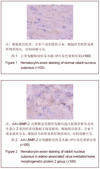
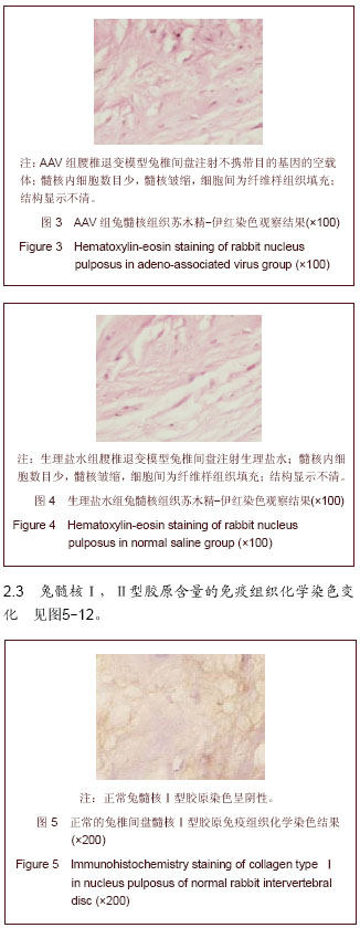
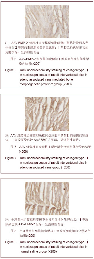
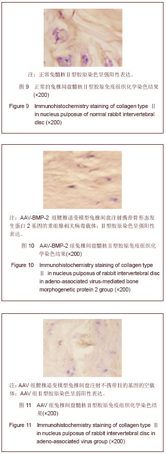
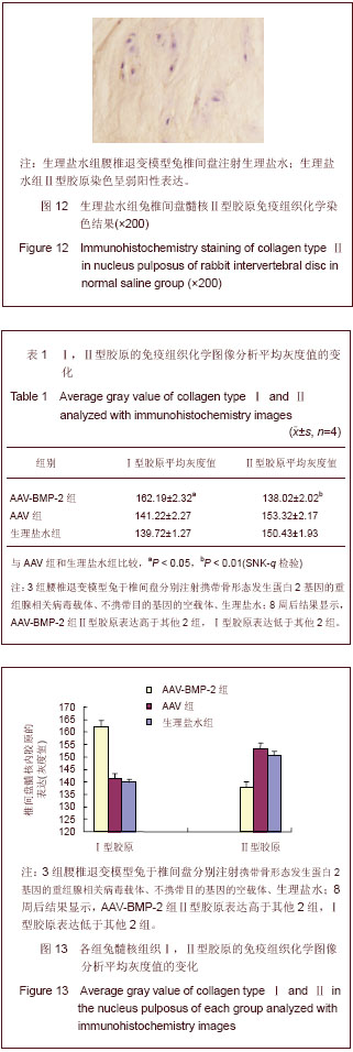
背景:研究表明,退变椎间盘细胞外基质的主要变化是Ⅱ型胶原和蛋白多糖含量的减少和Ⅰ型胶原含量的增加。
目的:观察腺相关病毒介导的骨形态发生蛋白2基因(AAV-BMP-2)对兔退变腰椎间盘内髓核Ⅰ,Ⅱ型胶原的影响。
方法:将12只新西兰大白兔L2-3,L3-4,L4-5,L5-6椎间盘针刺制造退变模型后随机分为3组,每组4只。其中AAV-BMP-2组兔椎间盘注射AAV-BMP-2,AAV组兔椎间盘注射不携带目的基因的空载体,生理盐水组兔椎间盘注射生理盐水,注射后第8周处死动物取其相应的椎间盘常规石蜡包埋,组织切片,以苏木精-伊红染色观察其髓核组织学变化,免疫组织化学法检测髓核组织Ⅰ型、Ⅱ型胶原表达并进行半定量分析。
结果与结论:普通苏木精-伊红染色结果显示AAV-BMP-2组椎间盘内髓核细胞数目较多,呈单个或者簇状分布,髓核结构清晰,无纤维样组织填充。而AAV组和生理盐水组的组织结构相似,髓核内细胞数目少,髓核皱缩干瘪,细胞间为纤维样组织填充且排列紊乱。免疫组织化学染色显示:AAV-BMP-2组椎间盘髓核内Ⅱ型胶原的表达均高于AAV组和生理盐水组(P < 0.05);AAV-BMP-2组椎间盘髓核内Ⅰ型胶原的表达均低于AAV组和生理盐水组(P < 0.05)。结果说明,体内转染AAV-BMP-2能抑制椎间盘髓核细胞表达Ⅰ型胶原,促进椎间盘髓核细胞表达合成Ⅱ型胶原,提示维持椎间盘内胶原的含量、种类,可维持椎间盘内组织学的结构和形态,稳定髓核细胞生长环境,延缓椎间盘的退变。

关键词: 骨关节植入物, 脊柱损伤基础实验, 骨形态发生蛋白2, 腺相关病毒, 椎间盘, 髓核组织, Ⅰ型胶原, Ⅱ型胶原, 省级基金

Abstract:

BACKGROUND: Studies have shown that the changes of extracellular matrix in degenerative intervertebral disc tissues mainly present as the decrease of collagen type Ⅱ and proteoglycan contents and the increase of collagen type Ⅰ content.
OBJECTIVE: To explore the effect of adeno-associated virus-mediated bone morphogenetic protein 2 gene on nucleus pulposus Ⅰ and Ⅱ collagen levels in rabbit degenerative intervertebral disc tissues.
METHODS: L2-3, L3-4, L4-5 and L5-6 lumbar discs of 12 New Zealand white rabbits were punctured to establish interverbral disc degeneration model. Subsequently, 12 rabbits were randomly divided into three groups, four rabbits in each group. The intervertebral discs in the adeno-associated virus-mediated bone morphogenetic protein 2 group were injected with the adeno-associated virus-mediated bone morphogenetic protein 2 gene, the intervertebral discs in the adeno-associated virus group were injected with adeno-associated virus only, while the discs in the normal saline group were injected with normal saline. All rabbits were sacrificed after injected for 8 weeks, and the L2-3, L3-4, L4-5 and L5-6 lumbar discs of each rabbit were collected, paraffin-embedded and sliced. The histological changes of nucleus pulposus were observed with hematoxylin-eosin staining, and the immunohistochemistry was used to detect the collagen type Ⅰ and Ⅱ expressions in nucleus pulposus. Semi-quantitative analysis was performed.
RESULTS AND CONCLUSION: Hematoxylin-eosin staining showed that the nucleus pulposus in the intervertebral disc tissues was less in the adeno-associated virus-mediated bone morphogenetic protein 2 group, the nucleus pulposus was in single or clustered distribution with clear nucleus structure and without fibrous tissue filling. The tissue structures of nucleus pulposus were the same in the adeno-associated virus group and normal saline group, the cell number in nucleus pulposus was small, the nucleus pulposus was shrunken and shriveled, and the cells were filled with fibrous tissue and arranged disorderly. Immunohistochemistry staining showed the expression of collage type Ⅰ in the intervertebral disc nucleus pulposus of adeno-associated virus-mediated bone morphogenetic protein 2 group was higher than that of the adeno-associated virus group and normal saline group (P < 0.05); the expression of collage typeⅠ in the intervertebral disc nucleus pulposus of adeno-associated virus-mediated bone morphogenetic protein 2 group was lower than that of the adeno-associated virus group and normal saline group (P < 0.05). The results indicate that adeno-associated virus-mediated bone morphogenetic protein 2 can inhibit the expression of collagen type Ⅰ in the intervertebral disc nucleus pulposus, promote the expression of collagen type Ⅱ. Maintaining the content of collagen in intervertebral disc can keep the histological structure and morphology of intervertebral disc, stabilize the environment for nucleus pulposus cell growth, and delay the intervertebral disc degeneration.

Key words: bone morphogenetic proteins, intervertebral disk, collagen type Ⅰ, collagen type Ⅱ

中图分类号: