中国组织工程研究 ›› 2018, Vol. 22 ›› Issue (35): 5681-5686.doi: 10.3969/j.issn.2095-4344.1016

• 骨与关节图像与影像 bone and joint imaging • 上一篇    下一篇

高频彩色多普勒超声显像评价健康成人胫神经及主要分支的特点和意义

袁 晨,景江新,刘纯红   

  1. 新疆医科大学第二附属医院超声科,新疆维吾尔自治区乌鲁木齐市   830063
  • 出版日期:2018-12-18 发布日期:2018-12-18
  • 通讯作者: 刘纯红,新疆医科大学第二附属医院超声科,新疆维吾尔自治区乌鲁木齐市 830063
  • 作者简介:袁晨,女,1993年生,甘肃省天水市人,回族,新疆医科大学第二附属医院在读硕士,医师,主要从事肌骨超声方面的研究。
  • 基金资助:

    新疆医科大学研究生创新创业项目(CXCY2017070)

Characteristics and significance of high-frequency color Doppler ultrasound in assessing tibial nerve and its main branches of healthy adults  

Yuan Chen, Jing Jiangxin, Liu Chunhong   

  1. Department of Ultrasound, the Second Affiliated Hospital of Xinjiang Medical University, Urumqi 830063, Xinjiang Uygur Autonomous Region, China
  • Online:2018-12-18 Published:2018-12-18
  • Contact: Liu Chunhong, Department of Ultrasound, the Second Affiliated Hospital of Xinjiang Medical University, Urumqi 830063, Xinjiang Uygur Autonomous Region, China
  • About author:Yuan Chen, Master candidate, Physician, Department of Ultrasound, the Second Affiliated Hospital of Xinjiang Medical University, Urumqi 830063, Xinjiang Uygur Autonomous Region, China
  • Supported by:

    the Graduate Innovation and Entrepreneurship Project of Xinjiang Medical University, No. CXCY2017070

摘要:

文章快速阅读:


文题释义:
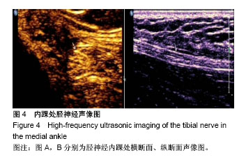
胫神经:是坐骨神经在腘窝的粗大分支之一,伴行腘血管经比目鱼肌腱弓深面至小腿与胫后动脉同行在内踝下方分出足底内侧神经和足底外侧神经,胫神经支配小腿后侧屈肌群和足底感觉,为足底提供自主感觉和运动神经纤维。
胫神经高频彩超显像:MRI虽然对软组织的高分辨率特点使得胫神经可以得到良好的显示效果并可直观量化分析胫神经的特点,但其扫描时间较长,花费高,不能动态扫查双侧对比,有相对禁忌证且数据处理较为复杂,临床应用不如高频彩超快速便捷易行。
 
摘要
背景:在过去的10年中神经肌肉超声已经成为周围神经疾病诊断的有效手段,国外仅有少量学者对胫神经全程进行研究,但目前国内尚未见超声学者对胫神经自腘窝上角至足底分支全程的详细报道。
目的:探究正常成人胫神经及其主要分支高频彩色多普勒超声(高频彩超)声像图特点及其可能的临床意义。
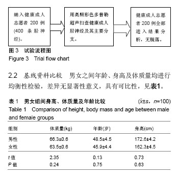
方法:用高频彩超探查200名健康成人志愿者400条胫神经及其主要分支,观察其声像图特点、走行、毗邻结构及血流信号显示情况;分别测量腘窝、内踝处胫神经及足底处胫神经分支的前后径、左右径及横截面积并进行统计学分析。
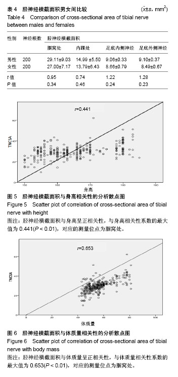
结果与结论:①高频彩超对200名志愿者胫神经全程显示率为100%,走行规律且声像图具备一定特点;②左右侧对比,胫神经在所选位点测值差异均无显著性意义(P > 0.05);③男女对比,男性胫神经在所选位点的横截面积均较女性稍大,但差异无显著性意义(P > 0.05);④胫神经横截面积与身高和体质量均呈正相关性(P < 0.01);⑤结果表明,高频彩超对胫神经及其主要分支具有良好的显示效果,可为基础解剖学研究提供可能的正常成人影像学数据。

中国组织工程研究杂志出版内容重点:人工关节;骨植入物;脊柱骨折;内固定;数字化骨科;组织工程
ORCID: 0000-0002-8503-6816(袁晨)

关键词: 高频彩色多普勒超声检查, 高频彩超, 胫神经, 足底内外侧神经, 声像图

Abstract:

BACKGROUND: In the past decades, neuromuscular ultrasound has become an effective method for the diagnosis of peripheral nerve diseases. Only a few scholars at abroad have studied the tibial nerve in detail. However, the details of armpit to the plantar branches of the tibial nerve are none reported in China.

OBJECTIVE: To investigate the features of high-frequency color Doppler ultrasound sonography of normal adult phrenic nerve and its main branches and its possible clinical significance.
METHODS: Ultrasonography was used to probe 200 healthy adult volunteers with 400 tibial nerves and their main branches to observe the characteristics of the sonographic features, walking, adjoining structures, and blood flow signal display. The anteroposterior diameter, lateral diameter and cross-sectional area of the iliac nerves and soles at the axillary and medial malleolus were measured.
RESULTS AND CONCLUSION: (1) High-frequency color Doppler ultrasound showed 100% display rate of sacral nerves in 200 healthy adult volunteers, and their walking and sonography had certain characters. (2) There was no significant difference between left and right tibial nerves at the selected sites (P > 0.05). (3) The cross-sectional area of male tibial nerve at the selected sites was slightly larger than that of the females (P > 0.05). (4) The cross-sectional area of tibial nerve was positively correlated with height and body mass (P < 0.01). (5) These results imply that high-frequency color ultrasound has a good display for the tibial nerve and its main branches, which can provide normal human imaging data for anatomical studies. 

中国组织工程研究杂志出版内容重点:人工关节;骨植入物;脊柱骨折;内固定;数字化骨科;组织工程

Key words: Ultrasonography, Doppler, Tibial Nerve, Tissue Engineering

中图分类号: