[1] BRUNET J, PIERRAT B, BADEL P. Review of Current Advances in the Mechanical Description and Quantification of Aortic Dissection Mechanisms. IEEE Rev Biomed Eng. 2021;14:240-255.
[2] HIBINO M, OTAKI Y, KOBEISSI E, et al. Blood Pressure, Hypertension, and the Risk of Aortic Dissection Incidence and Mortality: Results From the J-SCH Study, the UK Biobank Study, and a Meta-Analysis of Cohort Studies. Circulation. 2022;145(9):633-644.
[3] ERBEL R, ABOYANS V, BOILEAU C, et al. Corrigendum to: 2014 ESC Guidelines on the diagnosis and treatment of aortic diseases. Eur Heart J. 2015;36(41):2779.
[4] LOVATT S, WONG CW, SCHWARZ K, et al. Misdiagnosis of aortic dissection: A systematic review of the literature. Am J Emerg Med. 2022;53:16-22.
[5] HSU JY, LIU PP, LIU AB, et al. Long-Term Risks of Stroke in Patients With Type A Aortic Dissection: A Nationwide Cohort Study. J Am Heart Assoc. 2022;11(22):e027178.
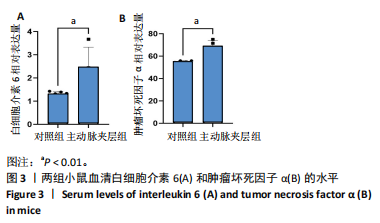
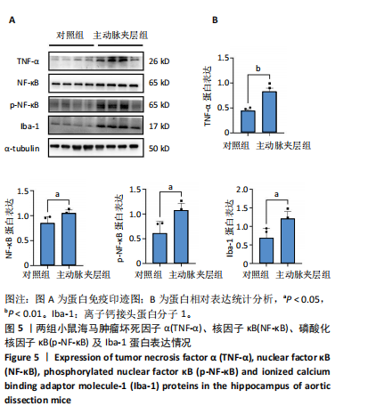
[6] 王宝珠,邵美华,马翔,等.30例主动脉夹层患者认知功能与血清C反应蛋白水平的相关性分析[J].新疆医科大学学报,2021,44(5): 581-584+590.
[7] TZILIVAKI A, TUKKER JJ, MAIER N, et al. Hippocampal GABAergic interneurons and memory. Neuron. 2023;111(20):3154-3175.
[8] AMANOLLAHI M, JAMEIE M, HEIDARI A, et al. The Dialogue Between Neuroinflammation and Adult Neurogenesis: Mechanisms Involved and Alterations in Neurological Diseases. Mol Neurobiol. 2023;60(2):923-959.
[9] GÖK E, DEVECI E. Histopathological evaluation of IBA-1, GFAP activity in the brain cortex of rats administered cadmium chloride. Arch Ital Biol. 2022;160(1-2):20-27.
[10] BALZANO T, ARENAS YM, DADSETAN S, et al. Sustained hyperammonemia induces TNF-a IN Purkinje neurons by activating the TNFR1-NF-κB pathway. J Neuroinflammation. 2020;17(1):70.
[11] ANILKUMAR S, WRIGHT-JIN E. NF-κB as an Inducible Regulator of Inflammation in the Central Nervous System. Cells. 2024;13(6):485.
[12] LIU H, WU X, LUO J, et al. Pterostilbene Attenuates Astrocytic Inflammation and Neuronal Oxidative Injury After Ischemia-Reperfusion by Inhibiting NF-κB Phosphorylation. Front Immunol. 2019;10:2408.
[13] LEE JC, TAE HJ, KIM IH, et al. Roles of HIF-1α, VEGF, and NF-κB in Ischemic Preconditioning-Mediated Neuroprotection of Hippocampal CA1 Pyramidal Neurons Against a Subsequent Transient Cerebral Ischemia. Mol Neurobiol. 2017;54(9):6984-6998.
[14] SAWADA H, BECKNER ZA, ITO S, et al. β-Aminopropionitrile- induced aortic aneurysm and dissection in mice. JVS Vasc Sci. 2022;3: 64-72.
[15] KHAJA MS, WILLIAMS DM. Aortic Dissection: Introduction. Tech Vasc Interv Radiol. 2021;24(2):100745.
[16] KAGEYAMA S, OHASHI T, YOSHIDA T, et al. Early mortality of emergency surgery for acute type A aortic dissection in octogenarians and nonagenarians: A multi-center retrospective study. J Thorac Cardiovasc Surg. 2024;167(1):65-75.e8.
[17] SHONO Y, AKAHOSHI T, MEZUKI S, et al. Clinical characteristics of type A acute aortic dissection with CNS symptom. Am J Emerg Med. 2017;35(12):1836-1838.
[18] LANA D, UGOLINI F, NOSI D, et al. The Emerging Role of the Interplay Among Astrocytes, Microglia, and Neurons in the Hippocampus in Health and Disease. Front Aging Neurosci. 2021;13:651973.
[19] WU Y, FAN L, WANG Y, et al. Isorhamnetin Alleviates High Glucose-Aggravated Inflammatory Response and Apoptosis in Oxygen-Glucose Deprivation and Reoxygenation-Induced HT22 Hippocampal
Neurons Through Akt/SIRT1/Nrf2/HO-1Signaling Pathway. Inflammation. 2021;44(5):1993-2005.
[20] QIANG M, TAN BH, HUO DS, et al. Changes in early endosomes in rat hippocampal CA1 neurons after transient global cerebral ischaemia. Folia Histochem Cytobiol. 2024;62(3):123-132.
[21] THADATHIL N, NICKLAS EH, MOHAMMED S, et al. Necroptosis increases with age in the brain and contributes to age-related neuroinflammation. Geroscience. 2021;43(5):2345-2361.
[22] TUCSEK Z, NOA VALCARCEL-ARES M, TARANTINI S, et al. Hypertension-induced synapse loss and impairment in synaptic plasticity in the mouse hippocampus mimics the aging phenotype: implications
for the pathogenesis of vascular cognitive impairment. Geroscience. 2017;39(4):385-406.
[23] THONG EHE, QUEK EJW, LOO JH, et al. Acute Myocardial Infarction and Risk of Cognitive Impairment and Dementia: A Review. Biology(Basel). 2023;12(8):1154.
[24] LI L, JIANG W, YU B, et al. Quercetin improves cerebral ischemia/reperfusion injury by promoting microglia/macrophages M2 polarization via regulating PI3K/Akt/NF-κB signaling pathway. Biomed
Pharmacother. 2023;168:115653.
[25] DENG L, GUO Y, LIU J, et al. miR-671-5p Attenuates Neuroinflammation via Suppressing NF-κB Expression in an Acute Ischemic Stroke Model. Neurochem Res. 2021;46(7):1801-1813.
[26] QI L, WU K, SHI S, et al.Thrombospondin-2 is upregulated in patients with aortic dissection and enhances ngiotensin II-induced smooth muscle cell apoptosis. Exp Ther Med. 2020;20(6):150. |