中国组织工程研究 ›› 2024, Vol. 28 ›› Issue (36): 5766-5772.doi: 10.12307/2024.680
• 骨与关节生物力学 bone and joint biomechanics • 上一篇 下一篇
颈椎钩突相关夹角增龄变化的特征与意义
张德洲1,王超群2,史 君3,李 琨4,5,张少杰4,5,马 渊5,侯二飞4,赵丹阳5,郝韵腾1,王思敏1,李筱贺4,王海燕4,李志军4,5,王 星4,5
- 1内蒙古医科大学研究生院,内蒙古自治区呼和浩特市 010110;2内蒙古医科大学附属医院影像科,内蒙古自治区呼和浩特市 010050;内蒙古医科大学基础医学院,3生理学教研室,4解剖学教研室,内蒙古自治区呼和浩特市 010110;5内蒙古医科大学基础医学院数字医学中心,内蒙古自治区呼和浩特市 010059
Characteristics and significance of age-related changes in cervical uncinate process-related angle
Zhang Dezhou1, Wang Chaoqun2, Shi Jun3, Li Kun4, 5, Zhang Shaojie4, 5, Ma Yuan5, Hou Erfei4, Zhao Danyang5, Hao Yunteng1, Wang Simin1, Li Xiaohe4, Wang Haiyan4, Li Zhijun4, 5, Wang Xing4, 5
- 1Graduate School of Inner Mongolia Medical University, Hohhot 010110, Inner Mongolia Autonomous Region, China; 2Department of Imaging, Affiliated Hospital of Inner Mongolia Medical University, Hohhot 010050, Inner Mongolia Autonomous Region, China; 3Department of Physiology, 4Department of Anatomy, School of Basic Medicine, Inner Mongolia Medical University, Hohhot 010110, Inner Mongolia Autonomous Region, China; 5Center for Digital Medicine, School of Basic Medicine, Inner Mongolia Medical University, Hohhot 010059, Inner Mongolia Autonomous Region, China
摘要:
文题释义:
钩椎关节:又称Luschka关节(Luschka joint),是由颈椎椎体上面侧方的骨性突起——钩突与相邻上位椎体下面侧方的斜坡关节面构成,具有限制椎体间侧方移位的作用。是由德国解剖学家Von Luschka于1858年首次观察发现并提出,后学术界以其名字命名了钩椎关节。钩突:位于椎体上面后外侧呈矢状位的嵴样隆起,与椎间孔、横突孔及椎间盘紧密相邻。该结构的增生、骨折等病变往往会压迫神经根、椎动脉、交感神经等结构,导致相应颈椎病症状的发生,因此针对该关节的研究也比较广泛。
背景:钩椎关节作为颈椎所特有的结构,其发生、发展和发育直接影响着颈椎的稳定性和活动度,也与颈椎病的发病密切相关,深入了解钩椎关节的发育特点对颈椎病的发病类型、诊断与治疗有着重要意义。
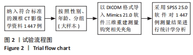
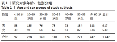
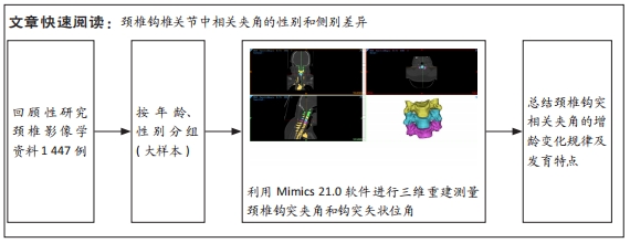
目的:利用影像学和三维重建技术对不同年龄段的颈椎钩突相关夹角进行大样本测量和观察,旨在揭示其随年龄和椎骨增长变化的特点,以及与颈椎稳定性之间的关系。方法:采用回顾性设计,将收集到的符合研究要求的1 447例完整颈椎节段CT影像学资料的原始数据以DICOM格式导入Mimics 21.0软件进行后处理,并进行钩突夹角、钩突矢状位角测量,按照性别、年龄、侧别分组。
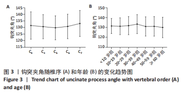
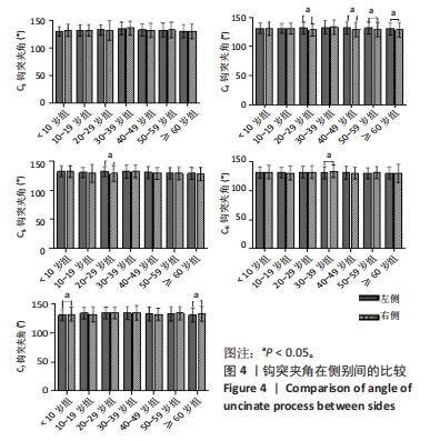
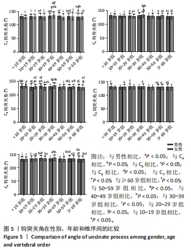
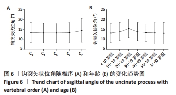
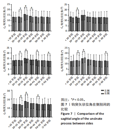
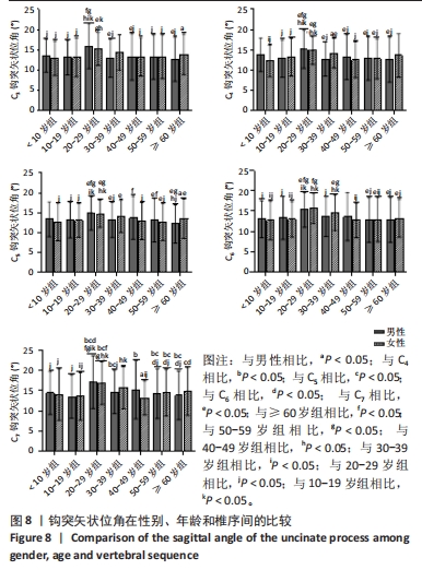
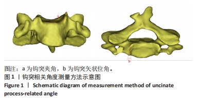
结果与结论:①钩突夹角随椎序的增加呈V字形增长,其最低峰值位于C5处;总体随年龄的增长呈尖峰状,其峰值多介于30-39岁年龄范围内;②钩突矢状位角随椎序的增加呈鱼钩样增长、总体随年龄的递增也呈尖峰状,其峰值多介于20-29岁年龄范围内;钩突夹角与钩突矢状位角在侧别和性别间仅部分存在显著性差异(P < 0.05);③提示钩突夹角随椎序递增呈V字形增长;钩突矢状位角随椎序递增鱼钩样增长,而2个夹角随年龄的增长总体呈尖峰状,钩突夹角在131°左右,可能与颈椎的稳定性密切相关;而钩突矢状位角在14°左右,可能对限制颈椎过度旋转有一定作用。
https://orcid.org/0000-0003-0059-4921 (王星)
中国组织工程研究杂志出版内容重点:人工关节;骨植入物;脊柱;骨折;内固定;数字化骨科;组织工程
中图分类号: