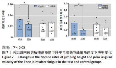
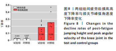
[1] HEJBOL EK, HARBO T, AGERGAARD J, et al. Myopathy as a cause of fatigue in long-term post-COVID-19 symptoms: evidence of skeletal muscle histopathology. Eur J Neurol. 2022;29: 2832-2841.
[2] KINDLER E, THIEL V. SARS-CoV and IFN: too little, too late. Cell Host Microbe. 2016;19(2):139-141.
[3] HARTENIAN E, NANDAKUMAR D, LARI A, et al. The molecular virology of coronaviruses. J Biol Chem. 2020;295(37):12910-12934.
[4] CHANNAPPANAVAR R, FEHR AR, VIJAY R, et al. Dysregulated type I interferon and inflammatory monocyte-macrophage responses cause lethal pneumonia in SARS-CoV-infected mice. Cell Host Microbe. 2016;19(2):181-193.
[5] COSTELA-RUIZ VJ, ILLESCAS-MONTES R, PUERTA-PUERTA JM, et al. SARS-CoV-2 infection: The role of cytokines in COVID-19 disease. Cytokine Growth Factor Rev. 2020;54:62-75.
[6] JOHNSON TA, JINNAH HA, KAMATANI N. Shortage of cellular ATP as a cause of diseases and strategies to enhance ATP. Front Pharmacol. 2019;10:98.
[7] LIU H, LIU Y, LI B. Predictive analysis of health/physical fitness in health-promoting lifestyle of adolescents. Front Public Health. 2021;9:691669.
[8] SOARES MN, EGGELBUSCH M, NADDAF E, et al. Skeletal muscle alterations in patients with acute Covid-19 and post-acute sequelae of Covid-19. J Cachexia Sarcopenia Muscle. 2022;13(1):11-22.
[9] MORO T, PAOLI A. When COVID-19 affects muscle: effects of quarantine in older adults. Eur. Transl Myol. 2020;30:9069.
[10] HARTLEY P, COSTELLO P, FENNER R, et al. Change in skeletal muscle associated with unplanned hospital admissions in adult patients: a systematic review and meta-analysis. PLoS One. 2019;14:e0210186.
[11] NARICI M, DE VITO G, FRANCHI M, et al. Impact of sedentarism due to the COVID-19 home confnement on neuromuscular, cardiovascular and metabolic health: physiological and pathophysiological implications and recommendations for physical and nutritional countermeasures. Eur J Sport Sci. 2022;12:1-22.
[12] LORENT N, WEYGAERDE YV, CLAEYS E, et al. Prospective longitudinal evaluation of hospitalised COVID-19 survivors 3 and 12 months after discharge. ERJ Open Res. 2022;8: 00004-2022.
[13] HUANG C, HUANG L, WANG Y, et al. 6-month consequences of COVID-19 in patients discharged from hospital: a cohort study. Lancet. 2021;397:220-232.
[14] STOFFELS AAF, VAN VOORTHUIZEN EL, VAN HEES HWH, et al. Longitudinal analysis of quadriceps muscle strength in patients with previous COVID-19 hospitalization and in patients with post-acute sequelae following mild COVID-19. nutrients. 2022;14(20):4319.
[15] MIKINES KJ, RICHTER EA, DELA F, et al. Seven days of bed rest decrease insulin action on glucose uptake in leg and whole body. J Appl Physiol .1991;70(3):1245-1254.
[16] 世界卫生组织 2019冠状病毒病(COVID-19)疫情报告 (2020-07-13). https://www.who.int/publications/i/item/WHO-2019-nCoV-therapeutics-2023.1.
[17] PUGA L, DINIS P, TEIXEIRA R, et al. Cardiovascular risk assessment after COVID-19 infection before resuming sports activities-practical flowchart and meta-analysis. Sociedade Brasileira de Cardiologia. 2021.
[18] CHEN C, ZHOU Y, WANG DW. SARS-CoV-2: a potential novel etiology of fulminant myocarditis. Herz. 2020;45(3):230-232.
[19] FAYOL A, LIVROZET M, BOUTOUYRIE P, et al. Cardiac performance in patients hospitalized with COVID-19: a 6 month follow-up study. ESC Heart Fail. 2021;8(3):2232-2239.
[20] DI STEFANO V, BATTAGLIA G, GIUSTINO V, et al. Signifificant reduction of physical activity in patients with neuromuscular disease during COVID-19 pandemic: the long-term consequences of quarantine. J Neurol. 2021;268:20-26.
[21] ALFAROUK KO, ALHOUFIE STS, HIFNY A, et al. Of mitochondrion and COVID-19. J Enzyme Inhib Med Chem. 2021;36(1):1258-1267.
[22] ZHANG C, HE H, WANG L, et al. Virus-triggered ATP release limits viral replication through facilitating IFN-β production in a P2X7-dependent manner. Immunol 2017;199(4):1372-1381.
[23] LUOMA RL, BUTLER MW, STAHLSCHMIDT ZR. Plasticity of immunity in response to eating. Exp Biol. 2016;219(13):1965-1968.
[24] TAGHIZADEH-HESARY F, AKBARI H. The powerful immune system against powerful COVID-19: a hypothesis. Med Hypotheses. 2020;140:109762.
[25] DA SILVEIRA MP, DA SILVA FAGUNDES KK, BIZUTI MR, et al. Physical exercise as a tool to help the immune system against COVID-19: an integrative review of the current literature. Clin Exp Med. 2021;21(1):15-28.
[26] CÁRDENAS C, MILLER RA, SMITH I, et al. Essential regulation of cell bioenergetics by constitutive InsP3 receptor Ca2+ transfer to mitochondria. Cell. 2010;142(2):270-283.
[27] KAREN B, GÁLVEZ NICOLÁS MS, ANDRADE CATALINA A, et al. Modulation of adaptive immunity and viral infections by ion channels. Front Physiol. 2021;12:736681.
[28] YAP J, TAI YK, FRHLICH J, et al. Ambient and supplemental magnetic fields promote myogenesisviaa TRPC1-mitochondrial axis: evidence of a magnetic mitohormetic mechanism. FASEB J. 2019;33(11):12853-12872.
[29] TAI YK, NG C, PURNAMAWATI K, et al. Magnetic fields modulate metabolism and gut microbiome in correlation with Pgc‐1α expression: follow‐up to an in vitro magnetic mitohormetic study. FASEB J. 2020;34(8):11143-11167.
[30] STEPHENSON MC, KRISHNA L, PANNIR SELVAN RM, et al. Magnetic field therapy enhances muscle mitochondrial bioenergetics and attenuates systemic ceramide levels following ACL reconstruction: southeast Asian randomized-controlled pilot trial. J Orthop Translat. 2022;35:99-112.
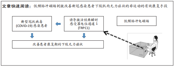
[31] 厉中山,王春露,刘洁,等.短期低频脉冲磁场诱导经典瞬时感受器电位通道1对肱二头肌最大自主收缩力与力量耐力的影响[J].中国组织工程研究,2023,27(11):1796-1804.
[32] 厉中山,白石,刘洁等.短期低频脉冲磁场诱导经典瞬时感受器电位通道1对局部肌肉肌力提升后的保持与衰减变化轨迹[J].中国组织工程研究,2023,27(23):3721-3727.
[33] DOMINGUEZ-NICOLAS SM, MANJARREZ E. Low-field thoracic magnetic stimulation increases peripheral oxygen saturation levels in coronavirus disease (COVID-19) patients: a single-blind, sham-controlled, crossover study. Medicine. 2021;40:27444.
[34] D PARATE, A FRANCO-OBREGÓN, HLICH J, et al. Enhancement of mesenchymal stem cell chondrogenesis with short-term low intensity pulsed electromagnetic fields. Sci Rep. 2017;7(1):9421.
[35] SARITAS EU, GOODWILL PW, ZHANG GZ, et al. Magnetostimulation limits in magnetic particle imaging. IEEE Trans Med Imaging. 2013;32(9):1600-1610.
[36] SCHMALE I, GLEICH B, SCHMIDT J, et al. Human PNS and SAR study in the frequency range from 24 to 162 kHz// Magnetic Particle Imaging (IWMPI), 2013 International Workshop on. IEEE. 2013.
[37] GRSER M, THIEBEN F, SZWARGULSKI P, et al. Human-sized magnetic particle imaging for brain applications. Nat Commun. 2019;10(1):1936.
[38] KONRAD P. The ABC of EMG. Noraxon USA, Inc. 2005.
[39] CLAUDINO JG, CRONIN J, MEZÊNCIO B, et al. The countermovement jump to monitor neuromuscular status: A meta-analysis. J Sci Med Sport. 2017;20(4):397-402.
[40] PETRIGNA L, KARSTEN B, MARCOLIN G, et al. A review of countermovement and squat jump testing methods in the context of public health examination in adolescence: reliability and feasibility of current testing procedures. Front Physiol. 2019;10:1384.
[41] STONE MH, STONE ME, SANDS WA, et al. Maximum strength and strength training: a relationship to endurance. Strength Condition J. 2006;28(3):44-53.
[42] CIFREK M, MEDVED V, TONKOVI S, et al. Surface EMG based muscle fatigue evaluation in biomechanics. Clin Biomech. 2009;24(4):327-340.
[43] ANDERSSON H, RAASTAD T, NILSSON J, et al. Neuromuscular fatigue and recovery in elite female soccer: effects of active recovery. Med Sci Sports Exerc. 2008;40(2):372-380.
[44] THOMAS K, DENT J, HOWATSON G, et al. Etiology and recovery of neuromuscular fatigue after simulated soccer match play. Med Sci Sports Exerc. 2017;49(5):955-964.
[45] IZQUIERDO M, AGUADO X, GONZALEZ R, et al. Maximal and explosive force production capacity and balance performance in men of different ages. Eur J Appl Physiol Occup Physiol. 1999;79(3):260-267.
[46] MARKOVIC G, DIZDAR D, JUKIC I, et al. Reliability and factorial validity of squat and countermovement jump tests. J Strength Condition Res. 2004;18(3):551-555.
[47] MAFFIULETTI N A, AAGAARD P, BLAZEVICH A J, et al. Rate of force development: physiological and methodological considerations. Eur J Appl Physiol. 2016;(6):116.
[48] ANDERSEN LL, AAGAARD P. Influence of maximal muscle strength and intrinsic muscle contractile properties on contractile rate of force development. Eur J Appl Physiol. 2006;96(1):46-52.
[49] HUGHES DC, ELLEFSEN S, BAAR K. Adaptations to endurance and strength training. Cold Spring Harbor Laboratory Press. 2018.
[50] VERSACE V, SEBASTIANELLI L, FERRAZZOLI D, et al. Case report: myopathy in critically Ill COVID-19 patients: a consequence of hyperinflammation?. Front Neurol. 2021;12(1): 625144.
[51] COSTA TJ, POTJE SR, FRAGA-SILVA TFC, et al. Mitochondrial DNA and TLR9 activation contribute to SARS-CoV-2-induced endothelial cell damage. Vascul Pharmacol. 2022;142: 106946.
[52] PILEGAARD H, SALTIN B, NEUFER PD. Exercise induces transient transcriptional activation of the PGC-1alpha gene in human skeletal muscle. J Physiol. 2003;546(3):851-858.
[53] JENINGA EH, SCHOONJANS K, AUWERX J. Reversible acetylation of PGC-1: connecting energy sensors and effectors to guarantee metabolic flexibility. Oncogene. 2010;29(33): 4617-4624.
[54] FRIEDMAN JR, NUNNARI J. Mitochondrial form and function. Nature. 2014;505(7483): 335-343.
[55] HARGREAVES M, SPRIET LL. Skeletal muscle energy metabolism during exercise. Nat Metab. 2020;2(9):817-828.
[56] FRY CS, KIRBY TJ, KOSMAC K, et al. Myogenic progenitor cells control extracellular matrix production by fibroblasts during skeletal muscle hypertrophy. Cell Stem Cell. 2017;20(1):56-69.
[57] HEJBOL EK, HARBO T, AGERGAARD J, et al. Myopathy as a cause of fatigue in long-term post-COVID-19 symptoms: evidence of skeletal muscle histopathology. Eur J Neurol. 2022;29:2832-2841.
[58] CHEN X, CAO R, ZHONG W. Host calcium channels and pumps in viral infections. Cells. 2019;9(1):94.
[59] CORMIE P, MCGUIGAN MR, NEWTON RU. Developing maximal neuromuscular power. Sports Med. 2011;41(2):125-146.
[60] CARLSEN RC, VILLARIN JJ. Membrane excitability and calcium homeostasis in exercising skeletal muscle. Am J Phys Med Rehabil. 2002;81(11 Suppl):S28-S39.
[61] HILLE B. Ionic Channels of Excitable Membranes, ed 2. Sunderland, MA, Sinauer Associates, 1992.
[62] ZANOU N. Role of TRPC1 channel in skeletal muscle function. Am J Physiol Cell Physiol 2010;298:C149-C162.
[63] HILL A, KUPALOV P. Anaerobic and aerobic activity in isolated muscle. Proc R Soc Lond B. 1929;105: 313-322.
[64] ALLEN DG. Skeletal muscle function: role of ionic changes in fatigue, damage and disease. Clin Exp Pharmacol Physiol. 2004;31:485-493.
[65] BEECH DJ, XU SZ, MCHUGH D, FLEMMING R. TRPC1 store-operated cationic channel subunit. Cell Calcium. 2003;33:433-440.
[66] CLAPHAM DE. TRP channels as cellular sensors. Nature. 2003;426(4):517-524.
[67] AMBUDKAR IS. TRPC1: a core component of store-operated calcium channels. Biochem Soc Trans. 2007;35:96-100.
[68] LYFENKO AD, DIRKSEN RT. Differential dependence of store-operated and excitation-coupled Ca2 entry in skeletal muscle on STIM1 and Orai1. J Physiol. 2008;586:4815-4824.
[69] JIN JM, BAI P, HE W, et al. Gender differences in patients with COVID-19: focus on severity and mortality. Front Public Health. 2020;8:152.
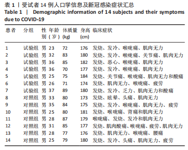
[70] GOODPASTER BH, CARLSON CL, VISSER M, et al. Attenuation of skeletal muscle and strength in the elderly: the Health ABC Study. J Appl Physiol. 2001;90(6):2157-2165. |