| [1] Kielbassa AM, Muller J, Gernhardt CR. Closing the gap between oral hygiene and minimally invasive dentistry: a review on the resin infiltration technique of incipient (proximal) enamel lesions. Quintessence Int. 2009;40(8):663-681.
[2] 樊明文.牙体牙髓病学[M].4版.北京:人民卫生出版社,2012.
[3] 岳松龄.现代龋病学[M].北京:科学技术文献出版社,2009.
[4] Borges BC, de Souza Borges J, de Araujo LS, et al. Update on nonsurgical, ultraconservative approaches to treat effectively non-cavitated caries lesions in permanent teeth. Eur J Dent. 2011;5(2):229-236.
[5] Brazzelli M, McKenzie L, Fielding S, et al. Systematic review of the effectiveness and cost-effectiveness of HealOzone for the treatment of occlusal pit/fissure caries and root caries. Health Technol Assess. 2006;10(16):iii-iv, ix-80.
[6] National Institutes of Health (U.S.). Diagnosis and management of dental caries throughout life. NIH Consens Statement. 2001;18(1):1-23.
[7] Dalli M, Çolak H, Mustafa Hamidi M. Minimal intervention concept: a new paradigm for operative dentistry. J Investig Clin Dent. 2012;3(3):167-175.
[8] Anusavice KJ. Present and future approaches for the control of caries. J Dent Educ. 2005;69(5):538-554.
[9] Simonsen RJ. Preventive resin restorations and sealants in light of current evidence. Dent Clin North Am. 2005;49(4): 815-823, vii.
[10] Reynolds EC. Calcium phosphate-based remineralization systems: scientific evidence? Aust Dent J. 2008;53(3): 268-273.
[11] Tyas MJ, Anusavice KJ, Frencken JE, et al. Minimal intervention dentistry--a review. FDI Commission Project 1-97. Int Dent J. 2000;50(1):1-12.
[12] Cochrane NJ, Cai F, Huq NL, et al. New approaches to enhanced remineralization of tooth enamel. J Dent Res. 2010; 89(11):1187-1197.
[13] Stahl J, Zandona AF. Rationale and protocol for the treatment of non-cavitated smooth surface carious lesions. Gen Dent. 2007;55(2):105-111.
[14] Buczkowska-Radlińska J. Factors that modify de- and remineralization in dental enamel from the aspect of caries susceptibility. Ann Acad Med Stetin. 1999;Suppl 47:1-89.
[15] Kim S, Kim EY, Jeong TS, et al. The evaluation of resin infiltration for masking labial enamel white spot lesions. Int J Paediatr Dent. 2011;21(4):241-248.
[16] 吕雪芹,朱玲.早期平滑面龋再矿化初探[J].口腔医学研究,2007, 23(5):585-587.
[17] Ekstrand KR, Bakhshandeh A, Martignon S. Treatment of proximal superficial caries lesions on primary molar teeth with resin infiltration and fluoride varnish versus fluoride varnish only: efficacy after 1 year. Caries Res. 2010;44(1):41-46.
[18] Rocha Gomes Torres C, Borges AB, Torres LM, et al. Effect of caries infiltration technique and fluoride therapy on the colour masking of white spot lesions. J Dent. 2011;39(3):202-207.
[19] 金燕,惠建华,矢小萍.3种溶液对早期釉质龋再矿化作用的扫描电镜观察[J].口腔医学,2009,29(11):592-594.
[20] Paris S, Meyer-Lueckel H, Mueller J, et al. Progression of sealed initial bovine enamel lesions under demineralizing conditions in vitro. Caries Res. 2006;40(2):124-129.
[21] Anttonen V, Seppä L, Hausen H. A follow-up study of the use of DIAGNOdent for monitoring fissure caries in children. Community Dent Oral Epidemiol. 2004;32(4):312-318.
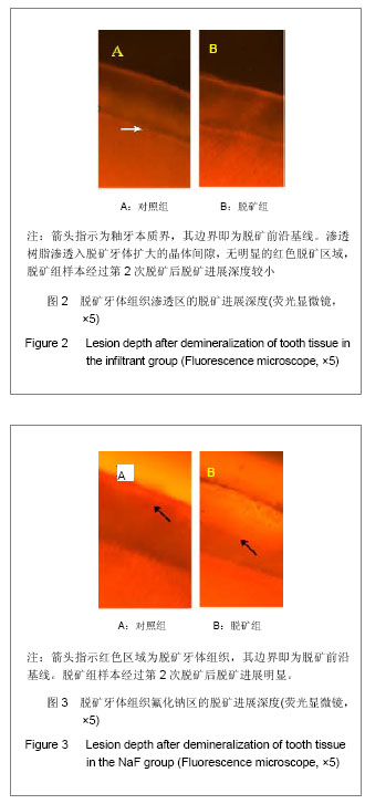
[22] 楚金普,郝玉庆,李继遥,等.两种脱矿体系制备人工龋损的病理学比较[J].中国比较医学杂志,2008,18(1):23-26.
[23] Amjad Z, Nancollas GH. Effect of fluoride on the growth of hydroxyapatite and human dental enamel. Caries Res. 1979; 13(5):250-258.
[24] Vogel GL, Mao Y, Chow LC, et al. Fluoride in plaque fluid, plaque, and saliva measured for 2 hours after a sodium fluoride monofluorophosphate rinse. Caries Res. 2000; 34(5):404-411.
[25] García-Godoy F, Hicks MJ. Maintaining the integrity of the enamel surface: the role of dental biofilm, saliva and preventive agents in enamel demineralization and remineralization. J Am Dent Assoc. 2008;139 Suppl:25S-34S.
[26] Kantovitz KR, Pascon FM, Nobre-dos-Santos M, et al. Review of the effects of infiltrants and sealers on non-cavitated enamel lesions. Oral Health Prev Dent. 2010; 8(3):295-305.
[27] Paris S, Meyer-Lueckel H, Cölfen H, et al. Penetration coefficients of commercially available and experimental composites intended to infiltrate enamel carious lesions. Dent Mater. 2007;23(6):742-748.
[28] Giachetti L, Bertini F, Bambi C, et al. A rational use of dental materials in posterior direct resin restorations in order to control polymerization shrinkage stress. Minerva Stomatol. 2007;56(3):129-138.
[29] Giachetti L, Scaminaci Russo D, Bambi C, et al. A review of polymerization shrinkage stress: current techniques for posterior direct resin restorations. J Contemp Dent Pract. 2006;7(4):79-88.
[30] Schmidlin PR, Zehnder M, Pasqualetti T, et al. Penetration of a bonding agent into de- and remineralized enamel in vitro.J Adhes Dent.2004;6(2):111-115
[31] Paris S, Bitter K, Naumann M, et al. Resin infiltration of proximal caries lesions differing in ICDAS codes. Eur J Oral Sci. 2011;119(2):182-186.
[32] Paris S, Meyer-Lueckel H, Cölfen H, et al. Resin infiltration of artificial enamel caries lesions with experimental light curing resins. Dent Mater J. 2007;26(4):582-588.
[33] Meyer-Lueckel H, Paris S. Infiltration of natural caries lesions with experimental resins differing in penetration coefficients and ethanol addition. Caries Res. 2010;44(4):408-414.
[34] Paris S, Meyer-Lueckel H. Inhibition of caries progression by resin infiltration in situ. Caries Res. 2010;44(1):47-54.
[35] Paris S, Hopfenmuller W, Meyer-Lueckel H. Resin infiltration of caries lesions: an efficacy randomized trial. J Dent Res. 2010;89(8):823-826.
[36] Wiegand A, Stawarczyk B, Kolakovic M, et al. Adhesive performance of a caries infiltrant on sound and demineralised enamel. J Dent.2011;39(2):117-121.
[37] Mueller J, Yang F, Neumann K, et al. Surface tridimensional topography analysis of materials and finishing procedures after resinous infiltration of subsurface bovine enamel lesions. Quintessence Int. 2011;42(2):135-147.
[38] 王芸,唐荣银.再矿化药物的研究进展[J].牙体牙髓牙周病学杂志,2005,15(9):523-525. |
.jpg)
.jpg)