Chinese Journal of Tissue Engineering Research ›› 2014, Vol. 18 ›› Issue (7): 1045-1050.doi: 10.3969/j.issn.2095-4344.2014.07.011
Previous Articles Next Articles
Recombinant human epidermal growth factor and basic fibroblast growth factor promote the proliferation of human corneal epithelial cells
Peng Yan-yang, Wu Wei, Zeng Li-na, Chen Xiao-hong, Lu Xiao-he
- Department of Ophthalmology, Zhujiang Hospital of Southern Medical University, Guangzhou 510282, Guangdong Province, China
-
Revised:2014-01-11Online:2014-02-12Published:2014-02-12 -
Contact:Lu Xiao-he, M.D., Doctoral supervisor, Professor, Department of Ophthalmology, Zhujiang Hospital of Southern Medical University, Guangzhou 510282, Guangdong Province, China -
About author:Peng Yan-yang, Studying for master’s degree, Department of Ophthalmology, Zhujiang Hospital of Southern Medical University, Guangzhou 510282, Guangdong Province, China
CLC Number:
Cite this article
Peng Yan-yang, Wu Wei, Zeng Li-na, Chen Xiao-hong, Lu Xiao-he . Recombinant human epidermal growth factor and basic fibroblast growth factor promote the proliferation of human corneal epithelial cells[J]. Chinese Journal of Tissue Engineering Research, 2014, 18(7): 1045-1050.
share this article

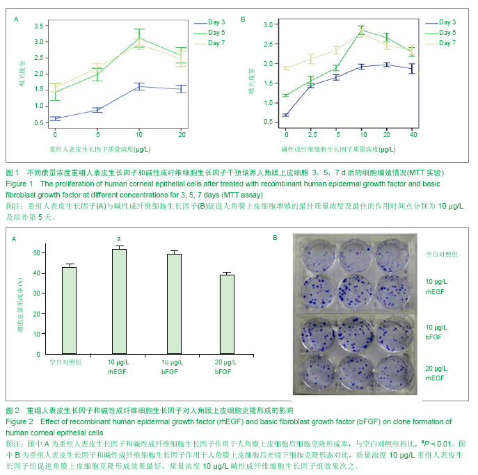
2.1 MTT细胞增殖实验结果 重组人表皮生长因子与碱性成纤维细胞生长因子促进人角膜上皮细胞增殖的最佳质量浓度及最佳的作用时间点分别为10 μg/L及培养第5天(图1)。质量浓度10 μg/L 重组人表皮生长因子和质量浓度10 μg/L 碱性成纤维细胞生长因子促进人角膜上皮细胞增殖能力均强于较空白对照组(P < 0.05)。 2.2 重组人表皮生长因子和碱性成纤维细胞生长因子对人角膜上皮细胞克隆形成的影响 质量浓度10 μg/L 重组人表皮生长因子,10 μg/L碱性成纤维细胞生长因子, 20 μg/L碱性成纤维细胞生长因子和空白对照组的Mean rank分别为10.67,7.83,2.67,4.83。经Kruskal-Wallis H检验,质量浓度10 μg/L重组人表皮生长因子组人角膜上皮细胞克隆数高于空白对照组(P < 0.01)。质量浓度10 μg/L重组人表皮生长因子组人角膜上皮细胞克隆形成率高于 10 μg/L碱性成纤维细胞生长因子组(P=0.02,图2)。结果提示,质量浓度10 μg/L重组人表皮生长因子组促进角膜上皮细胞克隆形成效果最好,质量浓度10 μg/L碱性成纤维细胞生长因子组效果次之。"
| [1] Williams KA, Irani YD, Klebe S. Novel therapeutic approaches for corneal disease. Discov Med. 2013;15(84):291-299.[2] Huo Y, Qiu WY, Pan Q, et al. Reactive oxygen species (ROS) are essential mediators in epidermal growth factor (EGF)-stimulated corneal epithelial cell proliferation, adhesion, migration, and wound healing. Exp Eye Res. 2009;89(6): 876-886.[3] Li H, Miao J, Zhao G, et al. Different expression patterns of growth factors in rat fetuses with spina bifida aperta after in utero mesenchymal stromal cell transplantation. Cytotherapy. 2013. [4] Suzuki K, Saito J, Yanai R, et al. Cell-matrix and cell-cell interactions during corneal epithelial wound healing. Prog Retin Eye Res. 2003;22(2):113-133. [5] Scholl S, Kirchhof J, Augustin A J. Antivascular endothelial growth factors in anterior segment diseases. Dev Ophthalmol. 2010;46:133-139.[6] Lyu J, Joo CK. Wnt-7a up-regulates matrix metalloproteinase-12 expression and promotes cell proliferation in corneal epithelial cells during wound healing. J Biol Chem. 2005;280(22):21653-21660.[7] Klenkler B, Sheardown H. Growth factors in the anterior segment: role in tissue maintenance, wound healing and ocular pathology. Exp Eye Res. 2004;79(5):677-688.[8] Yoshioka R, Shiraishi A, Kobayashi T, et al. Corneal epithelial wound healing impaired in keratinocyte-specific HB-EGF-deficient mice in vivo and in vitro. Invest Ophthalmol Vis Sci. 2010;51(11):5630-5639.[9] Hecquet C, Morisset S, Lorans G, et al. Effects of acidic and basic fibroblast growth factors on the proliferation of rabbit corneal cells. Curr Eye Res. 1990;9(5):429-433.[10] Bremnes RM, Camps C, Sirera R. Angiogenesis in non-small cell lung cancer: the prognostic impact of neoangiogenesis and the cytokines VEGF and bFGF in tumours and blood. Lung Cancer. 2006;51(2):143-158.[11] Liu J, Song G, Wang Z, et al. Establishment of a corneal epithelial cell line spontaneously derived from human limbal cells. Exp Eye Res. 2007;84(3):599-609.[12] Stockert J C, Blazquez-Castro A, Canete M, et al. MTT assay for cell viability: Intracellular localization of the formazan product is in lipid droplets. Acta Histochem. 2012;114(8):785-796.[13] Bennett NT, Schultz GS. Growth factors and wound healing: Part II. Role in normal and chronic wound healing. Am J Surg. 1993;166(1):74-81.[14] Thalmann-Goetsch A, Engelmann K, Bednarz J. Comparative study on the effects of different growth factors on migration of bovine corneal endothelial cells during wound healing. Acta Ophthalmol Scand. 1997;75(5):490-495.[15] Brown AC, Adams D, de Caestecker M, et al. FGF/EGF signaling regulates the renewal of early nephron progenitors during embryonic development. Development. 2011;138(23): 5099-5112.[16] Mie M, Sasaki S, Kobatake E. Construction of a bFGF-tethered multi-functional extracellular matrix protein through coiled-coil structures for neurite outgrowth induction. Biomed Mater. 2013;9(1):015004.[17] Zhou W, Zhao M, Zhao Y, et al. A fibrin gel loaded with chitosan nanoparticles for local delivery of rhEGF: preparation and in vitro release studies. J Mater Sci Mater Med. 2011; 22(5): 1221-1230.[18] Yanai R, Yamada N, Inui M, et al. Correlation of proliferative and anti-apoptotic effects of HGF, insulin, IGF-1, IGF-2, and EGF in SV40-transformed human corneal epithelial cells. Exp Eye Res. 2006;83(1):76-83. [19] Liu Z, Carvajal M, Carraway CA, et al. Expression of the receptor tyrosine kinases, epidermal growth factor receptor, ErbB2, and ErbB3, in human ocular surface epithelia. Cornea. 2001;20(1):81-85.[20] Wilson SE, He YG, Weng J, et al. Effect of epidermal growth factor, hepatocyte growth factor, and keratinocyte growth factor, on proliferation, motility and differentiation of human corneal epithelial cells. Exp Eye Res. 1994;59(6):665-678.[21] Stojanovic A, Chen X, Jin N, et al. Safety and efficacy of epithelium-on corneal collagen cross-linking using a multifactorial approach to achieve proper stromal riboflavin saturation. J Ophthalmol. 2012;2012:498435.[22] Gospodarowicz D, Ferrara N, Schweigerer L, et al. Structural characterization and biological functions of fibroblast growth factor. Endocr Rev. 1987;8(2):95-114.[23] 傅小兵,沈祖尧,陈玉林,等.碱性成纤维细胞生长因子与创面修 复-1024例多中心对照临床试验结果[J].中国修复重建外科杂志,1998,(4):22-24.[24] Gainza G, Aguirre J J, Pedraz JL, et al. rhEGF-loaded PLGA-Alginate microspheres enhance the healing of full-thickness excisional wounds in diabetised Wistar rats. Eur J Pharm Sci. 2013;50(3-4):243-252.[25] Wang Z, Zhong H, Yang Z, et al. Exogenous bFGF or TGFbeta accelerates healing of reconstructed dura by CO laser soldering in minipigs. Lasers Med Sci. 2013.[26] Xing B, Wu F, Li T, et al. Experimental study of comparing rhEGF with rhbetaFGF on improving the quality of wound healing. Int J Clin Exp Med. 2013;6(8):655-661.[27] Han KY, Fahd DC, Tshionyi M, et al. MT1-MMP modulates bFGF-induced VEGF-A expression in corneal fibroblasts. Protein Pept Lett. 2012;19(12):1334-1339.[28] Vadlapudi AD, Vadlapatla RK, Pal D, et al. Functional and molecular aspects of biotin uptake via SMVT in human corneal epithelial (HCEC) and retinal pigment epithelial (D407) cells. AAPS J. 2012;14(4):832-842.[29] Chandrasekher G, Pothula S, Maharaj G, et al. Differential effects of hepatocyte growth factor and keratinocyte growth factor on corneal epithelial cell cycle protein expression, cell survival, and growth. Mol Vis. 2014;20:24.[30] Wright B, Hopkinson A, Leyland M, et al. The secretome of alginate-encapsulated limbal epithelial stem cells modulates corneal epithelial cell proliferation. PLoS One. 2013;8(7): e70860.[31] Kitazawa K, Kawasaki S, Shinomiya K, et al. Establishment of a human corneal epithelial cell line lacking the functional TACSTD2 gene as an in vitro model for gelatinous drop-like dystrophy. Invest Ophthalmol Vis Sci. 2013;54(8):5701-5711.[32] Liu J, Lawrence BD, Liu A, et al. Silk fibroin as a biomaterial substrate for corneal epithelial cell sheet generation. Invest Ophthalmol Vis Sci. 2012;53(7):4130-4138.[33] Ghaffarieh A, Ghaffarpasand F, Dehghankhalili M, et al. Effect of transcutaneous electrical stimulation on rabbit corneal epithelial cell migration. Cornea. 2012;31(5): 559-563.[34] Wang Z, Bildin VN, Yang H, et al. Dependence of corneal epithelial cell proliferation on modulation of interactions between ERK1/2 and NKCC1. Cell Physiol Biochem. 2011; 28(4):703-714.[35] Tocce EJ, Broderick AH, Murphy KC, et al. Functionalization of reactive polymer multilayers with RGD and an antifouling motif: RGD density provides control over human corneal epithelial cell-substrate interactions. J Biomed Mater Res A. 2012;100(1):84-93.[36] Okada N, Kawakita T, Mishima K, et al. Clusterin promotes corneal epithelial cell growth through upregulation of hepatocyte growth factor by mesenchymal cells in vitro. Invest Ophthalmol Vis Sci. 2011;52(6): 2905-2910.[37] Ding L, Gao LJ, Gu PQ, et al. The role of eIF5A in epidermal growth factor-induced proliferation of corneal epithelial cell association with PI3-k/Akt activation.Mol Vis. 2011;17:16-22.[38] Fan TJ, Xu B, Zhao J, et al. Establishment of an untransfected human corneal epithelial cell line and its biocompatibility with denuded amniotic membrane. Int J Ophthalmol. 2011;4(3):228-234.[39] Imanishi J, Kamiyama K, Iguchi I, et al. Growth factors: importance in wound healing and maintenance of transparency of the cornea. Prog Retin Eye Res. 2000;19(1): 113-129.[40] Declair V. The importance of growth factors in wound healing. Ostomy Wound Manage.1999;45(4):64-74.[41] 班胜刚,李永均,卢荣强,等.重组人表皮生长因子治疗外伤性角膜上皮缺损[J].眼外伤职业眼病杂志,2005,(1):26-27. |
| [1] |
Xiang Haidong, Cheng Dongmei, Guo Han, Gao Qi .
Changes in the proliferation and angiogenesis of human dental pulp stem cells after treated with prostaglandin E1 combined with basic fibroblast growth factor [J]. Chinese Journal of Tissue Engineering Research, 2020, 24(25): 4006-4011. |
| [2] | Yang Fan, Liu Baoyi, Cao Meng, Zhu Xiaoshu, Zhang Yu, Qin Kairong, Zhao Dewei. Basic fibroblast growth factors protect chondrocytes by antagonizing extracellular inflammatory factors [J]. Chinese Journal of Tissue Engineering Research, 2020, 24(23): 3621-3626. |
| [3] | Liao Jianzhao, Zhang Xiaoyun, Zhang Xuan. Molecular signaling pathways in the occurrence and development of osteoarthritis [J]. Chinese Journal of Tissue Engineering Research, 2020, 24(21): 3394-3400. |
| [4] | Wang Lu, Yu Liang. Fibroblast growth factor 21 regulates browning of white fat: the role of exercise [J]. Chinese Journal of Tissue Engineering Research, 2020, 24(20): 3275-3280. |
| [5] | Li Hong, Wu Shaohua, Qiu Mingxing, Deng Qingfu, Lei Guolin, Li Qinglong. Mechanism underlying basic fibroblast growth factor and insulin-like growth factor-1 effects on proliferation and apoptosis of spermatogonial stem cells [J]. Chinese Journal of Tissue Engineering Research, 2020, 24(19): 3017-3022. |
| [6] | Qiu Xiaoyang, Wang Yuanyuan, Liu Chunpeng, Chen Hongcai, Wu Xuan, Zhan Xiaofen. Application of environment-friendly bio-tissue sample preparation kit in fluorescence in situ hybridization detection of HER2 protein 2-positive invasive breast cancer [J]. Chinese Journal of Tissue Engineering Research, 2020, 24(16): 2572-2577. |
| [7] | Liu Xing, Wei Xiaohan, Deng Jie, Li Zhongming. Resveratrol repairs skeletal muscle injury by up-regulating basic fibroblast growth factor and insulin-like growth factor 1 [J]. Chinese Journal of Tissue Engineering Research, 2020, 24(14): 2184-2191. |
| [8] | Zhang Wenjing, Wang Jia, Tian Mengting, Han Xiangzhen, Zhou Qiqi, He Huiyu. Effects of bone morphogenetic protein 2 and basic fibroblast growth factor 2 on proliferation and osteogenic differentiation of rat bone marrow mesenchymal stem cell sheet [J]. Chinese Journal of Tissue Engineering Research, 2020, 24(1): 65-71. |
| [9] | Lin Min, Wei Weili, He Yimi, Chen Zhikui. Construction of a gene-loaded multifunctional polymer microbubble targeting breast cancer cells [J]. Chinese Journal of Tissue Engineering Research, 2019, 23(34): 5468-5472. |
| [10] | Liu Jiajia, Li Ping, Deng Qi, Liu Huijuan. Overexpression of heparin-binding epidermal growth factor-like growth factor in Prrx1+ bone marrow mesenchymal stem cells inhibits mouse skeletal development [J]. Chinese Journal of Tissue Engineering Research, 2019, 23(29): 4623-4628. |
| [11] | Sang Peng, Liu Yi. Scleraxis combined with basic fibroblast growth factor promotes the differentiation of human amniotic mesenchymal stem cells into human ligament fibroblasts in vitro [J]. Chinese Journal of Tissue Engineering Research, 2019, 23(29): 4644-4650. |
| [12] | Zhu Xu, Shen Jun, Yang Rongkun, Wang Yong, Liu Yang, Sun Quan, He Zhuying, Chen Jiuyi. Mechanism of traditional Chinese medicine Shixiang plaster in promoting the repair of soft tissue defects [J]. Chinese Journal of Tissue Engineering Research, 2019, 23(27): 4350-4355. |
| [13] | Wang Tong, Liu Yang, Zhu Yetao. Combined use of exogenous nerve growth factor and basic fibroblast growth factor promotes proliferation of endogenous brain cells in a rat model of severe traumatic brain injury [J]. Chinese Journal of Tissue Engineering Research, 2019, 23(25): 3998-4003. |
| [14] | Wu Huala, Zhou Xiangxiang, Zhong Yulan, Gan Xin. Basic fibroblast growth factor-transfected bone marrow mesenchymal stem cell transplantation for chronic obstructive pulmonary disease [J]. Chinese Journal of Tissue Engineering Research, 2019, 23(21): 3378-3385. |
| [15] | Cui Tingting, Qiu Zewen, Gui Lin, Zhong Weijian, Ma Guowu. Effect of gelatin sponge/beta-tricalcium phosphate composite with different doses of basic fibroblast growth factor on bone regeneration [J]. Chinese Journal of Tissue Engineering Research, 2019, 23(2): 190-195. |
| Viewed | ||||||
|
Full text |
|
|||||
|
Abstract |
|
|||||