Chinese Journal of Tissue Engineering Research
Previous Articles Next Articles
Insight into bone tissue engineering scaffold materials and their vascularization
Zhao Tian-yuan, Sun Hong
- Department of Pathology, North China Coal Medical University, Tangshan 063100, Hebei Province, China
-
Received:2013-03-11Revised:2013-03-18Online:2013-09-17Published:2013-09-17 -
Contact:Sun Hong, Professor, Department of Pathology, North China Coal Medical University, Tangshan 063100, Hebei Province, China 1767471161@qq.com -
About author:Zhao Tian-yuan★, Studying for master’s degree, Department of Pathology, North China Coal Medical University, Tangshan 063100, Hebei Province, China 15081919626@163.com -
Supported by:the National Natural Science Foundation of China, No. 81101448*; the Natural Science Foundation of Hebei Province, No. C2011401006*
CLC Number:
Cite this article
Zhao Tian-yuan, Sun Hong. Insight into bone tissue engineering scaffold materials and their vascularization[J]. Chinese Journal of Tissue Engineering Research, doi: 10.3969/j.issn.2095-4344.2013.38.020.
share this article
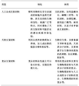
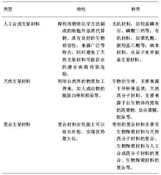
2.1 种子细胞 利用骨组织工程技术修复缺损的骨组织,就需要由种子细胞生长发育来实现。理想的种子细胞应是具有成骨潜能的细胞,需满足以下要求:①细胞易于获取且安全可靠。②体外培养时生长活跃,具备较强的传代能力,可以得到足够数量的细胞并可定向分化为成骨细胞。③能够适应体外和体内的培养环境且保持成骨活性[2]。④成骨迅速,无致瘤性倾向。 目前种子细胞主要有以下几个来源:骨髓间充质干细胞、骨膜成骨细胞、脂肪干细胞、胚胎干细胞等。 2.1.1 骨髓间充质干细胞 众多实验者广泛采用骨髓间充质干细胞进行研究。间充质干细胞是最有应用前景的种子细胞,其来源广泛,易于获取,具备快速增殖的能力,短时间即可得到大量细胞,并且可以根据实验的需要诱导分化成不同的细胞[3],如体外培养时,在培养基中添加地塞米松、β-甘油磷酸钠等诱导剂即可向成骨细胞转化。袁捷等[4]利用自体诱导的骨髓间充质干细胞成功修复了犬下颌骨骨缺损。但也有研究表明,体外培养间充质干细胞传代最大值为24-40代,以后细胞则会出现衰老和生长抑制现象[5]。 2.1.2 脂肪来源和脐血来源干细胞 作为骨组织工程的种子细胞,脂肪来源和脐血来源干细胞[6]:①来源广泛,且获取时造成的创伤更小。②具有更强的增殖能力。鞠秀丽等[7]将脐带血间充质干细胞成功诱导分化为骨细胞。Zuk等[8]在人体脂肪组织中成功分离出具有多能干细胞的脂肪细胞,这种细胞在体外经诱导可向脂肪细胞、成骨细胞、软骨细胞等分化。但是脂肪来源和脐带血来源干细胞也有自身不能忽视的不足:脂肪来源和脐带血来源干细胞属于成体干细胞范畴,因此就决定了其不具备无限增殖和分化潜能。再者在体外长时间培养的脂肪来源干细胞有自发向恶性转化的倾向,因此应用时必须要更加谨慎的考虑其安全性。也越来越得到研究者的关注。两者有着优于骨髓间充质干细胞的特点 2.1.3 胚胎干细胞 胚胎干细胞具有全能分化的能力,是胚胎发育早期囊胚内细胞团中尚未分化的细胞。其作为组织工程种子细胞的优势为:①能够长期保持未分化性增殖。②能够诱导出内、中、外胚层的所有细胞系。Zuk等[8]成功应用添加β-甘油磷酸、地塞米松、维生素C的培养液将胚胎干细胞诱导成为成骨细胞。但现今骨组织工程研究的胚胎干细胞均是异体来源,其在体外可以近乎无限增殖,因此达到了种子细胞高增殖潜能的要求,但是其免疫排斥反应却是无法解决的难题。再者胚胎干细胞涉及伦理道德问题,因此来源受到了极大的限制。 就目前研究形势来看,常用种子细胞不能同时具备高增殖能力和低免疫原性这两大要求。但值得肯定的是,随着细胞培养技术水平的不断提高,各种生长因子的广泛深入研究,以及通过基因技术改变细胞性状使得机体免疫耐受等方法的应用,使得种子细胞能够在体外大量增殖且又不具备免疫原性终将成为可能。与此同时,更加适合的种子细胞来源仍需要人们更多的研究和探索。 2.2 生长因子 诱导成骨的生长因子在骨组织工程技术中发挥着重要的调节作用,它参与着细胞的整个生命活动,如细胞分裂、基质合成、组织分化等。目前已发现的具备诱导成骨作用的生长因子主要包括骨形态发生蛋白、胰岛素样生长因子、转化生长因子β、血小板衍生生长因子、血管内皮细胞生长因子等。 2.2.1 骨形态发生蛋白 骨形态发生蛋白主要来源于成骨细胞,是正常胚胎期牙、骨组织及成年后骨修复的关键诱导因子。它也是目前已知的惟一一种可以促使间充质干细胞向骨或软骨细胞分化的细胞因子。在诱导成骨方面骨形态发生蛋白所起到的作用主要是增强碱性磷酸酶活性,促使mRNA和骨钙素表达水平提高,促进钙在细胞外基质的沉着。Friedlaender等[9]利用骨形态发生蛋白7成功在人体治疗122例顽固性胫骨骨不连病例。为了让骨形态发生蛋白更好地发挥成骨作用,研究者把焦点聚焦于在局部稳定持续释放骨形态发生蛋白,这样能够不断地促进种子细胞向成骨方面分化。基因转染技术可利用载体将骨形态发生蛋白基因转到至种子细胞,使二者基因整合,达到使种子细胞高效稳定持续表达骨形态发生蛋白的目的。 2.2.2 转化生长因子β 转化生长因子β为多功能的多肽生长因子,对骨组织、免疫系统和结缔组织等多种细胞均有一定程度的调节作用。其主要功能是:促进细胞合成DNA,刺激软骨形成和软骨内成骨,且能够调控成骨细胞的游走和聚集。与骨形态发生蛋白有一定的协调作用。但是转化生长因子β存在两面性,表现为低浓度促进细胞增殖,高浓度抑制细胞生长。 2.2.3 血小板衍生生长因子 血小板衍生生长因子是广泛存在于骨组织中的一类细胞因子。血小板衍生生长因子的主要特点是促进多种细胞增殖和胶原蛋白的形成。Lee等[10]构建血小板衍生生长因子和支架材料复合物用以修复兔颅骨缺损,研究表明缺损处骨修复速度明显加快。但是在体外长期应用血小板衍生生长因子却能抑制成骨细胞的分化,主要表现为降低碱性磷酸酶活性和骨钙素的表达,以及抑制钙化结节的合成等。 生长因子是修复骨缺损的必备要素,但是生长因子不能单一存在,必须同作为载体的支架材料相结合才能发挥其应有的功效,因此将生长因子和适宜的支架材料结合起来能更好地促进种子细胞的生长和定向分化。 2.3 支架材料 目前支架材料的选择已成为骨组织工程学的研究焦点之一。骨组织工程对于支架材料有诸多的特性要求。理想的支架材料应具备的特性包 括[11]:①良好的生物相容性。材料本身及其降解产物应无不良反应,不引起炎症反应,且要有利于细胞的黏附、增殖、生长和分化。②良好的生物降解性。当支架材料的支架作用完成后,其应该能被人体降解并被人体吸收。支架的降解速度应与组织细胞的生长速度保持动态平衡。③具有三维立体多孔结构。一般要求支架材料具有一定的孔隙率,孔隙率应达 90% 以上,以利于营养和氧气的吸收和代谢产物的排出,以及神经和血管的生长。④良好的材料-细胞界面。材料要为细胞的黏附和增殖提供良好的界面,以促进细胞的生长,特别是能激活细胞特异基因,保证正常细胞表型的表达。⑤可塑性和一定的机械强度。支架材料应能预先加工成与缺损区相匹配的形态结构,并能够为新生组织提供良好的机械支撑。⑥支架材料易于消毒、保存,且消毒后支架材料的内部结构完整无破坏。 目前按照材料的来源可将支架材料分为人工合成支架材料和天然支架材料两大类。 2.3.1 人工合成的支架材料 即利用物理化学方法制成的细胞外基质代替物,具有良好的生物相容性,来源广泛等特点,同时避免了天然支架材料可能存在的潜在疾病传染危险。①无机材料:如羟基磷灰石、磷酸三钙等。羟基磷灰石同骨的无机成分相近,因此具有良好的生物相容性和骨传导性;但其存在韧性差、难降解且单一成分无法塑型等缺点。Morishita等[12]将骨髓间充质干细胞接种于羟基磷灰石构成的支架材料上,6个月后骨缺损处得以修复。②有机材料:如聚乳酸、据羟基乙酸等。这类材料具有良好的生物相容性且易于塑型,但机械强度不足,降解产物为酸性,易引起炎症反应和免疫反应,因此应用受到限制。Meyer等[13]将猪自体骨植入聚乳酸乙醇酸支架材料中复合培养后,再植回猪下颌骨缺损处,缺损处得以修复。③纳米材料:从原子水平制备支架材料,具备以下特点:支架材料的结构单元在纳米数量级;各个结构单元的相互作用力或强或弱;支架材料的界面多。随着技术的发展,纳米材料已被广泛应用起来。姚晖等[14]制备出纳米尺寸的羟基磷灰石/胶原复合人工骨材料,并已成功修复家兔颅颌缺损。 2.3.2 天然支架材料 利用自然界的物质加工得来,如人或动物的脱蛋白骨和胶原等。①生物衍生骨:主要来源于异种骨基质。其来源广泛,易于获取,且具有良好的生物相容性、生物降解性及和正常骨组织接近的三维孔道系统生理构造。骨基质的网状结构利于种子细胞的黏附和增殖,并为种子细胞提供生长的内部空间和表面微环境。但其免疫原性仍是难以克服的难题。②天然高分子材料:主要来源于自生物体内提取的高聚物,如壳聚糖、胶原等。壳聚糖自然界惟一一种带正电荷的碱性多糖,具有无毒性、无刺激性、良好的生物相容性、良好的生物可吸收性、优异的药物活性、可降解性等优点[15],是较为理想的细胞外基质材料。有资料显示,由于壳聚糖本身含有大量的氨基、羟基等基团,化学性质活泼,能与成骨细胞膜负电成分呈现非特异相互作用,并可调控生物活性分子释放,具有促进前成骨细胞的分化、加速骨形成的作用,并且在体内可被降解为氨基葡萄糖,被人体吸 收[16-17]。另外壳聚糖具有生物功能性亲水表面,能够促进血管内皮细胞的生长,还具有抗肿瘤活性、消炎、杀菌等特性,而且对于受损机体能诱生特殊细胞,促进缺损部分愈合[18]。但壳聚糖单独作为支架材料也有一定的局限性,如机械强度差、不稳固,只能在酸性溶液中制备等,所以目前多将壳聚糖与其他材料复合制备支架材料。Cruz等[19]将山羊的骨髓间充质干细胞种植于壳聚糖支架材料上,细胞生长增殖良好,并向成骨细胞方向分化。 2.3.3 复合材料 大量实验和临床研究表明单一类型的支架材料不能满足修复骨组织缺损的需要,因此就需要利用先进的手段将两种或多种不同材料结合起来形成复合材料以满足实际需求。复合材料在性能上可以取长补短,实现优势最大化。常用的复合材料主要有生物陶瓷材料与天然高分子材料的复合、生物陶瓷材料与人工合成高分子材料的复合、生物陶瓷材料的复合等。Chen等[20]将聚羟基乙酸和聚乳酸形成的共聚物浸入胶原溶液中,形成的复合材料在亲水性和细胞黏附性方面都有所提高。 骨组织工程支架材料:"
| [1] Cancedda R, Giannoni P, Mastrogiacomo M. A tissue engineering approach to bone repair in large animal models and in clinical practice.Biomaterials. 2007;28(29): 4240-4250. [2] Reddi AH. Morphogenesis and tissue engineering of bone and carti-lage: inductive signals, stem cells, and biomaterials. Tissue Eng.2000;6(4): 351-359. [3] Kassem M.Stem cells: potential therapy for age-related diseases.Ann N Y Acad Sci.2006;1067:436-442. [4] 袁捷,祝联,王敏,等. 自体骨髓基质干细胞-珊瑚羟基磷灰石修复下颌骨节段缺损的实验研究[J].中华口腔医学杂志,2006,41(2): 94-97. [5] Banfi A,Muraglia A,Dozin B,et al.Proliferation kinetics and differentiation potential of ex vivo expanded human bone marrow stromal cells: Implications for their use in cell therapy. Exp Hematol.2001;28(6):707-715. [6] Kern S,Eichler H,Stoeve J,et al.Comparative analysis of mesenchymal stem cells from bone marrow, umbilical cord blood, or adipose tissue.Stem Cells. 2006;24(5): 1294-1301. [7] 鞠秀丽,黄志伟,时庆,等. 体外扩增脐血间充质干细胞的生物学特性和诱导分化能力的研究[J].中华儿科杂志,2005,43(7): 499-502. [8] Zuk PA,Zhu M,Mizuno H,et al. Multilineage cells from human adipose tissue: implications for cell-based therapies.Tissue Eng.2001;7(2):211. [9] Friedlaender GE,Perry CR,Cole JD,et al.Osteogenic protein-1 (bone morphogenetic protein-7) in the treatment of tibial nonunions.J Bone Joint Surg Am.2001;83-A Suppl 1(Pt 2): S151-S158. [10] Lee SJ,Park YJ,Park SN,et al. Molded porous poly (L- lactide) membranes for guided bone regeneration with enhanced effects by controlled growth factor release.J Bined Mater Res. 2001;55(3):295-303. [11] Lacroix D,Chateau A,Ginebra MP,et al.Microrfinite elementmodel of bone tissue engineeting scaffolds. Biomaterials. 2006;27(30):5326-5334. [12] Morishita T,Honoki K,Ohgushi H,et al. Tissue engineering approach to the treatment of bone tumors: three cases of cells.Artif Organs.2006;30(2):115-118. [13] Meyer U,Buchter A,Hohoff A,et al.Image-based extracorporeal tissue engineering of individualized bone constructs.Int J Oral Maxillofac Implants.2005;20(6): 882-890. [14] 姚晖,杜昶,杨韶华,等.纳米羟晶/胶原仿生骨修复家兔颅颌骨缺损的实验研究[J].透析与人工器官,2000,11(2):5-8. [15] 丁珊,李立华,周长忍.PLA/液晶复合膜的制备及其性能研究[J].生物医学工程学杂志,2002,19(3):383-385. [16] Chen RN,Wang GM,Chen CH,et al.Development of N, O-(carboxymethyl)chitosan/collagen matrixes as a wound dressing.Biomacromolecules.2006;7(4):1058-1064. [17] Wittaya AS,Prahsarn C.Development and in vitro evaluation of chitosan-polysaccharides composite wound dressings.Int J Pharm.2006;313(1-2):123-128. [18] 章文苑,旅娅婧.纳米羟基磷灰石/Ⅰ型胶原/壳聚糖复合支架材料的制备与优化[J].生物骨科材料与临床研究,2011,8(3):1-4. [19] Cruz DM,Gomes M,Reis RL,et al. Differentiation of mesenchymal stem cells in chitosan scaffold with double micro and macroporosity.J Biomed Mater Res.2010;95A(4): 1182-1193. [20] Chen G,Sato T,Ushida T,et al.Tissue engineering of cartilage using a hybrid scaffold of synthetic polymer and collagen. Tissue Eng.2004;10(3-4):323-330. [21] 杨志明,樊征夫.组织工程化人工骨血管化研究[J].中华显微外科杂志,2002, 25(2): 119-122. [22] 徐红珍,苏俭生.组织工程骨的血管化研究进展[J].中华临床医师杂志,2010,4(4):456-458. [23] Unger RE,Sartoris A,Peters K,et al.Tissue-like self-assembly in cocultures of endothelial cells and osteoblasts and the formation of microcapillary-like structures on three-dimensional porous biomaterials. Biomaterials.2007; 28(27):3965-3976. [24] Kaigler D,Krebsbach PH,Polverini PJ,et al.Transplanted endothelial cells enhance orthotopic bone regeneration.J Dent Res.2006;85(7):633-637. [25] Usami K,Mizuno H,Okada K,et al.Composite implantation of mesenchymal stem cells with endothelial progenitor cells enhances tissue- engineered bone formation.J Biomed Mater Res A.2009;90(3):730-741. [26] Liu L,Ratner BD,Sage EH,et al.Endothelial cell migration on surface-density gradients of fibronectin, VEGF, or both proteins.Langmuir. 2007;23(22):11168-11173. [27] 金丹,陈滨,裴国献,等. 筋膜瓣促组织工程骨再血管化及山羊长段骨缺损的修复[J].中华实验外科杂志,2005,22(3): 269-271. [28] Blidisel A,Mǎciuceanu B,Jiga L,et al.Endoscopy-assisted harvesting and free latissimus dorsi muscle flap transfer in reconstructive microsurgery.Chirurgia (Bucur).2008;103(1): 67-72. [29] Zhao M,Zhou J,Li X,et al.Repair of bone defect with vascularized tissue engineered bone graft seeded with mesenchymal stem cells in rabbits.Microsurgery.2011;31(2) : 130-137. [30] Wang XT,Liu PY,Tang JB. PDGF gene therapy enhances expression of VEGF nad bFGF genes and activates the NF- kB gene in signal pathways in ischemic flaps. Plast Reconstr Surg.2006;117(1):129-137. [31] 何清义,李强,温立升,等.组织工程骨血管化的研究进展[J].中国矫形外科杂志, 2005,13(19):1499-1501. [32] Kale S,Biermann S,Edwards C,et al.Three-dimensional cellular development is essential for ex vivo formation of human bone.Nat Biotechnol.2000; 18(9):954-958. [33] 翦新春,成洪泉,许春姣.天然生物衍生支架材料在骨组织工程中的应用进展[J]. 口腔颌面外科杂志,2003,13(3):239-242. [34] Murphy CM,Haugh MG,O’Brien FJ.The effect of mean pore size on cell attachment, proliferation and migration in collagen-glycosaminoglycan scaffolds for bone tissue engineering. Biomaterials.2010;31(3):461-466. [35] Kim K,Dean D,Mikos AG,et al.Effect of initial cell seeding density on early osteogenic signal expression of rat bone marrow stromal cells cultured on cross-linked poly(propylene fumarate) disks. Biomacromolecules.2009;10(7):1810-1817. [36] Karageorgiou V,Kaplan D.Porosity of 3D biomaterial scaffolds and osteogenesis. Biomaterials.2005;26(27): 5474-5491. [37] Byrne EM,Farrell E,McMahon LA,et al.Gene expression by marrow stromal cells in a porous collagen-glycosaminoglycan scaffold is affected by pore size and mechanical stimulation.J Mater Sci Mater Med.2008;19(11):3455-3463. [38] Kasten P,Beyen I,Niemeyer P,et al.Porosity and pore size of betatricalcium phosphate scaffold can influence protein production and osteogenic differentiation of human mesenchymal stem cells: an in vitro and in vivo study. Acta Biomater.2008;4(6):1904-1915. [39] Sundelacruz S,Kaplan DL.Stem cell- and scaffoldbased tissue engineering approaches to osteochondral regenerative medicine.Semin Cell Dev Biol.2009;20(6):646-655. [40] Betz MW,Yeatts AB,Richbourg W,et al.Macroporous hydrogels upregulate osteogenic signal expression and promote bone regeneration. Biomacromolecules.2010; 11(5): 1160-1168. [41] Yang SF,Leong KF,Du ZH,et al.The design of scaffolds for use in tissue engineering. Part 1. Traditional factors.Tissue Eng. 2001;7(6):679-689. [42] Uebersax L,Hagenmuller H,Hofmann S,et al.Effect of scaffold design on bone morphology in vitro. Tissue Eng.2006; 12(12): 3417-3429. [43] Rose FR,Cyster LA,Grant DM,et al.In vitro assessment of cell penetration into porous hydroxyapatite scaffolds with a central aligned channel. Biomaterials.2004;25(24):5507-5514. [44] Shor L,Guceri S,Wen X,et al.Fabrication of three-dimensional polycaprolactone/hydroxyapatite tissue scaffolds and osteoblast-scaffold interactions in vitro. Biomaterials.2007; 28(35):5291-5297. |
| [1] | Zhang Tongtong, Wang Zhonghua, Wen Jie, Song Yuxin, Liu Lin. Application of three-dimensional printing model in surgical resection and reconstruction of cervical tumor [J]. Chinese Journal of Tissue Engineering Research, 2021, 25(9): 1335-1339. |
| [2] | Zeng Yanhua, Hao Yanlei. In vitro culture and purification of Schwann cells: a systematic review [J]. Chinese Journal of Tissue Engineering Research, 2021, 25(7): 1135-1141. |
| [3] | Xu Dongzi, Zhang Ting, Ouyang Zhaolian. The global competitive situation of cardiac tissue engineering based on patent analysis [J]. Chinese Journal of Tissue Engineering Research, 2021, 25(5): 807-812. |
| [4] | Wu Zijian, Hu Zhaoduan, Xie Youqiong, Wang Feng, Li Jia, Li Bocun, Cai Guowei, Peng Rui. Three-dimensional printing technology and bone tissue engineering research: literature metrology and visual analysis of research hotspots [J]. Chinese Journal of Tissue Engineering Research, 2021, 25(4): 564-569. |
| [5] | Chang Wenliao, Zhao Jie, Sun Xiaoliang, Wang Kun, Wu Guofeng, Zhou Jian, Li Shuxiang, Sun Han. Material selection, theoretical design and biomimetic function of artificial periosteum [J]. Chinese Journal of Tissue Engineering Research, 2021, 25(4): 600-606. |
| [6] | Liu Fei, Cui Yutao, Liu He. Advantages and problems of local antibiotic delivery system in the treatment of osteomyelitis [J]. Chinese Journal of Tissue Engineering Research, 2021, 25(4): 614-620. |
| [7] | Li Xiaozhuang, Duan Hao, Wang Weizhou, Tang Zhihong, Wang Yanghao, He Fei. Application of bone tissue engineering materials in the treatment of bone defect diseases in vivo [J]. Chinese Journal of Tissue Engineering Research, 2021, 25(4): 626-631. |
| [8] | Zhang Zhenkun, Li Zhe, Li Ya, Wang Yingying, Wang Yaping, Zhou Xinkui, Ma Shanshan, Guan Fangxia. Application of alginate based hydrogels/dressings in wound healing: sustained, dynamic and sequential release [J]. Chinese Journal of Tissue Engineering Research, 2021, 25(4): 638-643. |
| [9] | Chen Jiana, Qiu Yanling, Nie Minhai, Liu Xuqian. Tissue engineering scaffolds in repairing oral and maxillofacial soft tissue defects [J]. Chinese Journal of Tissue Engineering Research, 2021, 25(4): 644-650. |
| [10] | Xing Hao, Zhang Yonghong, Wang Dong. Advantages and disadvantages of repairing large-segment bone defect [J]. Chinese Journal of Tissue Engineering Research, 2021, 25(3): 426-430. |
| [11] | Chen Siqi, Xian Debin, Xu Rongsheng, Qin Zhongjie, Zhang Lei, Xia Delin. Effects of bone marrow mesenchymal stem cells and human umbilical vein endothelial cells combined with hydroxyapatite-tricalcium phosphate scaffolds on early angiogenesis in skull defect repair in rats [J]. Chinese Journal of Tissue Engineering Research, 2021, 25(22): 3458-3465. |
| [12] | Wang Hao, Chen Mingxue, Li Junkang, Luo Xujiang, Peng Liqing, Li Huo, Huang Bo, Tian Guangzhao, Liu Shuyun, Sui Xiang, Huang Jingxiang, Guo Quanyi, Lu Xiaobo. Decellularized porcine skin matrix for tissue-engineered meniscus scaffold [J]. Chinese Journal of Tissue Engineering Research, 2021, 25(22): 3473-3478. |
| [13] | Mo Jianling, He Shaoru, Feng Bowen, Jian Minqiao, Zhang Xiaohui, Liu Caisheng, Liang Yijing, Liu Yumei, Chen Liang, Zhou Haiyu, Liu Yanhui. Forming prevascularized cell sheets and the expression of angiogenesis-related factors [J]. Chinese Journal of Tissue Engineering Research, 2021, 25(22): 3479-3486. |
| [14] | Liu Chang, Li Datong, Liu Yuan, Kong Lingbo, Guo Rui, Yang Lixue, Hao Dingjun, He Baorong. Poor efficacy after vertebral augmentation surgery of acute symptomatic thoracolumbar osteoporotic compression fracture: relationship with bone cement, bone mineral density, and adjacent fractures [J]. Chinese Journal of Tissue Engineering Research, 2021, 25(22): 3510-3516. |
| [15] | Liu Liyong, Zhou Lei. Research and development status and development trend of hydrogel in tissue engineering based on patent information [J]. Chinese Journal of Tissue Engineering Research, 2021, 25(22): 3527-3533. |
| Viewed | ||||||
|
Full text |
|
|||||
|
Abstract |
|
|||||