Chinese Journal of Tissue Engineering Research ›› 2025, Vol. 29 ›› Issue (34): 7353-7361.doi: 10.12307/2025.887
Previous Articles Next Articles
Effect and mechanism of polystyrene microplastics on prostate in male mice
Pan Chun1, 2, Fan Zhencheng2, Hong Runyang2, Shi Yujie2, Chen Hao1, 2
- 1Affiliated Hospital of Yangzhou University, Yangzhou 225000, Jiangsu Province, China; 2Yangzhou University School of Medicine-Institute of Translational Medicine, Yangzhou 225000, Jiangsu Province, China
-
Received:2024-07-05Accepted:2024-09-21Online:2025-12-08Published:2025-01-17 -
Contact:Chen Hao, PhD, Professor, Doctoral supervisor, Affiliated Hospital of Yangzhou University, Yangzhou 225000, Jiangsu Province, China; Yangzhou University School of Medicine-Institute of Translational Medicine, Yangzhou 225000, Jiangsu Province, China -
About author:Pan Chun, PhD, Lecturer, Affiliated Hospital of Yangzhou University, Yangzhou 225000, Jiangsu Province, China; Yangzhou University School of Medicine-Institute of Translational Medicine, Yangzhou 225000, Jiangsu Province, China Fan Zhencheng, Yangzhou University School of Medicine-Institute of Translational Medicine, Yangzhou 225000, Jiangsu Province, China -
Supported by:National Natural Science Foundation of China, No. 32301416 (to PC); Yangzhou Lv Yang Jin Feng Program, No. YZLVJFJH2022YXBS154 (to PC); National Natural Science Foundation of China, No. 82172468, 82372436 (to CH)
CLC Number:
Cite this article
Pan Chun, Fan Zhencheng, Hong Runyang, Shi Yujie, Chen Hao. Effect and mechanism of polystyrene microplastics on prostate in male mice[J]. Chinese Journal of Tissue Engineering Research, 2025, 29(34): 7353-7361.
share this article
Add to citation manager EndNote|Reference Manager|ProCite|BibTeX|RefWorks

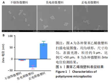
2.1 实验动物数量分析 48只小鼠全部进入结果分析。 2.2 聚苯乙烯微塑料表征结果 在经尿道前列腺切除术患者的前列腺中检出不同类型的微塑料,微塑料粒径范围在2.5-26.0 μm之间,呈现出颗粒、球体或纤维状结构[20]。在人类尿液标本中检测到的微塑料粒径在4-15 μm之间,呈不规则状[31]。据报道,在日常饮用的自来水和瓶装水中小颗粒微塑料颗粒数通常更高[32]。更小尺寸的微塑料更容易通过生物膜渗透到组织中,在器官中积累,从而产生更毒的生理危害[33];此外,前期研究在探讨聚苯乙烯微塑料的器官毒性时,5 μm是常用的聚苯乙烯微塑料实验尺寸[34-35],基于此,此次实验选择5 μm的聚苯乙烯微塑料作为研究对象。 扫描电镜观察显示不修饰微塑料、负电荷微塑料和正电荷微塑料均为球形,尺寸均匀,表面光滑,粒径约5 μm,与人体前列腺组织检测出的微塑料粒径及形状接近,见图1A。其中不修饰微塑料和磺酸基修饰微塑料的Zeta电位呈负电荷,而氨基修饰微塑料的Zeta电位则呈正电荷,见图1B,符合实验要求。"
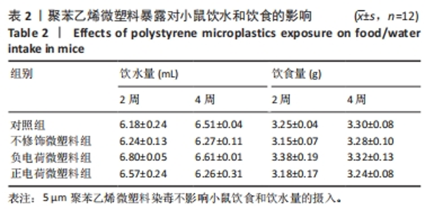
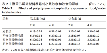
2.3 聚苯乙烯微塑料暴露对小鼠每日饮食量和饮水量的影响 为了进一步统计聚苯乙烯微塑料染毒对小鼠饮食和饮水量的影响,定期统计小鼠饮水和饮食变化,结果显示5 μm聚苯乙烯微塑料染毒不影响小鼠饮食和饮水量的摄入,见表2。"
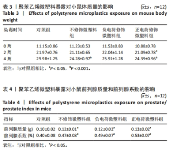
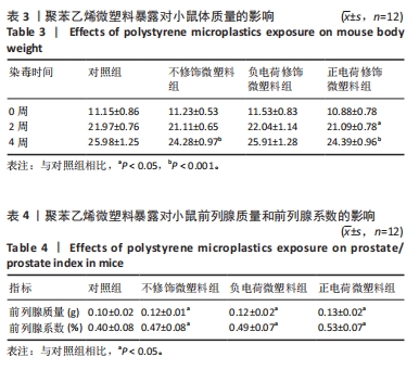
2.4 聚苯乙烯微塑料暴露对小鼠体质量、前列腺质量和前列腺系数的影响 如表3所示,4组小鼠初始体质量比较差异无显著性意义(P > 0.05);随着5 μm聚苯乙烯微塑料染毒时间的延长,不修饰微塑料组和正电荷微塑料组小鼠体质量受到明显抑制作用。如表4所示,与对照组比较,其他3组小鼠前列腺质量及前列腺系数均明显升高(P < 0.05),其中正电荷微塑料组小鼠前列腺质量及前列腺系数变化最为显著。"
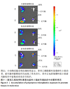
2.5 聚苯乙烯微塑料在小鼠泌尿系统中的分布 不同电荷修饰荧光聚苯乙烯微塑料进入雄性小鼠泌尿系统的情况,见图2所示。结果显示,聚苯乙烯微塑料染毒小鼠的胃、前列腺和膀胱组织均出现了荧光信号,正电荷微塑料组小鼠前列腺组织中富集的荧光信号较多,确认5 μm聚苯乙烯微塑料可以进入小鼠前列腺组织并在前列腺组织中蓄积。"


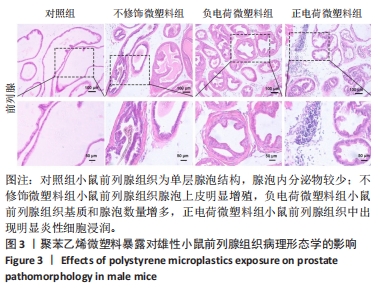
2.6 聚苯乙烯微塑料暴露对小鼠前列腺病理形态的影响 苏木精-伊红染色结果显示,对照组小鼠前列腺组织为单层腺泡结构,腺泡内分泌物较少;不修饰微塑料组小鼠前列腺组织腺泡上皮明显增殖,负电荷微塑料组小鼠前列腺组织基质和腺泡数量增多,正电荷微塑料组小鼠前列腺组织中出现明显炎性细胞浸润,见图3。"
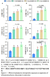
2.7 聚苯乙烯微塑料暴露对小鼠前列腺中炎性因子表达的影响 各组小鼠前列腺组织中炎性因子mRNA表达,见图4A。qPCR检测结果显示,与对照组相比,其他3组小鼠前列腺组织中白细胞介素6、白细胞介素1β及肿瘤坏死因子α mRNA表达均升高(P < 0.05,P < 0.01,P < 0.001),其中正电荷微塑料组炎性因子mRNA表达升高最显著。ELISA检测结果显示,与对照组相比,其他3组小鼠前列腺组织中3种炎性因子质量浓度均升高(P < 0.05,P < 0.001),其中正电荷微塑料组炎性因子升高最明显,见图4B。以上结果提示5 μm聚苯乙烯微塑料暴露可引起雄性小鼠前列腺内炎症反应,正电荷微塑料组炎症因子变化最为显著,毒性效果最强。"

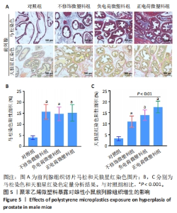
2.8 聚苯乙烯微塑料暴露对小鼠前列腺增生的影响 各组小鼠前列腺组织马松和天狼星红染色结果,见图5。染色结果显示,与对照组相比,其他3组小鼠前列腺基质区域胶原沉积显著、纤维化面积明显增加,其中正电荷微塑料组小鼠前列腺中天狼星红染色面积显著大于不修饰微塑料组。"

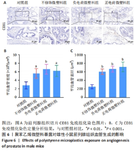
2.9 聚苯乙烯微塑料暴露对小鼠前列腺中血管生成的影响 在前列腺上皮和间质增殖的过程中,必然伴随着血管的增多,增加的血管为前列腺上皮和基质细胞提供氧气和营养,从而促进疾病进展[36]。免疫组化染色结果显示,与对照组比较,其他3组小鼠前列腺组织中CD31表达显著升高,见图6A,说明微塑料暴露可以促进前列腺组织中血管的生成。进一步的定量分析结果显示,与对照组相比,其他3组小鼠前列腺组织中血管密度与血管面积均增加,见图6B,C。"

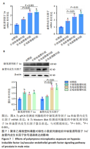
2.10 聚苯乙烯微塑料暴露对小鼠前列腺中缺氧诱导因子1α/血管内皮生长因子信号通路的影响 缺氧诱导因子1α是血管生成的启动因子,可通过诱导内皮细胞特异性生长因子血管内皮生长因子的表达,促进血管生成[37]。 qPCR与Western blot检测结果显示,与对照组相比,其他3组小鼠前列腺组织中缺氧诱导因子1α、血管内皮生长因子的 mRNA和蛋白表达均升高;与不修饰微塑料组相比,正电荷微塑料组、负电荷微塑料组小鼠前列腺组织中血管内皮生长因子mRNA表达均升高,血管内皮生长因子蛋白表达升高,见图7。说明5 μm聚苯乙烯微塑料暴露可以激活前列腺组织中缺氧诱导因子1α/血管内皮生长因子信号通路,电荷修饰聚苯乙烯微塑料对前列腺组织的毒性作用效果相对更明显。"

| [1] JAMBECK JR, GEYER R, WILCOX C, et al. Marine pollution. Plastic waste inputs from land into the ocean. Science. 2015;347(6223):768-771. [2] SCHMID C, COZZARINI L, ZAMBELLO E. Microplastic’s story. Mar Pollut Bull. 2021;162:111820. [3] BOWLEY J, BAKER-AUSTIN C, PORTER A, et al. Oceanic Hitchhikers-Assessing Pathogen Risks from Marine Microplastic. Trends Microbiol. 2021;29(2):107-116. [4] ZEB A, LIU W, ALI N, et al. Microplastic pollution in terrestrial ecosystems: Global implications and sustainable solutions. J Hazard Mater. 2024;461:132636. [5] THOMPSON RC, OLSEN Y, MITCHELL RP, et al. Lost at sea: where is all the plastic? Science. 2004;304(5672):838. [6] WALLER CL, GRIFFITHS HJ, WALUDA CM, et al. Microplastics in the Antarctic marine system: An emerging area of research. Sci Total Environ. 2017;598:220-227. [7] MOOS NV, BURKHARDT-HOLM P, KOHLER A. Uptake and effects of microplastics on cells and tissue of the blue mussel Mytilus edulis L. after an experimental exposure. Environ Sci Technol. 2012;46(20): 11327-11335. [8] WATTS AJ, LEWIS C, GOODHEAD RM, et al. Uptake and retention of microplastics by the shore crab Carcinus maenas. Environ Sci Technol. 2014;48(15):8823-8830. [9] BROWNE MA, DISSANAYAKE A, GALLOWAY TS, et al. Ingested microscopic plastic translocates to the circulatory system of the mussel, Mytilus edulis (L). Environ Sci Technol. 2008;42(13):5026-5031. [10] BHAGAT J, ZANG L, NISHIMURA N, et al. Zebrafish: An emerging model to study microplastic and nanoplastic toxicity. Sci Total Environ. 2020;728:138707. [11] XU X, ZHANG L, XUE Y, et al. Microplastic pollution characteristic in surface water and freshwater fish of Gehu Lake, China. Environ Sci Pollut Res Int. 2021;28(47):67203-67213. [12] HARLEY-NYANG D, MEMON FA, BAQUERO AO, et al. Variation in microplastic concentration, characteristics and distribution in sewage sludge & biosolids around the world. Sci Total Environ. 2023; 891:164068. [13] ZHAO B, REHATI P, YANG Z, et al. The potential toxicity of microplastics on human health. Sci Total Environ. 2024;912:168946. [14] LESLIE HA, VELZEN MJM, BRANDSMA SH, et al. Discovery and quantification of plastic particle pollution in human blood. Environ Int. 2022;163:107199. [15] MAZZILLI R, RUCCI C, VAIARELLI A, et al. Male factor infertility and assisted reproductive technologies: indications, minimum access criteria and outcomes. J Endocrinol Invest. 2023;46(6):1079-1085. [16] WARREN BD, AHN SH, BRITTAIN KS, et al. Multiple Lesions Contribute to Infertility in Males Lacking Autoimmune Regulator. Am J Pathol. 2021;191(9):1592-1609. [17] JIN H, MA T, SHA X, et al. Polystyrene microplastics induced male reproductive toxicity in mice. J Hazard Mater. 2021;401:123430. [18] HOU B, WANG F, LIU T, et al. Reproductive toxicity of polystyrene microplastics: In vivo experimental study on testicular toxicity in mice. J Hazard Mater. 2021;405:124028. [19] VERZE P, CAI T, LORENZETTI S. The role of the prostate in male fertility, health and disease. Nat Rev Urol. 2016;13(7):379-386. [20] DEMIRELLI E, TEPE Y, OGUZ U, et al. The first reported values of microplastics in prostate. BMC Urol. 2024;24(1):106. [21] GAO D, ZHANG C, GUO H, et al. Low-dose polystyrene microplastics exposure impairs fertility in male mice with high-fat diet-induced obesity by affecting prostate function. Environ Pollut. 2024;346: 123567. [22] YAN J, PAN Y, HE J, et al. Toxic vascular effects of polystyrene microplastic exposure. Sci Total Environ. 2023;905:167215. [23] LI N, YANG H, DONG Y, et al. Prevalence and implications of microplastic contaminants in general human seminal fluid: A Raman spectroscopic study. Sci Total Environ. 2024;937:173522. [24] DANSO IK, WOO JH, BAEK SH, et al. Pulmonary toxicity assessment of polypropylene, polystyrene, and polyethylene microplastic fragments in mice. Toxicol Res. 2024;40(2):313-323. [25] YANG Y, YANG J, WU WM, et al. Biodegradation and Mineralization of Polystyrene by Plastic-Eating Mealworms: Part 2. Role of Gut Microorganisms. Environ Sci Technol. 2015;49(20):12087-12093. [26] SIDDIQUI SA, SINGH S, BAHMID NA, et al. Polystyrene microplastic particles in the food chain: Characteristics and toxicity - A review. Sci Total Environ. 2023;892:164531. [27] LIU Z, ZHUAN Q, ZHANG L, et al. Polystyrene microplastics induced female reproductive toxicity in mice. J Hazard Mater. 2022;424(Pt C): 127629. [28] SUN XD, YUAN XZ, JIA Y, et al. Differentially charged nanoplastics demonstrate distinct accumulation in Arabidopsis thaliana. Nat Nanotechnol. 2020;15(9):755-760. [29] ZHU K, JIA H, JIANG W, et al. The First Observation of the Formation of Persistent Aminoxyl Radicals and Reactive Nitrogen Species on Photoirradiated Nitrogen-Containing Microplastics. Environ Sci Technol. 2022;56(2):779-789. [30] LOOS C, SYROVETS T, MUSYANOVYCH A, et al. Functionalized polystyrene nanoparticles as a platform for studying bio-nano interactions. Beilstein J Nanotechnol. 2014;5:2403-2412. [31] PIRONTI C, NOTARSTEFANO V, RICCIARDI M, et al. First Evidence of Microplastics in Human Urine, a Preliminary Study of Intake in the Human Body. Toxics. 2022;11(1):40. [32] VETHAAK AD, LEGLER J. Microplastics and human health. Science. 2021;371(6530):672-674. [33] XU D, MA Y, HAN X, et al. Systematic toxicity evaluation of polystyrene nanoplastics on mice and molecular mechanism investigation about their internalization into Caco-2 cells. J Hazard Mater. 2021;417: 126092. [34] KANG H, HUANG D, ZHANG W, et al. Inhaled polystyrene microplastics impaired lung function through pulmonary flora/TLR4-mediated iron homeostasis imbalance. Sci Total Environ. 2024;946:174300. [35] YIN K, WANG D, ZHANG Y, et al. Polystyrene microplastics promote liver inflammation by inducing the formation of macrophages extracellular traps. J Hazard Mater. 2023;452:131236. [36] FU T, SULLIVAN DP, GONZALEZ AM, et al. Mechanotransduction via endothelial adhesion molecule CD31 initiates transmigration and reveals a role for VEGFR2 in diapedesis. Immunity. 2023;56(10):2311-2324 e2316. [37] SONG S, ZHANG G, CHEN X, et al. HIF-1alpha increases the osteogenic capacity of ADSCs by coupling angiogenesis and osteogenesis via the HIF-1alpha/VEGF/AKT/mTOR signaling pathway. J Nanobiotechnology. 2023;21(1):257. [38] JIN H, YAN M, PAN C, et al. Chronic exposure to polystyrene microplastics induced male reproductive toxicity and decreased testosterone levels via the LH-mediated LHR/cAMP/PKA/StAR pathway. Part Fibre Toxicol. 2022;19(1):13. [39] XIE X, DENG T, DUAN J, et al. Exposure to polystyrene microplastics causes reproductive toxicity through oxidative stress and activation of the p38 MAPK signaling pathway. Ecotoxicol Environ Saf. 2020;190: 110133. [40] XU D, MA Y, PENG C, et al. Differently surface-labeled polystyrene nanoplastics at an environmentally relevant concentration induced Crohn’s ileitis-like features via triggering intestinal epithelial cell necroptosis. Environ Int. 2023;176:107968. [41] PAN C, CHEN Y, XU T, et al. Chronic exposure to microcystin-leucine-arginine promoted proliferation of prostate epithelial cells resulting in benign prostatic hyperplasia. Environ Pollut. 2018;242(Pt B):1535-1545. [42] WANG K, HUANG D, ZHOU P, et al. Bisphenol A exposure triggers the malignant transformation of prostatic hyperplasia in beagle dogs via cfa-miR-204/KRAS axis. Ecotoxicol Environ Saf. 2022;235:113430. [43] HALIMU G, ZHANG Q, LIU L, et al. Toxic effects of nanoplastics with different sizes and surface charges on epithelial-to-mesenchymal transition in A549 cells and the potential toxicological mechanism. J Hazard Mater. 2022;430:128485. [44] GOODMAN CM, MCCUSKER CD, YILMAZ T, et al. Toxicity of gold nanoparticles functionalized with cationic and anionic side chains. Bioconjug Chem. 2004;15(4):897-900. [45] LI Y, XU M, ZHANG Z, et al. In vitro study on the toxicity of nanoplastics with different charges to murine splenic lymphocytes. J Hazard Mater. 2022;424(Pt B):127508. [46] PAN C, WU Y, HU S, et al. Polystyrene microplastics arrest skeletal growth in puberty through accelerating osteoblast senescence. Environ Pollut. 2023;322:121217. [47] CHUGHTAI B, FORDE JC, THOMAS DD, et al. Benign prostatic hyperplasia. Nat Rev Dis Primers. 2016;2:16031. [48] AFIFY H, ABO-YOUSSEF AM, ABDEL-RAHMAN HM, et al. The modulatory effects of cinnamaldehyde on uric acid level and IL-6/JAK1/STAT3 signaling as a promising therapeutic strategy against benign prostatic hyperplasia. Toxicol Appl Pharmacol. 2020;402:115122. [49] LIEDTKE V, STOCKLE M, JUNKER K, et al. Benign prostatic hyperplasia - A novel autoimmune disease with a potential therapy consequence? Autoimmun Rev. 2024;23(3):103511. [50] BLOOM SI, ISLAM MT, LESNIEWSKI LA, et al. Mechanisms and consequences of endothelial cell senescence. Nat Rev Cardiol. 2023; 20(1):38-51. [51] TRUJILLO-ROJAS L, FERNANDEZ-NOVELL JM, BLANCO-PRIETO O, et al. The onset of age-related benign prostatic hyperplasia is concomitant with increased serum and prostatic expression of VEGF in rats: Potential role of VEGF as a marker for early prostatic alterations. Theriogenology. 2022;183:69-78. [52] KIM EY, JIN BR, CHUNG TW, et al. 6-sialyllactose ameliorates dihydrotestosterone-induced benign prostatic hyperplasia through suppressing VEGF-mediated angiogenesis. BMB Rep. 2019; 52(9):560-565. [53] WU X, GU Y, LI L. The anti-hyperplasia, anti-oxidative and anti-inflammatory properties of Qing Ye Dan and swertiamarin in testosterone-induced benign prostatic hyperplasia in rats. Toxicol Lett. 2017;265:9-16. |
| [1] | Wu Yanting, Li Yu, Liao Jinfeng. Magnesium oxide nanoparticles regulate osteogenesis- and angiogenesis-related gene expressions to promote bone defect healing [J]. Chinese Journal of Tissue Engineering Research, 2026, 30(8): 1885-1895. |
| [2] | Hou Chaowen, Li Zhaojin, Kong Jianda, Zhang Shuli. Main physiological changes in skeletal muscle aging and the multimechanism regulatory role of exercise [J]. Chinese Journal of Tissue Engineering Research, 2026, 30(6): 1464-1475. |
| [3] | You Huijuan, Wu Shuzhen, Rong Rong, Chen Liyuan, Zhao Yuqing, Wang Qinglu, Ou Xiaowei, Yang Fengying. Macrophage autophagy in lung diseases: two-sided effects [J]. Chinese Journal of Tissue Engineering Research, 2026, 30(6): 1516-1526. |
| [4] | Liu Kexin, , Hao Kaimin, Zhuang Wenyue, , Li Zhengyi. Autophagy-related gene expression in pulmonary fibrosis models: bioinformatic analysis and experimental validation [J]. Chinese Journal of Tissue Engineering Research, 2026, 30(5): 1129-1138. |
| [5] | Zhang Di, Zhao Jun, Ma Guangyue, Sun Hui, Jiang Rong. Mechanism of depression-like behavior in chronic social defeat stress mice based on high-throughput sequencing [J]. Chinese Journal of Tissue Engineering Research, 2026, 30(5): 1139-1146. |
| [6] | Li Haojing, Wang Xin, Song Chenglin, Zhang Shengnan, Chen Yunxin. Therapeutic efficacy of extracorporeal shock wave therapy in the upper trapezius muscle area combined with exercise control training in patients with chronic non-specific neck pain [J]. Chinese Journal of Tissue Engineering Research, 2026, 30(5): 1162-1170. |
| [7] | Liu Yu, Lei Senlin, Zhou Jintao, Liu Hui, Li Xianhui. Mechanisms by which aerobic and resistance exercises improve obesity-related cognitive impairment [J]. Chinese Journal of Tissue Engineering Research, 2026, 30(5): 1171-1183. |
| [8] | Yu Huifen, Mo Licun, Cheng Leping. The position and role of 5-hydroxytryptamine in the repair of tissue injury [J]. Chinese Journal of Tissue Engineering Research, 2026, 30(5): 1196-1206. |
| [9] | Wen Xiaolong, Weng Xiquan, Feng Yao, Cao Wenyan, Liu Yuqian, Wang Haitao. Effects of inflammation on serum hepcidin and iron metabolism related parameters in patients with type 2 diabetes mellitus: a meta-analysis [J]. Chinese Journal of Tissue Engineering Research, 2026, 30(5): 1294-1301. |
| [10] | Yin Yongcheng, Zhao Xiangrui, Yang Zhijie, Li Zheng, Li Fang, Ning Bin. Effect and mechanism of peroxiredoxin 1 in microglial inflammation after spinal cord injury [J]. Chinese Journal of Tissue Engineering Research, 2026, 30(5): 1106-1113. |
| [11] | Cao Wenqi, Feng Xiuzhi, Zhao Yi, Wang Zhimin, Chen Yiran, Yang Xiao, Ren Yanling. Effect of macrophage polarization on osteogenesis-angiogenesis coupling in type 2 diabetic osteoporosis [J]. Chinese Journal of Tissue Engineering Research, 2026, 30(4): 917-925. |
| [12] | Yang Xiao, Bai Yuehui, Zhao Tiantian, Wang Donghao, Zhao Chen, Yuan Shuo. Cartilage degeneration in temporomandibular joint osteoarthritis: mechanisms and regenerative challenges [J]. Chinese Journal of Tissue Engineering Research, 2026, 30(4): 926-935. |
| [13] | Yu Shiyu, Yu Sutong, Xu Yang, Zhen Xiangyan, Han Fengxuan. Advances in research and application of tissue engineering therapeutic strategies in oral submucous fibrosis [J]. Chinese Journal of Tissue Engineering Research, 2026, 30(4): 936-948. |
| [14] | Chen Yixian, Chen Chen, Lu Liheng, Tang Jinpeng, Yu Xiaowei. Triptolide in the treatment of osteoarthritis: network pharmacology analysis and animal model validation [J]. Chinese Journal of Tissue Engineering Research, 2026, 30(4): 805-815. |
| [15] | Zhang Qian, Wang Fuxia, Wang Wen, Zhang Kun. Characteristic analysis of nanogel composite system and its application strategies in visualization of diagnostic imaging and therapy [J]. Chinese Journal of Tissue Engineering Research, 2026, 30(2): 480-488. |
| Viewed | ||||||
|
Full text |
|
|||||
|
Abstract |
|
|||||