Chinese Journal of Tissue Engineering Research ›› 2025, Vol. 29 ›› Issue (34): 7353-7361.doi: 10.12307/2025.887
Previous Articles Next Articles
Effect and mechanism of polystyrene microplastics on prostate in male mice
Pan Chun1, 2, Fan Zhencheng2, Hong Runyang2, Shi Yujie2, Chen Hao1, 2
- 1Affiliated Hospital of Yangzhou University, Yangzhou 225000, Jiangsu Province, China; 2Yangzhou University School of Medicine-Institute of Translational Medicine, Yangzhou 225000, Jiangsu Province, China
-
Received:2024-07-05Accepted:2024-09-21Online:2025-12-08Published:2025-01-17 -
Contact:Chen Hao, PhD, Professor, Doctoral supervisor, Affiliated Hospital of Yangzhou University, Yangzhou 225000, Jiangsu Province, China; Yangzhou University School of Medicine-Institute of Translational Medicine, Yangzhou 225000, Jiangsu Province, China -
About author:Pan Chun, PhD, Lecturer, Affiliated Hospital of Yangzhou University, Yangzhou 225000, Jiangsu Province, China; Yangzhou University School of Medicine-Institute of Translational Medicine, Yangzhou 225000, Jiangsu Province, China Fan Zhencheng, Yangzhou University School of Medicine-Institute of Translational Medicine, Yangzhou 225000, Jiangsu Province, China -
Supported by:National Natural Science Foundation of China, No. 32301416 (to PC); Yangzhou Lv Yang Jin Feng Program, No. YZLVJFJH2022YXBS154 (to PC); National Natural Science Foundation of China, No. 82172468, 82372436 (to CH)
CLC Number:
Cite this article
Pan Chun, Fan Zhencheng, Hong Runyang, Shi Yujie, Chen Hao. Effect and mechanism of polystyrene microplastics on prostate in male mice[J]. Chinese Journal of Tissue Engineering Research, 2025, 29(34): 7353-7361.
share this article
Add to citation manager EndNote|Reference Manager|ProCite|BibTeX|RefWorks
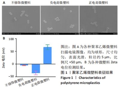
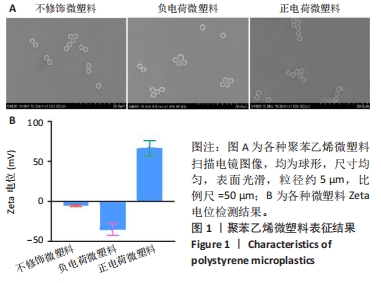
2.1 实验动物数量分析 48只小鼠全部进入结果分析。 2.2 聚苯乙烯微塑料表征结果 在经尿道前列腺切除术患者的前列腺中检出不同类型的微塑料,微塑料粒径范围在2.5-26.0 μm之间,呈现出颗粒、球体或纤维状结构[20]。在人类尿液标本中检测到的微塑料粒径在4-15 μm之间,呈不规则状[31]。据报道,在日常饮用的自来水和瓶装水中小颗粒微塑料颗粒数通常更高[32]。更小尺寸的微塑料更容易通过生物膜渗透到组织中,在器官中积累,从而产生更毒的生理危害[33];此外,前期研究在探讨聚苯乙烯微塑料的器官毒性时,5 μm是常用的聚苯乙烯微塑料实验尺寸[34-35],基于此,此次实验选择5 μm的聚苯乙烯微塑料作为研究对象。 扫描电镜观察显示不修饰微塑料、负电荷微塑料和正电荷微塑料均为球形,尺寸均匀,表面光滑,粒径约5 μm,与人体前列腺组织检测出的微塑料粒径及形状接近,见图1A。其中不修饰微塑料和磺酸基修饰微塑料的Zeta电位呈负电荷,而氨基修饰微塑料的Zeta电位则呈正电荷,见图1B,符合实验要求。"
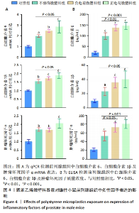
2.7 聚苯乙烯微塑料暴露对小鼠前列腺中炎性因子表达的影响 各组小鼠前列腺组织中炎性因子mRNA表达,见图4A。qPCR检测结果显示,与对照组相比,其他3组小鼠前列腺组织中白细胞介素6、白细胞介素1β及肿瘤坏死因子α mRNA表达均升高(P < 0.05,P < 0.01,P < 0.001),其中正电荷微塑料组炎性因子mRNA表达升高最显著。ELISA检测结果显示,与对照组相比,其他3组小鼠前列腺组织中3种炎性因子质量浓度均升高(P < 0.05,P < 0.001),其中正电荷微塑料组炎性因子升高最明显,见图4B。以上结果提示5 μm聚苯乙烯微塑料暴露可引起雄性小鼠前列腺内炎症反应,正电荷微塑料组炎症因子变化最为显著,毒性效果最强。"

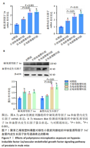
2.10 聚苯乙烯微塑料暴露对小鼠前列腺中缺氧诱导因子1α/血管内皮生长因子信号通路的影响 缺氧诱导因子1α是血管生成的启动因子,可通过诱导内皮细胞特异性生长因子血管内皮生长因子的表达,促进血管生成[37]。 qPCR与Western blot检测结果显示,与对照组相比,其他3组小鼠前列腺组织中缺氧诱导因子1α、血管内皮生长因子的 mRNA和蛋白表达均升高;与不修饰微塑料组相比,正电荷微塑料组、负电荷微塑料组小鼠前列腺组织中血管内皮生长因子mRNA表达均升高,血管内皮生长因子蛋白表达升高,见图7。说明5 μm聚苯乙烯微塑料暴露可以激活前列腺组织中缺氧诱导因子1α/血管内皮生长因子信号通路,电荷修饰聚苯乙烯微塑料对前列腺组织的毒性作用效果相对更明显。"

| [1] JAMBECK JR, GEYER R, WILCOX C, et al. Marine pollution. Plastic waste inputs from land into the ocean. Science. 2015;347(6223):768-771. [2] SCHMID C, COZZARINI L, ZAMBELLO E. Microplastic’s story. Mar Pollut Bull. 2021;162:111820. [3] BOWLEY J, BAKER-AUSTIN C, PORTER A, et al. Oceanic Hitchhikers-Assessing Pathogen Risks from Marine Microplastic. Trends Microbiol. 2021;29(2):107-116. [4] ZEB A, LIU W, ALI N, et al. Microplastic pollution in terrestrial ecosystems: Global implications and sustainable solutions. J Hazard Mater. 2024;461:132636. [5] THOMPSON RC, OLSEN Y, MITCHELL RP, et al. Lost at sea: where is all the plastic? Science. 2004;304(5672):838. [6] WALLER CL, GRIFFITHS HJ, WALUDA CM, et al. Microplastics in the Antarctic marine system: An emerging area of research. Sci Total Environ. 2017;598:220-227. [7] MOOS NV, BURKHARDT-HOLM P, KOHLER A. Uptake and effects of microplastics on cells and tissue of the blue mussel Mytilus edulis L. after an experimental exposure. Environ Sci Technol. 2012;46(20): 11327-11335. [8] WATTS AJ, LEWIS C, GOODHEAD RM, et al. Uptake and retention of microplastics by the shore crab Carcinus maenas. Environ Sci Technol. 2014;48(15):8823-8830. [9] BROWNE MA, DISSANAYAKE A, GALLOWAY TS, et al. Ingested microscopic plastic translocates to the circulatory system of the mussel, Mytilus edulis (L). Environ Sci Technol. 2008;42(13):5026-5031. [10] BHAGAT J, ZANG L, NISHIMURA N, et al. Zebrafish: An emerging model to study microplastic and nanoplastic toxicity. Sci Total Environ. 2020;728:138707. [11] XU X, ZHANG L, XUE Y, et al. Microplastic pollution characteristic in surface water and freshwater fish of Gehu Lake, China. Environ Sci Pollut Res Int. 2021;28(47):67203-67213. [12] HARLEY-NYANG D, MEMON FA, BAQUERO AO, et al. Variation in microplastic concentration, characteristics and distribution in sewage sludge & biosolids around the world. Sci Total Environ. 2023; 891:164068. [13] ZHAO B, REHATI P, YANG Z, et al. The potential toxicity of microplastics on human health. Sci Total Environ. 2024;912:168946. [14] LESLIE HA, VELZEN MJM, BRANDSMA SH, et al. Discovery and quantification of plastic particle pollution in human blood. Environ Int. 2022;163:107199. [15] MAZZILLI R, RUCCI C, VAIARELLI A, et al. Male factor infertility and assisted reproductive technologies: indications, minimum access criteria and outcomes. J Endocrinol Invest. 2023;46(6):1079-1085. [16] WARREN BD, AHN SH, BRITTAIN KS, et al. Multiple Lesions Contribute to Infertility in Males Lacking Autoimmune Regulator. Am J Pathol. 2021;191(9):1592-1609. [17] JIN H, MA T, SHA X, et al. Polystyrene microplastics induced male reproductive toxicity in mice. J Hazard Mater. 2021;401:123430. [18] HOU B, WANG F, LIU T, et al. Reproductive toxicity of polystyrene microplastics: In vivo experimental study on testicular toxicity in mice. J Hazard Mater. 2021;405:124028. [19] VERZE P, CAI T, LORENZETTI S. The role of the prostate in male fertility, health and disease. Nat Rev Urol. 2016;13(7):379-386. [20] DEMIRELLI E, TEPE Y, OGUZ U, et al. The first reported values of microplastics in prostate. BMC Urol. 2024;24(1):106. [21] GAO D, ZHANG C, GUO H, et al. Low-dose polystyrene microplastics exposure impairs fertility in male mice with high-fat diet-induced obesity by affecting prostate function. Environ Pollut. 2024;346: 123567. [22] YAN J, PAN Y, HE J, et al. Toxic vascular effects of polystyrene microplastic exposure. Sci Total Environ. 2023;905:167215. [23] LI N, YANG H, DONG Y, et al. Prevalence and implications of microplastic contaminants in general human seminal fluid: A Raman spectroscopic study. Sci Total Environ. 2024;937:173522. [24] DANSO IK, WOO JH, BAEK SH, et al. Pulmonary toxicity assessment of polypropylene, polystyrene, and polyethylene microplastic fragments in mice. Toxicol Res. 2024;40(2):313-323. [25] YANG Y, YANG J, WU WM, et al. Biodegradation and Mineralization of Polystyrene by Plastic-Eating Mealworms: Part 2. Role of Gut Microorganisms. Environ Sci Technol. 2015;49(20):12087-12093. [26] SIDDIQUI SA, SINGH S, BAHMID NA, et al. Polystyrene microplastic particles in the food chain: Characteristics and toxicity - A review. Sci Total Environ. 2023;892:164531. [27] LIU Z, ZHUAN Q, ZHANG L, et al. Polystyrene microplastics induced female reproductive toxicity in mice. J Hazard Mater. 2022;424(Pt C): 127629. [28] SUN XD, YUAN XZ, JIA Y, et al. Differentially charged nanoplastics demonstrate distinct accumulation in Arabidopsis thaliana. Nat Nanotechnol. 2020;15(9):755-760. [29] ZHU K, JIA H, JIANG W, et al. The First Observation of the Formation of Persistent Aminoxyl Radicals and Reactive Nitrogen Species on Photoirradiated Nitrogen-Containing Microplastics. Environ Sci Technol. 2022;56(2):779-789. [30] LOOS C, SYROVETS T, MUSYANOVYCH A, et al. Functionalized polystyrene nanoparticles as a platform for studying bio-nano interactions. Beilstein J Nanotechnol. 2014;5:2403-2412. [31] PIRONTI C, NOTARSTEFANO V, RICCIARDI M, et al. First Evidence of Microplastics in Human Urine, a Preliminary Study of Intake in the Human Body. Toxics. 2022;11(1):40. [32] VETHAAK AD, LEGLER J. Microplastics and human health. Science. 2021;371(6530):672-674. [33] XU D, MA Y, HAN X, et al. Systematic toxicity evaluation of polystyrene nanoplastics on mice and molecular mechanism investigation about their internalization into Caco-2 cells. J Hazard Mater. 2021;417: 126092. [34] KANG H, HUANG D, ZHANG W, et al. Inhaled polystyrene microplastics impaired lung function through pulmonary flora/TLR4-mediated iron homeostasis imbalance. Sci Total Environ. 2024;946:174300. [35] YIN K, WANG D, ZHANG Y, et al. Polystyrene microplastics promote liver inflammation by inducing the formation of macrophages extracellular traps. J Hazard Mater. 2023;452:131236. [36] FU T, SULLIVAN DP, GONZALEZ AM, et al. Mechanotransduction via endothelial adhesion molecule CD31 initiates transmigration and reveals a role for VEGFR2 in diapedesis. Immunity. 2023;56(10):2311-2324 e2316. [37] SONG S, ZHANG G, CHEN X, et al. HIF-1alpha increases the osteogenic capacity of ADSCs by coupling angiogenesis and osteogenesis via the HIF-1alpha/VEGF/AKT/mTOR signaling pathway. J Nanobiotechnology. 2023;21(1):257. [38] JIN H, YAN M, PAN C, et al. Chronic exposure to polystyrene microplastics induced male reproductive toxicity and decreased testosterone levels via the LH-mediated LHR/cAMP/PKA/StAR pathway. Part Fibre Toxicol. 2022;19(1):13. [39] XIE X, DENG T, DUAN J, et al. Exposure to polystyrene microplastics causes reproductive toxicity through oxidative stress and activation of the p38 MAPK signaling pathway. Ecotoxicol Environ Saf. 2020;190: 110133. [40] XU D, MA Y, PENG C, et al. Differently surface-labeled polystyrene nanoplastics at an environmentally relevant concentration induced Crohn’s ileitis-like features via triggering intestinal epithelial cell necroptosis. Environ Int. 2023;176:107968. [41] PAN C, CHEN Y, XU T, et al. Chronic exposure to microcystin-leucine-arginine promoted proliferation of prostate epithelial cells resulting in benign prostatic hyperplasia. Environ Pollut. 2018;242(Pt B):1535-1545. [42] WANG K, HUANG D, ZHOU P, et al. Bisphenol A exposure triggers the malignant transformation of prostatic hyperplasia in beagle dogs via cfa-miR-204/KRAS axis. Ecotoxicol Environ Saf. 2022;235:113430. [43] HALIMU G, ZHANG Q, LIU L, et al. Toxic effects of nanoplastics with different sizes and surface charges on epithelial-to-mesenchymal transition in A549 cells and the potential toxicological mechanism. J Hazard Mater. 2022;430:128485. [44] GOODMAN CM, MCCUSKER CD, YILMAZ T, et al. Toxicity of gold nanoparticles functionalized with cationic and anionic side chains. Bioconjug Chem. 2004;15(4):897-900. [45] LI Y, XU M, ZHANG Z, et al. In vitro study on the toxicity of nanoplastics with different charges to murine splenic lymphocytes. J Hazard Mater. 2022;424(Pt B):127508. [46] PAN C, WU Y, HU S, et al. Polystyrene microplastics arrest skeletal growth in puberty through accelerating osteoblast senescence. Environ Pollut. 2023;322:121217. [47] CHUGHTAI B, FORDE JC, THOMAS DD, et al. Benign prostatic hyperplasia. Nat Rev Dis Primers. 2016;2:16031. [48] AFIFY H, ABO-YOUSSEF AM, ABDEL-RAHMAN HM, et al. The modulatory effects of cinnamaldehyde on uric acid level and IL-6/JAK1/STAT3 signaling as a promising therapeutic strategy against benign prostatic hyperplasia. Toxicol Appl Pharmacol. 2020;402:115122. [49] LIEDTKE V, STOCKLE M, JUNKER K, et al. Benign prostatic hyperplasia - A novel autoimmune disease with a potential therapy consequence? Autoimmun Rev. 2024;23(3):103511. [50] BLOOM SI, ISLAM MT, LESNIEWSKI LA, et al. Mechanisms and consequences of endothelial cell senescence. Nat Rev Cardiol. 2023; 20(1):38-51. [51] TRUJILLO-ROJAS L, FERNANDEZ-NOVELL JM, BLANCO-PRIETO O, et al. The onset of age-related benign prostatic hyperplasia is concomitant with increased serum and prostatic expression of VEGF in rats: Potential role of VEGF as a marker for early prostatic alterations. Theriogenology. 2022;183:69-78. [52] KIM EY, JIN BR, CHUNG TW, et al. 6-sialyllactose ameliorates dihydrotestosterone-induced benign prostatic hyperplasia through suppressing VEGF-mediated angiogenesis. BMB Rep. 2019; 52(9):560-565. [53] WU X, GU Y, LI L. The anti-hyperplasia, anti-oxidative and anti-inflammatory properties of Qing Ye Dan and swertiamarin in testosterone-induced benign prostatic hyperplasia in rats. Toxicol Lett. 2017;265:9-16. |
| [1] | Li Zikai, Zhang Chengcheng, Xiong Jiaying, Yang Xirui, Yang Jing, Shi Haishan. Potential effects of ornidazole on intracanal vascularization in endodontic regeneration [J]. Chinese Journal of Tissue Engineering Research, 2025, 29(在线): 1-7. |
| [2] | Yu Shuai, Liu Jiawei, Zhu Bin, Pan Tan, Li Xinglong, Sun Guangfeng, Yu Haiyang, Ding Ya, Wang Hongliang. Hot issues and application prospects of small molecule drugs in treatment of osteoarthritis [J]. Chinese Journal of Tissue Engineering Research, 2025, 29(9): 1913-1922. |
| [3] | Li Kaiying, Wei Xiaoge, Song Fei, Yang Nan, Zhao Zhenning, Wang Yan, Mu Jing, Ma Huisheng. Mechanism of Lijin manipulation regulating scar formation in skeletal muscle injury repair in rabbits [J]. Chinese Journal of Tissue Engineering Research, 2025, 29(8): 1600-1608. |
| [4] | Yu Jingbang, Wu Yayun. Regulatory effect of non-coding RNA in pulmonary fibrosis [J]. Chinese Journal of Tissue Engineering Research, 2025, 29(8): 1659-1666. |
| [5] | Lou Guo, Zhang Min, Fu Changxi. Exercise preconditioning for eight weeks enhances therapeutic effect of adipose-derived stem cells in rats with myocardial infarction [J]. Chinese Journal of Tissue Engineering Research, 2025, 29(7): 1363-1370. |
| [6] | Wang Yuru, Li Siyuan, Xu Ye, Zhang Yumeng, Liu Yang, Hao Huiqin. Effects of wogonin on joint inflammation in collagen-induced arthritis rats via the endoplasmic reticulum stress pathway [J]. Chinese Journal of Tissue Engineering Research, 2025, 29(5): 1026-1035. |
| [7] | Bai Jing, Zhang Xue, Ren Yan, Li Yuehui, Tian Xiaoyu. Effect of lncRNA-TNFRSF13C on hypoxia-inducible factor 1alpha in periodontal cells by modulation of #br# miR-1246 #br# [J]. Chinese Journal of Tissue Engineering Research, 2025, 29(5): 928-935. |
| [8] | Zhi Fang, Zhu Manhua, Xiong Wei, Lin Xingzhen. Analgesic effect of acupuncture in a rat model of lumbar disc herniation [J]. Chinese Journal of Tissue Engineering Research, 2025, 29(5): 936-941. |
| [9] | Han Haihui, Meng Xiaohu, Xu Bo, Ran Le, Shi Qi, Xiao Lianbo. Effect of fibroblast growth factor receptor 1 inhibitor on bone destruction in rats with collagen-induced arthritis [J]. Chinese Journal of Tissue Engineering Research, 2025, 29(5): 968-977. |
| [10] | Zheng Yitong, Wang Yongxin, Liu Wen, Amujite, Qin Hu. Action mechanism of intrathecal transplantation of human umbilical cord mesenchymal stem cell-derived exosomes for repair of spinal cord injury under neuroendoscopy [J]. Chinese Journal of Tissue Engineering Research, 2025, 29(36): 7743-7751. |
| [11] | Yu Hui, Yang Yang, Wei Ting, Li Wenli, Luo Wenqian, Liu Bin. Gadd45b alleviates white matter damage in chronic ischemic rats by modulating astrocyte phenotype [J]. Chinese Journal of Tissue Engineering Research, 2025, 29(36): 7797-7803. |
| [12] | Zhang Fei, Zuo Jun. Inhibition of hypertrophic scar in rats by beta-sitosterol-laden mesoporous silica nanoparticles [J]. Chinese Journal of Tissue Engineering Research, 2025, 29(34): 7301-7309. |
| [13] | Zhao Jianwei, Li Xunsheng, Lyu Jinpeng, Zhou Jue, Jiang Yidi, Yue Zhigang, Sun Hongmei. Deer antler stem cell exosome composite hydrogel promotes the repair of burned skin [J]. Chinese Journal of Tissue Engineering Research, 2025, 29(34): 7344-7352. |
| [14] | Wang Guanyuan, Li Wenli, Liu Jinglin, Zhang Jie. Development and application of fully automated intelligent intravenous medication dispensing robot ML300 in Pharmacy Intravenous Admixture Services [J]. Chinese Journal of Tissue Engineering Research, 2025, 29(34): 7362-7368. |
| [15] | Chen Jing, Zhang Nan, Meng Qinghua, Bao Chunyu. Material characterization of finite element computational models of knee joints at different ages [J]. Chinese Journal of Tissue Engineering Research, 2025, 29(34): 7369-7375. |
| Viewed | ||||||
|
Full text |
|
|||||
|
Abstract |
|
|||||
