Chinese Journal of Tissue Engineering Research
Tissue-engineered tendon construction using bone marrow mesenchymal stem cells induced by bone morphogenetic protein 12
Cai Rong-hui, Liu Kang
- Zhongshan People’s Hospital, Zhongshan 528400, Guangdong Province, China
-
Received:2012-10-09Revised:2012-11-20Online:2013-07-02Published:2013-07-02 -
About author:Cai Rong-hui★, Master, Associate chief physician, Zhongshan People’s Hospital, Zhongshan 528400, Guangdong Province, China caironghui88@163.com
CLC Number:
Cite this article
Cai Rong-hui, Liu Kang. Tissue-engineered tendon construction using bone marrow mesenchymal stem cells induced by bone morphogenetic protein 12[J]. Chinese Journal of Tissue Engineering Research, doi: 10.3969/j.issn.2095-4344.2013.27.001.
share this article
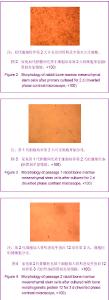
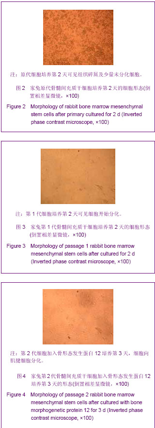
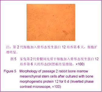
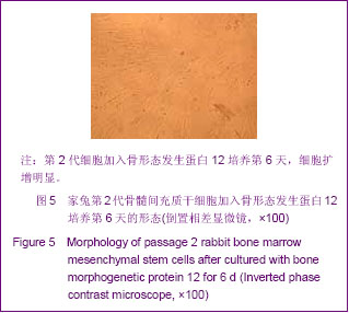
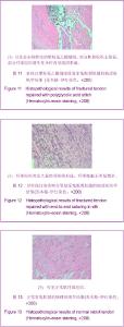
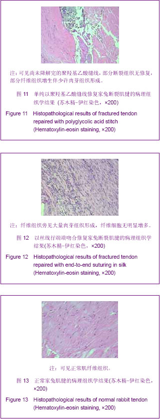
2.1 实验动物数量分析 造模未造成动物死亡,最终为12只兔。干细胞培养阶段分成2组,组织化工程肌腱构建时分成3组。 2.2 骨髓间充质干细胞体外扩增生长情况 细胞传至第2代加入骨形态发生蛋白12后,扩增明显,生长加快,细胞向肌腱细胞分化,见图2-5。"
"
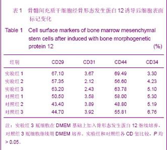
2.3 骨髓间充质干细胞经骨形态发生蛋白12诱导后细胞表面标记CD值 结果见表1。"
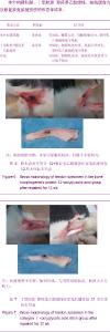
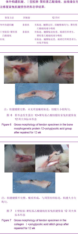
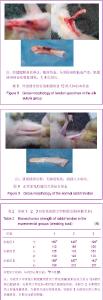
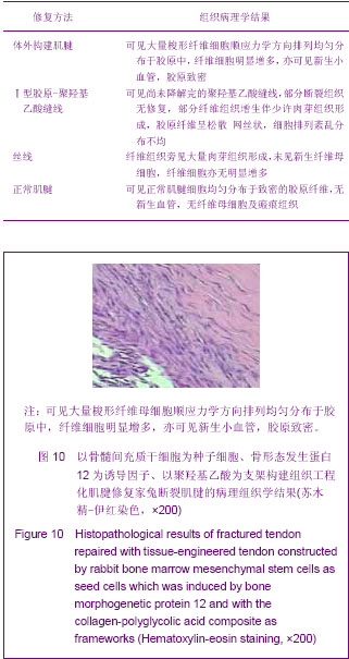
流式细胞仪检测细胞表面标记CD29,CD31,CD34,CD44,结果显示无论是实验组(DMEM+骨形态发生蛋白12),还是对照组(DMEM),其CD44和CD29阳性标记均出现单峰,CD31和CD34表达阴性,证明所培养的骨髓间充质干细胞是均一的细胞群,不含造血细胞、内皮细胞等其他细胞。应用SPSS 16.0软件,对实验组和对照组各CD值进行统计学分析,P均 > 0.05。 从上述分析结果看,实验组与对照组CD29值统计学分析,P > 0.05,证明两组差异无显著性意义。CD31,CD44,CD34统计学分析,P 也> 0.05,这结果说明骨形态发生蛋白12不会改变骨髓间充质干细胞表面标志,只会使骨髓间充质干细胞增殖分化加速。 2.4 组织化工程肌腱镜下形态学、力学及组织病理学观察结果 见图6-13及表2。 体外构建肌腱、Ⅰ型胶原-聚羟基乙酸缝线、丝线缝合方法修复家兔肌腱损伤的形态学结果:"
"
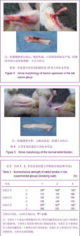
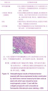
应用SPSS 16.0软件分别对实验组1、实验组2、实验组3结果进行配对t 检验,其中实验组1、实验组3左、右侧相比,P < 0.05;实验组2左、右侧相比,P > 0.05。 从上述结果可以看出,实验组1中,骨形态发生蛋白12+聚羟基乙酸重建肌腱的力学强度明显优于Ⅰ型胶原-聚羟基乙酸组;而实验组2中骨形态发生蛋白12+聚羟基乙酸重建肌腱的力学强度与丝线缝合组差异无显著性意义,这可能与丝线刺激纤维肉芽组织增生,使断端接触面积增大,从而增强了肌腱抗牵拉能力有关;至于实验组3中骨形态发生蛋白12+聚羟基乙酸重建肌腱的力学强度与正常肌腱强度差异存在显著性意义,说明组织工程重建肌腱还有待进一步改进,以增强抗牵拉力,达到完美修复肌腱损伤的程度。 体外构建肌腱、Ⅰ型胶原-聚羟基乙酸缝线、丝线缝合方法修复家兔肌腱12周时组织病理学结果:"
"
| [1] Flynn JE,Wilson JT,Child CG,et al.Heterogenous andautogenous-tendon transplants.An experimental study ofpreserved bovine-tendon transplants in dogs andautogenous-tendon transplants in dogs.J Bone Joint Surg Am.1960;42-A:91-110.[2] Jenkins DH,McKibbin B.The role of flexible carbon-fibre implantsas tendon and ligament substitutes in clinical practice.Apreliminary report.J Bone Joint Surg Br.1980; 62-B(4):497-499.[3] Friedenstein AJ,Chailakhyan RK,Gerasimov UV.Bone marrowosteogenic stem cells:in vitro cultivation and transplantation in diffusionchambers. Cell Tissue Kinet. 1987; 20(3):263-272.[4] Pittenger MF,Mackay AM,Beck SC,et al.Multilineage potential ofadult human mesenchymal stem cells. Science. 1999;284 (5411):143-147.[5] Garner WL, McDonald JA, Koo M, et al. Identification of the collagen-producing cells in healing flexor tendons. Plast Reconstr Surg. 1989;83(5):875-879. [6] Gelberman RH, Amiel D, Harwood F. Genetic expression for type I procollagen in the early stages of flexor tendon healing.J Hand Surg [Am]. 1992;17(3):551-558. [7] Khan U, Edwards JC, McGrouther DA. Patterns of cellular activation after tendon injury. J Hand Surg [Br].1996; 21(6): 813-820.[8] Lunberg G, Rank F, Heinau B.Intrinsic tendon healing. A newexperimental model.Scand J Plast Reconstr Surg. 1985; 19(2): 113-117.[9] Lister GD. Cyclic stress analysis of flexor tendon repair. J Hand Surg(Am).1991;16:701.[10] Lundorg G. Tendon healing: Intrisinc mechanisms Tendon Surgery in the hand. The CV Mosby Company.1987: 54-60.[11] Manske PR, Bridwell K, Lesker PA. Nutrient pathways to lexortendons of chickens using tritiated prohne. J Hand Surg. 1978;3(4):352-357.[12] Haynesworth SE, Baber MA, Caplan AL. Cell surface antigens on human marrow-derived mesenchymal cells are detected by monoclonalantibodies. Bone. 1992;13(1):69-80.[13] Pittenger MF, Mackay AM, Beck SC, et al. Multilineage potential of adult human mesenchymal stem cells. Science. 1999;284 (5411):143-147.[14] Kramer J,Hegea C, Cuan K, et al. Embryonic 8lelll cell derivedehondrngenfo differentiation in vito aefivatioa by BMP22 and BMP24. Meeh Dev. 2000;92:193.[15] Cetrulo CL Jr.Cord-blood mesenchymal stem cells and tis-sue engineering. Stem Cell Rev. 2006;2(2):163-168.[16] Kim DW,Chung YJ,Kim TG,et al.Cotransplantation ofthird-party mesenchymal stromal cells can alleviate sin-gle-donor predominance and increase engraftment fromdouble cord transplantation. Blood.2004; 103(5): 1941-1948.[17] 董志宁,孙奋勇,潘秋辉,等. rhbFGF转染人皮肤成纤维细胞的研究[J].生物医学工程研究,2004,34(1):33-37. [18] Sellers RS, Zhang R, Glasson SS, et al. Repair of articular cartilage defects one year after treatment with recombinant human bone morphogenetic protein-2 (rhBMP-2). J Bone Joint Surg Am. 2000;82(2): 151-160.[19] Lou J, Tu Y, Lud W. Effect of bone morphogenetic protein-12 genetransfer on mesenchymal progenitor cell. Clin Orthop. 1999;369:339.[20] Worster AA, Nixon AJ, Brower-Toland BD, et al. Effect of transforming growth factor beta1 on chondrogenic differentiation of cultured equine mesenchymal stem cells. Am J Vet Res. 2000;61(9):1003-1010.[21] Sahoo S,Cho-Hong JG,Siew-Lok T. Development of hybrid polymerscaffolds for potential applications in ligament and tendon tissueengineering. Biomed Mater. 2007;2(3):169-173.[22] Cen L,Liu W,Cui L,et al.Collagen tissue engineering:developmentof novel biomaterials and applications. Pediatr Res. 2008;63(5):492-496.[23] Yokoya S,Mochizuki Y,Nagata Y,et al.Tendon-bone insertion repairand regeneration using polyglycolic acid sheet in the rabbit rotator cuffinjury model.Am J Sports Med. 2008;36(7): 1298-1309.[24] Sahoo S,Ouyang H,Goh JC,et al.Characterization of a novelpolymeric scaffold for potential application in tendon/ligament tissueengineering.Tissue Eng. 2006;12(1): 91-99.[25] 潘海涛,郑启新,郭晓东.Ⅰ型胶原在PLGA-[ASP-PEG]表面修饰对兔骨髓间充质干细胞生物力学的影响[J].中华创伤骨科杂志, 2006,8(10):938-943.[26] 曲彦隆,杨志明,朱伟南,等.衍生肌腱支架材料的细胞相容性研究[J]. 中华骨科杂志,2006,26(12):842-845. [27] 周悦婷,项舟,阳富春,等.生物衍生材料构建组织工程肌腱体内植入的实验研究[J].中国修复重建外科杂志,2003,2(2):152-156.[28] Richmond JC,Manseau CJ,Patz R,et al. Anterior cmciatere construction using adacron ligamentprosthesis A longterm study. Am J Sports Med. 1992;20:24 -28.[29] 曹德君,翟华玲,刘伟,等.体外构建组织工程化肌腱的初步研究[J].中华外科杂志, 2004,42(2):110-113.[30] Harris MT,Butler DL,Boivin GP,et al.Mesenchymal stemcells usedfor rabbit tendon repair can form ectopic bone and express alkalinephosphatase activity in constructs. J Orthop Res. 2004;22(5):998-1003 |
| [1] | Pu Rui, Chen Ziyang, Yuan Lingyan. Characteristics and effects of exosomes from different cell sources in cardioprotection [J]. Chinese Journal of Tissue Engineering Research, 2021, 25(在线): 1-. |
| [2] | Xu Feng, Kang Hui, Wei Tanjun, Xi Jintao. Biomechanical analysis of different fixation methods of pedicle screws for thoracolumbar fracture [J]. Chinese Journal of Tissue Engineering Research, 2021, 25(9): 1313-1317. |
| [3] | Zhang Tongtong, Wang Zhonghua, Wen Jie, Song Yuxin, Liu Lin. Application of three-dimensional printing model in surgical resection and reconstruction of cervical tumor [J]. Chinese Journal of Tissue Engineering Research, 2021, 25(9): 1335-1339. |
| [4] | Chen Xinmin, Li Wenbiao, Xiong Kaikai, Xiong Xiaoyan, Zheng Liqin, Li Musheng, Zheng Yongze, Lin Ziling. Type A3.3 femoral intertrochanteric fracture with augmented proximal femoral nail anti-rotation in the elderly: finite element analysis of the optimal amount of bone cement [J]. Chinese Journal of Tissue Engineering Research, 2021, 25(9): 1404-1409. |
| [5] | Zhou Jihui, Li Xinzhi, Zhou You, Huang Wei, Chen Wenyao. Multiple problems in the selection of implants for patellar fracture [J]. Chinese Journal of Tissue Engineering Research, 2021, 25(9): 1440-1445. |
| [6] | Wu Xun, Meng Juanhong, Zhang Jianyun, Wang Liang. Concentrated growth factors in the repair of a full-thickness condylar cartilage defect in a rabbit [J]. Chinese Journal of Tissue Engineering Research, 2021, 25(8): 1166-1171. |
| [7] | Shen Jinbo, Zhang Lin. Micro-injury of the Achilles tendon caused by acute exhaustive exercise in rats: ultrastructural changes and mechanism [J]. Chinese Journal of Tissue Engineering Research, 2021, 25(8): 1190-1195. |
| [8] | Zhang Xiumei, Zhai Yunkai, Zhao Jie, Zhao Meng. Research hotspots of organoid models in recent 10 years: a search in domestic and foreign databases [J]. Chinese Journal of Tissue Engineering Research, 2021, 25(8): 1249-1255. |
| [9] | Wang Zhengdong, Huang Na, Chen Jingxian, Zheng Zuobing, Hu Xinyu, Li Mei, Su Xiao, Su Xuesen, Yan Nan. Inhibitory effects of sodium butyrate on microglial activation and expression of inflammatory factors induced by fluorosis [J]. Chinese Journal of Tissue Engineering Research, 2021, 25(7): 1075-1080. |
| [10] | Wang Xianyao, Guan Yalin, Liu Zhongshan. Strategies for improving the therapeutic efficacy of mesenchymal stem cells in the treatment of nonhealing wounds [J]. Chinese Journal of Tissue Engineering Research, 2021, 25(7): 1081-1087. |
| [11] | Liao Chengcheng, An Jiaxing, Tan Zhangxue, Wang Qian, Liu Jianguo. Therapeutic target and application prospects of oral squamous cell carcinoma stem cells [J]. Chinese Journal of Tissue Engineering Research, 2021, 25(7): 1096-1103. |
| [12] | Xie Wenjia, Xia Tianjiao, Zhou Qingyun, Liu Yujia, Gu Xiaoping. Role of microglia-mediated neuronal injury in neurodegenerative diseases [J]. Chinese Journal of Tissue Engineering Research, 2021, 25(7): 1109-1115. |
| [13] | Li Shanshan, Guo Xiaoxiao, You Ran, Yang Xiufen, Zhao Lu, Chen Xi, Wang Yanling. Photoreceptor cell replacement therapy for retinal degeneration diseases [J]. Chinese Journal of Tissue Engineering Research, 2021, 25(7): 1116-1121. |
| [14] | Jiao Hui, Zhang Yining, Song Yuqing, Lin Yu, Wang Xiuli. Advances in research and application of breast cancer organoids [J]. Chinese Journal of Tissue Engineering Research, 2021, 25(7): 1122-1128. |
| [15] | Wang Shiqi, Zhang Jinsheng. Effects of Chinese medicine on proliferation, differentiation and aging of bone marrow mesenchymal stem cells regulating ischemia-hypoxia microenvironment [J]. Chinese Journal of Tissue Engineering Research, 2021, 25(7): 1129-1134. |
| Viewed | ||||||
|
Full text |
|
|||||
|
Abstract |
|
|||||