Chinese Journal of Tissue Engineering Research ›› 2024, Vol. 28 ›› Issue (1): 50-55.doi: 10.12307/2023.908
Previous Articles Next Articles
Human umbilical cord mesenchymal stem cell-derived exosomes reduce the permeability of blood-spinal cord barrier after spinal cord injury
Zheng Mingkui, Xue Chenhui, Guan Xiaoming, Ma Xun
- Third Hospital of Shanxi Medical University, Shanxi Bethune Hospital, Shanxi Academy of Medical Sciences, Tongji Shanxi Hospital, Taiyuan 030032, Shanxi Province, China
-
Received:2022-12-26Accepted:2023-02-04Online:2024-01-08Published:2023-06-28 -
Contact:Ma Xun, Professor, Doctoral supervisor, Chief physician, Third Hospital of Shanxi Medical University, Shanxi Bethune Hospital, Shanxi Academy of Medical Sciences, Tongji Shanxi Hospital, Taiyuan 030032, Shanxi Province, China Guan Xiaoming, MD, Associate chief physician, Third Hospital of Shanxi Medical University, Shanxi Bethune Hospital, Shanxi Academy of Medical Sciences, Tongji Shanxi Hospital, Taiyuan 030032, Shanxi Province, China -
About author:Zheng Mingkui, Master candidate, Third Hospital of Shanxi Medical University, Shanxi Bethune Hospital, Shanxi Academy of Medical Sciences, Tongji Shanxi Hospital, Taiyuan 030032, Shanxi Province, China Xue Chenhui, Doctoral candidate, Third Hospital of Shanxi Medical University, Shanxi Bethune Hospital, Shanxi Academy of Medical Sciences, Tongji Shanxi Hospital, Taiyuan 030032, Shanxi Province, China -
Supported by:Shanxi Natural Science Foundation, No. 201901D111410 (to GXM); Outstanding Youth Initiation Fund of Shanxi Bethune Hospital, No. 2019YJ07 (to GXM)
CLC Number:
Cite this article
Zheng Mingkui, Xue Chenhui, Guan Xiaoming, Ma Xun. Human umbilical cord mesenchymal stem cell-derived exosomes reduce the permeability of blood-spinal cord barrier after spinal cord injury[J]. Chinese Journal of Tissue Engineering Research, 2024, 28(1): 50-55.
share this article
Add to citation manager EndNote|Reference Manager|ProCite|BibTeX|RefWorks
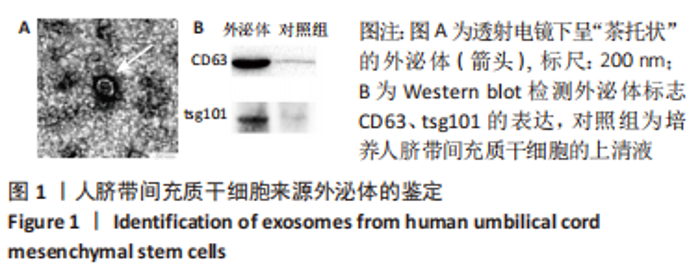
2.1 人脐带间充质干细胞外泌体的鉴定结果 透射电子显微镜下观察到人脐带间充质干细胞来源外泌体呈椭圆形或“茶托”形囊泡,具有完整的外形结构,见图1A;Western blot 检测到外泌体颗粒阳性表达CD63、tsg101,见图1B。"


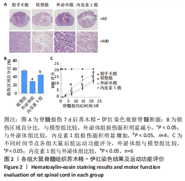
2.2 大鼠脊髓组织苏木精-伊红染色结果及运动功能评价 脊髓损伤后7 d,病理检测结果显示,假手术组脊髓组织分布均匀、结构致密,细胞核圆形,核仁清晰,无明显坏死;模型组可见细胞溶解并消失,局部形成空泡、坏死、出血;外泌体组脊髓组织损伤情况较模型组明显减轻;内皮素1组损伤情况与模型组比较无明显差别,见图2A,B;采用BBB评分评估大鼠运动功能恢复情况,外泌体组与模型组相比,在损伤后3-21 d运动功能显著增强(P < 0.05);内皮素1组运动功能与外泌体组有显著差别(P < 0.05),见图2C。假手术组、模型组、外泌体组比较可以得出外泌体对大鼠脊髓损伤有治疗作用;外泌体组、内皮素1组比较可知内皮素1抵消了外泌体治疗脊髓损伤的作用。"
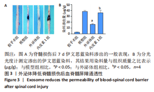
2.3 血脊髓屏障通透性 在脊髓损伤后第7天,用伊文思蓝染色检测血脊髓屏障的通透性,见图3。假手术组染料几乎无外渗,模型组和内皮素1组染料外渗显著增加,外泌体组染料外渗量较模型组明显减少(P < 0.05),外泌体治疗后可以降低血脊髓屏障的通透性,但局部注射内皮素1后,外泌体治疗效果显著减弱(P < 0.05)。"

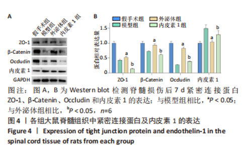
2.4 脊髓紧密连接蛋白及内皮素1表达 通过Western blot检测损伤段脊髓组织中紧密连接蛋白ZO-1、β-Catenin、Occludin及内皮素1的表达量,见图4。"


与假手术组比较,模型组紧密连接蛋白ZO-1、β-Catenin、Occludin表达量显著降低,内皮素1表达量显著增加,表明内皮素1及紧密连接蛋白之间可能存在交互。与模型组比较,外泌体组紧密连接蛋白表达明显升高、内皮素1表达明显降低(P < 0.05),这些结果表明,外泌体可以增加紧密连接蛋白的表达,降低内皮素1的表达。局部组织预先注射内皮素1后再注射外泌体,紧密连接蛋白的表达水平明显低于外泌体组(P < 0.05),说明外泌体促进紧密连接蛋白的表达作用受到内皮素1的抑制。"

| [1] KUMAR R, LIM J, MEKARY RA, et al. Traumatic Spinal Injury: Global Epidemiology and Worldwide Volume. World Neurosurg. 2018;113: e345-e363. [2] FAN B, WEI Z, YAO X, et al. Microenvironment Imbalance of Spinal Cord Injury. Cell Transplant. 2018;27(6):853-866. [3] TAKAHASHI A, NAKAJIMA H, UCHIDA K, et al. Comparison of Mesenchymal Stromal Cells Isolated from Murine Adipose Tissue and Bone Marrow in the Treatment of Spinal Cord Injury. Cell Transplant. 2018;27(7):1126-1139. [4] MATSUSHITA T, LANKFORD KL, ARROYO EJ, et al. Diffuse and persistent blood-spinal cord barrier disruption after contusive spinal cord injury rapidly recovers following intravenous infusion of bone marrow mesenchymal stem cells. Exp Neurol. 2015;267:152-164. [5] ABBASZADEH HA, NIKNAZAR S, DARABI S, et al. Stem cell transplantation and functional recovery after spinal cord injury: a systematic review and meta-analysis. Anat Cell Biol. 2018;51(3):180-188. [6] WANG C, SHI D, SONG X, et al. Calpain inhibitor attenuates ER stress-induced apoptosis in injured spinal cord after bone mesenchymal stem cells transplantation. Neurochem Int. 2016;97:15-25. [7] 张勇,马迅,孙麟,等.人脐带间充质干细胞来源外泌体减轻大鼠脊髓星形胶质细胞氧糖剥夺/复氧损伤所致的水肿[J].中国组织工程研究,2019,23(25):4011-4017. [8] SUN G, LI G, LI D, et al. hucMSC derived exosomes promote functional recovery in spinal cord injury mice via attenuating inflammation. Mater Sci Eng C Mater Biol Appl. 2018;89:194-204. [9] LIU W, WANG Y, GONG F, et al. Exosomes Derived from Bone Mesenchymal Stem Cells Repair Traumatic Spinal Cord Injury by Suppressing the Activation of A1 Neurotoxic Reactive Astrocytes. J Neurotrauma. 2019;36(3):469-484. [10] MCKENZIE AL, HALL JJ, AIHARA N, et al. Immunolocalization of endothelin in the traumatized spinal cord: relationship to blood-spinal cord barrier breakdown. J Neurotrauma. 1995;12(3):257-268. [11] SALZMAN SK, ACOSTA R, BECK G, et al. Spinal endothelin content is elevated after moderate local trauma in the rat to levels associated with locomotor dysfunction after intrathecal injection. J Neurotrauma. 1996;13(2):93-101. [12] 姜亮,刘忠军,党耕町.内皮素在急性脊髓损伤中的变化[J].中华骨科杂志,1998,18(10):594-597. [13] 刘明明,程建,马勇,等.脊髓损伤后血脊髓屏障病变机制研究进展[J].安徽医科大学学报,2015,50(12):1827-1830. [14] 彭新生,李佛保,潘滔,等.内皮素与脊髓损伤后血脊屏障损害的关系[J].中国脊柱脊髓杂志,2000,10(5):280-282. [15] REN ZW, ZHOU JG, XIONG ZK, et al. Effect of exosomes derived from MiR-133b-modified ADSCs on the recovery of neurological function after SCI. Eur Rev Med Pharmacol Sci. 2019;23(1):52-60. [16] HUANG JH, YIN XM, XU Y, et al. Systemic Administration of Exosomes Released from Mesenchymal Stromal Cells Attenuates Apoptosis, Inflammation, and Promotes Angiogenesis after Spinal Cord Injury in Rats. J Neurotrauma. 2017;34(24):3388-3396. [17] KORNILOV R, PUHKA M, MANNERSTRÖM B, et al. Efficient ultrafiltration-based protocol to deplete extracellular vesicles from fetal bovine serum. J Extracell Vesicles. 2018;7(1):1422674. [18] HU Y, RAO SS, WANG ZX, et al. Exosomes from human umbilical cord blood accelerate cutaneous wound healing through miR-21-3p-mediated promotion of angiogenesis and fibroblast function. Theranostics. 2018;8(1):169-184. [19] FRANZEN CA, BLACKWELL RH, TODOROVIC V, et al. Urothelial cells undergo epithelial-to-mesenchymal transition after exposure to muscle invasive bladder cancer exosomes. Oncogenesis. 2015;4(8):e163. [20] EKSTRÖM EJ, BERGENFELZ C, VON BÜLOW V, et al. WNT5A induces release of exosomes containing pro-angiogenic and immunosuppressive factors from malignant melanoma cells. Mol Cancer. 2014;13:88. [21] 周燕,王琳,裴双,等.骨髓间充质干细胞外泌体可减少脊髓损伤后A1型星形胶质细胞的活化[J].中国组织工程研究,2019,23(21): 3294-3301. [22] RONG Y, LIU W, LV C, et al. Neural stem cell small extracellular vesicle-based delivery of 14-3-3t reduces apoptosis and neuroinflammation following traumatic spinal cord injury by enhancing autophagy by targeting Beclin-1. Aging (Albany NY). 2019;11(18):7723-7745. [23] BASSO DM, BEATTIE MS, BRESNAHAN JC. A sensitive and reliable locomotor rating scale for open field testing in rats. J Neurotrauma. 1995;12(1):1-21. [24] 胡伟,谢兴奇,屠冠军.骨髓间充质干细胞来源外泌体改善脊髓损伤后血脊髓屏障的完整性[J].中国组织工程研究,2022,26(7):992-998. [25] JIN LY, LI J, WANG KF, et al. Blood-Spinal Cord Barrier in Spinal Cord Injury: A Review. J Neurotrauma. 2021;38(9):1203-1224. [26] MANNELLO F, GAZZANELLI G. Tissue inhibitors of metalloproteinases and programmed cell death: conundrums, controversies and potential implications. Apoptosis. 2001;6(6):479-482. [27] BARTANUSZ V, JEZOVA D, ALAJAJIAN B, et al. The blood-spinal cord barrier: morphology and clinical implications. Ann Neurol. 2011;70(2):194-206. [28] FIGLEY SA, KHOSRAVI R, LEGASTO JM, et al. Characterization of vascular disruption and blood-spinal cord barrier permeability following traumatic spinal cord injury. J Neurotrauma. 2014;31(6):541-552. [29] ZHENG B, YE L, ZHOU Y, et al. Epidermal growth factor attenuates blood-spinal cord barrier disruption via PI3K/Akt/Rac1 pathway after acute spinal cord injury. J Cell Mol Med. 2016;20(6):1062-1075. [30] YE LB, YU XC, XIA QH, et al. Erratum to: Regulation of Caveolin-1 and Junction Proteins by bFGF Contributes to the Integrity of Blood-Spinal Cord Barrier and Functional Recovery. Neurotherapeutics. 2017;14(3):828-829. [31] FlACK JA, SHARMA KD, XIE JY. Delving into the recent advancements of spinal cord injury treatment: a review of recent progress. Neural Regen Res. 2022;17(2):283-291. [32] CHEN X, LAN X, ROCHE I, et al. Caffeine protects against MPTP-induced blood-brain barrier dysfunction in mouse striatum. J Neurochem. 2008;107(4):1147-1157. [33] LIU C, YANG TH, LI HD, et al. Exosomes from bone marrow mesenchymal stem cells are a potential treatment for ischemic stroke. Neural Regen Res. 2023;18(10):2246-2251. [34] YU B, ZHANG X, LI X. Exosomes derived from mesenchymal stem cells. Int J Mol Sci. 2014;15(3):4142-4157. [35] VILLARROYA-BELTRI C, BAIXAULI F, GUTIÉRREZ-VÁZQUEZ C, et al. Sorting it out: regulation of exosome loading. Semin Cancer Biol. 2014;28:3-13. [36] VLASSOV AV, MAGDALENO S, SETTERQUIST R, et al. Exosomes: current knowledge of their composition, biological functions, and diagnostic and therapeutic potentials. Biochim Biophys Acta. 2012;1820(7):940-948. [37] HUTSON TH, DI GIOVANNI S. The translational landscape in spinal cord injury: focus on neuroplasticity and regeneration. Nat Rev Neurol. 2019;15(12):732-745. [38] LIEBNER S, CZUPALLA CJ, WOLBURG H. Current concepts of blood-brain barrier development. Int J Dev Biol. 2011;55(4-5):467-476. [39] LEE JY, KIM HS, CHOI HY, et al. Fluoxetine inhibits matrix metalloprotease activation and prevents disruption of blood-spinal cord barrier after spinal cord injury. Brain. 2012;135(Pt 8):2375-2389. [40] PARK CS, LEE JY, CHOI HY, et al. Protocatechuic acid improves functional recovery after spinal cord injury by attenuating blood-spinal cord barrier disruption and hemorrhage in rats. Neurochem Int. 2019;124:181-192. [41] ZHANG Q, WANG J, GU Z, et al. Effect of lycopene on the blood-spinal cord barrier after spinal cord injury in mice. Biosci Trends. 2016;10(4): 288-293. [42] YU DS, WANG YS, BI YL, et al. Salvianolic acid A ameliorates the integrity of blood-spinal cord barrier via miR-101/Cul3/Nrf2/HO-1 signaling pathway. Brain Res. 2017;1657:279-287. [43] GUO J, LI Y, HE Z, et al. Targeting endothelin receptors A and B attenuates the inflammatory response and improves locomotor function following spinal cord injury in mice. Int J Mol Med. 2014;34(1):74-82. [44] SINESCU C, POPA F, GRIGOREAN VT, et al. Molecular basis of vascular events following spinal cord injury. J Med Life. 2010;3(3):254-261. [45] MICHINAGA S, KIMURA A, HATANAKA S, et al. Delayed Administration of BQ788, an ETB Antagonist, after Experimental Traumatic Brain Injury Promotes Recovery of Blood-Brain Barrier Function and a Reduction of Cerebral Edema in Mice. J Neurotrauma. 2018;35(13):1481-1494. [46] HAO Y, MIAO J, LIU W, et al. Mesenchymal Stem Cell-Derived Exosomes Carry MicroRNA-125a to Protect Against Diabetic Nephropathy by Targeting Histone Deacetylase 1 and Downregulating Endothelin-1. Diabetes Metab Syndr Obes. 2021;14:1405-1418. [47] ZHANG Y, CHEN L, LI Z, et al. Endothelin-1, over-expressed in SOD1G93A mice, aggravates injury of NSC34-hSOD1G93A cells through complicated molecular mechanism revealed by quantitative proteomics analysis. Front Cell Neurosci. 2022;16:1069617. [48] A JC, LI ZY, LONG QF, et al. MiR-379-5p improved locomotor function recovery after spinal cord injury in rats by reducing endothelin 1 and inhibiting astrocytes expression. Eur Rev Med Pharmacol Sci. 2019; 23(22):9738-9745. |
| [1] | Peng Yingnan, Bian Zhilei, Zhang Suping, Li Li, Cao Weijie, Wan Dingming. Effect of CD34+ cell dose on haploidentical hematopoietic stem cell transplantation for treating malignant hematological diseases [J]. Chinese Journal of Tissue Engineering Research, 2024, 28(1): 1-6. |
| [2] | Wei Yurou, Tian Jiaqing, He Xianshun, Zhan Zhiwei, Wei Tengfei, Lin Tianye, He Wei, Wei Qiushi. Effect of lentiviral silencing of Piezo1 on osteogenic differentiation and TAZ expression in human bone marrow mesenchymal stem cells [J]. Chinese Journal of Tissue Engineering Research, 2024, 28(1): 12-19. |
| [3] | Wang Xianfeng, Wang Kun, Sun Han, Sun Xiaoliang, Yan Litao. Mechanism underlying exosomal lncRNA H19 derived from umbilical cord mesenchymal stem cells promotes cartilage injury repair [J]. Chinese Journal of Tissue Engineering Research, 2024, 28(1): 20-25. |
| [4] | Zhang Yuanshu, He Xu, Xue Yuan, Jin Yesheng, Wang Kai, Shi Qin, Rui Yongjun. Irisin alleviates palmitic acid-induced osteogenic inhibition in bone marrow mesenchymal stem cells [J]. Chinese Journal of Tissue Engineering Research, 2024, 28(1): 26-31. |
| [5] | He Lijun, Qi Xiaojuan. Adipose-derived mesenchymal stem cells overexpressing bone morphogenetic protein 2 promote alveolar bone defect repair in osteoporosis rats [J]. Chinese Journal of Tissue Engineering Research, 2024, 28(1): 32-37. |
| [6] | Zhou Minghua, Hu Xiaoyu. LncRNA SNHG4 regulates miR-152-3p during osteoblastic differentiation of periodontal ligament stem cells [J]. Chinese Journal of Tissue Engineering Research, 2024, 28(1): 38-43. |
| [7] | Zheng Rongjiong, Deng Zerun, Han Dan, Sun Lihua. Mechanism underlying rat hepatocyte apoptosis regulated by exosomes derived from bone marrow mesenchymal stem cells [J]. Chinese Journal of Tissue Engineering Research, 2024, 28(1): 44-49. |
| [8] | Sun Jing, Liao Jian, Sun Jiangling, Cheng Ping, Feng Hongchao. Recombinant human growth hormone promotes osteogenic differentiation of human dental pulp stem cells [J]. Chinese Journal of Tissue Engineering Research, 2024, 28(1): 56-61. |
| [9] | Ai Fangfang, Xiao Hongyan, Wang Fang, Zhu Yongzhao, Ma Lijun. Reversal effect of Lycium barbarum polysaccharide in combination with oxaliplatin on drug resistance of colon cancer stem cells [J]. Chinese Journal of Tissue Engineering Research, 2024, 28(1): 74-79. |
| [10] | Chen Guanting, Zhang Linqi, Li Qingru. Research hot spots and trends of exosomes in theranostic application for chronic kidney disease [J]. Chinese Journal of Tissue Engineering Research, 2024, 28(1): 86-92. |
| [11] | Fan Yongjing, Wang Shu, Jin Wulong. Characteristics, advantages and application of osteogenic differentiation of jaw bone marrow mesenchymal stem cells [J]. Chinese Journal of Tissue Engineering Research, 2024, 28(1): 100-106. |
| [12] | Huang Yongbin, Wang Tao, Lou Yuanyi, Pang Jingqun, Chen Guanghua. Application prospect of mesenchymal stem cells in promoting muscle tissue repair [J]. Chinese Journal of Tissue Engineering Research, 2024, 28(1): 107-112. |
| [13] | Ying Chunmiao, Pan Xiaolong, Liu Feixiang, Chen Na, Fan Feiyan, Zhang Yunke. Effect of traditional Chinese medicine and compounds for supplementing qi and activating blood circulation and inducing resuscitation on regulating stem cells to promote nerve repair of acute ischemic stroke [J]. Chinese Journal of Tissue Engineering Research, 2024, 28(1): 121-130. |
| [14] | Long Qingxi, Zhang Pingshu, Liu Qing, Ou Ya, Zhang Lili, Yuan Xiaodong. Single-cell RNA sequencing reveals the heterogeneity of astrocytes [J]. Chinese Journal of Tissue Engineering Research, 2024, 28(1): 139-146. |
| [15] | Nong Fuxiang, Jiang Zhixiong, Li Yinghao, Xu Wencong, Shi Zhilan, Luo Hui, Zhang Qinglang, Zhong Shuang, Tang Meiwen. Bone cement augmented proximal femoral nail antirotation for type A3.3 intertrochanteric femoral fracturalysis [J]. Chinese Journal of Tissue Engineering Research, 2023, 27(在线): 1-10. |
| Viewed | ||||||
|
Full text |
|
|||||
|
Abstract |
|
|||||