Chinese Journal of Tissue Engineering Research ›› 2023, Vol. 27 ›› Issue (25): 3977-3983.doi: 10.12307/2023.506
Previous Articles Next Articles
Local injection of ginsenoside Rg1 nanoparticles in the treatment of myocardial infarction in rats
Cao Congcong1, Ling Gengfei2, Yang Chunhua3
- 1Zhoukou Vocational and Technical College, Zhoukou 466000, Henan Province, China; 2Department of Critical Care, 3Department of Cardiovascular Medicine, Zhoukou Central Hospital, Zhoukou 466000, Henan Province, China
-
Received:2022-07-08Accepted:2022-08-19Online:2023-09-08Published:2023-01-17 -
Contact:Yang Chunhua, Master, Associate chief physician, Department of Cardiovascular Medicine, Zhoukou Central Hospital, Zhoukou 466000, Henan Province, China -
About author:Cao Congcong, Lecturer, Zhoukou Vocational and Technical College, Zhoukou 466000, Henan Province, China
CLC Number:
Cite this article
Cao Congcong, Ling Gengfei, Yang Chunhua. Local injection of ginsenoside Rg1 nanoparticles in the treatment of myocardial infarction in rats[J]. Chinese Journal of Tissue Engineering Research, 2023, 27(25): 3977-3983.
share this article
Add to citation manager EndNote|Reference Manager|ProCite|BibTeX|RefWorks
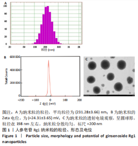
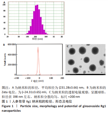
2.1 人参皂苷Rg1纳米粒的粒径、形态及电位 由检测结果可知,人参皂苷Rg1纳米粒的平均粒径为(231.28±3.66) nm(图1A),Zeta电位(-24.31±3.65) mV(图1B),透射电镜下可见该纳米粒呈圆球形,粒径在198 nm左右,纳米粒分散均匀,无团聚(图1C)。"
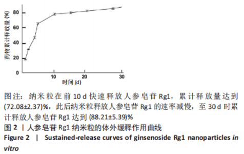
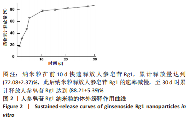
2.3 人参皂苷Rg1纳米粒的体外缓释作用 人参皂苷Rg1纳米粒的体外缓释作用曲线见图2,结果可见:纳米粒在前10 d快速释放人参皂苷Rg1,累计释放量达到(72.08±2.37)%,此后纳米粒释放人参皂苷Rg1的速率减慢,至30 d时累计释放人参皂苷Rg1达到(88.21±5.39)%。"
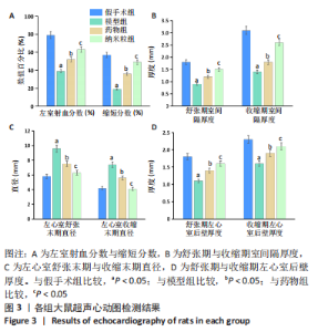
2.4 人参皂苷Rg1纳米粒的动物体内应用 2.4.1 实验动物数量分析 造模过程中无大鼠死亡,因此,实验纳入的64只大鼠全部进入结果分析。 2.4.2 超声心动图检测结果 见图3。"
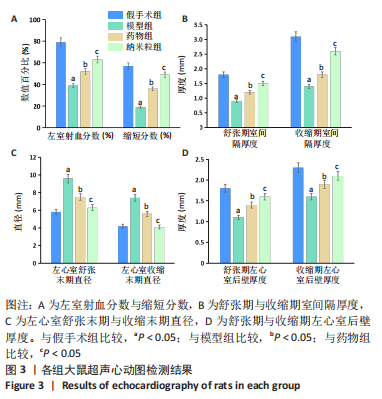

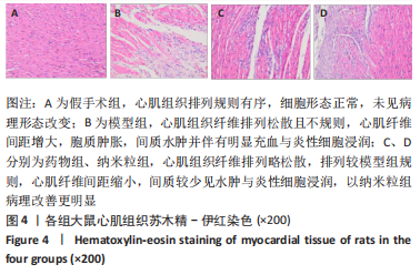
术后28 d,与假手术组相比,模型组大鼠左心室射血分数、缩短分数、舒张期室间隔厚度、收缩期室间隔厚度、舒张期左心室后壁厚度、收缩期左心室后壁厚度减少(P < 0.05),左心室舒张末期直径、左心室收缩末期直径增加(P < 0.05)。与模型组比较,药物组、纳米粒组大鼠左心室射血分数、缩短分数、舒张期室间隔厚度、收缩期室间隔厚度、舒张期左心室后壁厚度、收缩期左心室后壁厚度增加(P < 0.05),左心室舒张末期直径、左心室收缩末期直径减少(P < 0.05)。与药物组比较,纳米粒组大鼠的左心室射血分数、缩短分数、舒张期室间隔厚度、收缩期室间隔厚度、舒张期左心室后壁厚度、收缩期左心室后壁厚度增加(P < 0.05),左心室舒张末期直径、左心室收缩末期直径减少(P < 0.05)。 2.4.3 心肌组织学分析 术后28 d,苏木精-伊红染色显示,假手术组大鼠心肌组织排列规则有序,细胞形态正常,未见病理形态改变;模型组大鼠心肌组织纤维排列松散且不规则,心肌纤维间距增大,胞质肿胀,间质水肿并伴有明显充血与炎性细胞浸润;药物组、纳米粒组大鼠心肌组织纤维排列略松散,排列较模型组规则,心肌纤维间距缩小,间质较少见水肿与炎性细胞浸润,其中以纳米粒组病理改善更明显,见图4。"

Masson染色显示,假手术组心肌纤维排列规则有序;与假手术组比较,模型组大鼠心肌纤维排列紊乱,部分纤维断裂,缺血中心及周围可见大量胶原沉积;药物组、纳米粒组大鼠心肌纤维排列较规则,缺血中心及周围胶原沉积明显减少,其中以纳米粒组胶原沉积更少,见图5。"
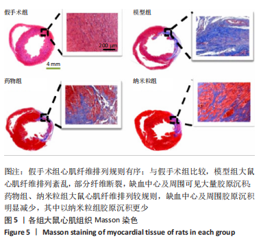
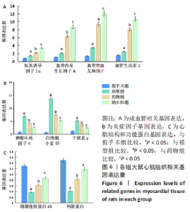
胶原面积定量分析结果显示,假手术组、模型组、药物组、纳米粒组大鼠心肌组织胶原面积百分比分别为(5.31±0.38)%,(72.57±6.97)%,(53.02±3.88)%,(39.74±7.19)%;其中,模型组胶原面积百分比大于假手术组(P < 0.05),药物组胶原面积百分比小于模型组(P < 0.05),纳米粒组胶原面积百分比小于药物组(P < 0.05)。 2.4.4 RT-qPCR检测结果 各组心肌组织各基因表达量检测结果,见图6。"
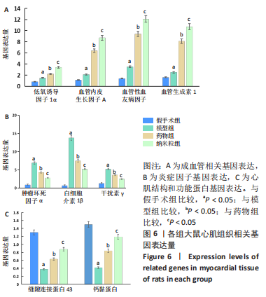

检测结果显示,与假手术组比较,模型组缝隙连接蛋白43、钙黏蛋白的mRNA表达量明显降低(P < 0.05),低氧诱导因子1α、血管性血友病因子、血管内皮生长因子A、血管生成素1、肿瘤坏死因子α、白细胞介素1β、干扰素γ的mRNA表达量明显升高(P < 0.05)。与模型组比较,药物组低氧诱导因子1α、血管性血友病因子、血管内皮生长因子A、血管生成素1、缝隙连接蛋白43、钙黏蛋白的mRNA表达量明显升高(P < 0.05),肿瘤坏死因子α、白细胞介素1β、干扰素γ的mRNA表达量明显降低(P < 0.05)。与药物组比较,纳米粒组低氧诱导因子1α、血管性血友病因子、血管内皮生长因子A、血管生成素1、缝隙连接蛋白43、钙黏蛋白的mRNA表达量明显升高(P < 0.05),肿瘤坏死因子α、白细胞介素1β、干扰素γ的mRNA表达量明显降低(P < 0.05)。 2.4.5 纳米粒的生物相容性 由动物实验结果可知,人参皂苷Rg1纳米粒具有良好的生物相容性。"

| [1] CHEN R, XIAO Y, CHEN M, et al. A traditional Chinese medicine therapy for coronary heart disease after percutaneous coronary intervention: a meta-analysis of randomized, double-blind, placebo-controlled trials. Biosci Rep. 2018;38(5):BSR20180973. [2] 中国心血管健康与疾病报告2019 概要 [J].心脑血管病防治,2020, 20(5):437-450. [3] YANAMANDALA M, ZHU W, GARRY DJ, et al. Overcoming the roadblocks to cardiac cell therapy using tissue engineering. J Am Coll Cardiol. 2017; 70(6):766-775. [4] BLANCO-FERNANDEZ B, CASTAÑO O, MATEOS-TIMONEDA MÁ, et al. Nanotechnology Approaches in Chronic Wound Healing. Adv Wound Care (New Rochelle). 2021;10(5):234-256. [5] LUO Y, WANG Q, ZHANG Y. Biopolymer-Based Nanotechnology Approaches To Deliver Bioactive Compounds for Food Applications: A Perspective on the Past, Present, and Future. J Agric Food Chem. 2020;68(46):12993-13000. [6] BAYDA S, ADEEL M, TUCCINARDI T, et al.The History of Nanoscience and Nanotechnology: From Chemical-Physical Applications to Nanomedicine. Molecules. 2019;25(1):112. [7] TIBURCIUS S, KRISHNAN K, YANG JH, et al. Silica-Based Nanoparticles as Drug Delivery Vehicles for Prostate Cancer Treatment. Chem Rec. 2021;21(6):1535-1568. [8] TALAT A, KHAN AU. Patents in chemotherapy: nanoparticles as drug-delivery vehicles. Pharm Pat Anal. 2020;9(4):117-119. [9] LUO M, YAN D, SUN Q, et al. Ginsenoside Rg1 attenuates cardiomyocyte apoptosis and inflammation via the TLR4/NF-kB/NLRP3 pathway. J Cell Biochem. 2020;121(4):2994-3004. [10] LU ML, WANG J, SUN Y, et al. Ginsenoside Rg1 attenuates mechanical stress-induced cardiac injury via calcium sensing receptor-related pathway.J Ginseng Res. 2021;45(6):683-694. [11] HUANG L, CAI HA, ZHANG MS, et al. Ginsenoside Rg1 promoted the wound healing in diabetic foot ulcers via miR-489-3p/Sirt1 axis. J Pharmacol Sci. 2021;147(3):271-283. [12] 金岩,刘闺男.人参皂苷Rg1对急性心肌梗死大鼠血管新生的作用[J].中国医科大学学报,2007,36(5):517-519. [13] MARUYAMA K, NAEMURA K, YOSHIHARA K, et al. Surgical protocol for permanent ligation of the left anterior descending coronary artery in mice to generate a model of myocardial infarction. STAR Protoc. 2021;2(3):100775. [14] BORGHI C, BENTIVENGA C, COSENTINO ER. Uric acid and risk of myocardial infarction. A dynamic duo. Int J Cardiol. 2020;320:23-24. [15] CRITCHER CR, SIEGEL M. Re-examining the Association Between E-Cigarette Use and Myocardial Infarction: A Cautionary Tale. Am J Prev Med. 2021;61(4):474-482. [16] PEET C, IVETIC A, BROMAGE DI, et al. Cardiac monocytes and macrophages after myocardial infarction. Cardiovasc Res. 2020;116(6): 1101-1112. [17] TOMOAIA R, BEYER RS, SIMU G, et al. Understanding the role of echocardiography in remodeling after acute myocardial infarction and development of heart failure with preserved ejection fraction. Ultrason. 2019;21(1):69-76. [18] JENČA D, MELENOVSKÝ V, STEHLIK J, et al. Heart failure after myocardial infarction: incidence and predictors. ESC Heart Fail. 2021;8(1):222-237. [19] RUDDOX V, SANDVEN I, MUNKHAUGEN J, et al. Atrial fibrillation and the risk for myocardial infarction, all-cause mortality and heart failure: A systematic review and meta-analysis. Eur J Prev Cardiol. 2017;24(14): 1555-1566. [20] TOMOAIA R, BEYER RS, SIMU G, et al. Understanding the role of echocardiography in remodeling after acute myocardial infarction and development of heart failure with preserved ejection fraction.Med Ultrason. 2019;21(1):69-76. [21] KOC L, ONDRUS T, FILA P, et al. Right ventricular myocardial infarction in the era of primary percutaneous coronary intervention. Bratisl Lek Listy. 2021;122(10):700-707. [22] XIAO M, LI Y, GUAN X. Community-Based Physical Rehabilitation After Percutaneous Coronary Intervention for Acute Myocardial Infarction. Tex Heart Inst J. 2021;48(2):e197103. [23] FOKIN AA, KIREEV KA, NETISANOV SV. [Coronary artery bypass grafting in non-ST-segment elevation acute myocardial infarction]. Angiol Sosud Khir. 2020;26(3):142-149. [24] WU X, REBOLL MR, KORF-KLINGEBIEL M, et al. Angiogenesis after acute myocardial infarction. Cardiovasc Res. 2021;117(5):1257-1273. [25] MAHTTA D, SUDHAKAR D, KONERU S, et al. Targeting Inflammation After Myocardial Infarction. Curr Cardiol Rep. 2020;22(10):110. [26] DENG Y, ZHANG X, SHEN H, et al. Application of the Nano-Drug Delivery System in Treatment of Cardiovascular Diseases. Front Bioeng Biotechnol. 2020;7:489. [27] FORINI F, CANALE P, NICOLINI G, et al. Mitochondria-Targeted Drug Delivery in Cardiovascular Disease: A Long Road to Nano-Cardio Medicine. Pharmaceutics. 2020;12(11):1122. [28] FU Y, TAN L, MENG L, et al. Therapeutic Effects of Paclitaxel Loaded Polyethylene Glycol-Polylactic Acid-Glycolic Acid Copolymer Nanoparticles on Pancreatic Cancer in Rats. J Nanosci Nanotechnol. 2020;20(12):7271-7275. [29] HUA Y, SU Y, ZHANG H, et al. Poly(lactic-co-glycolic acid) microsphere production based on quality by design: a review. Drug Deliv. 2021;28(1): 1342-1355. [30] SONG F, CHEN L, LIN R, et al. Synthesis of carboxy-polyethylene glycol-amine (CA (PEG)n ) and [1-14 C]-CA (PEG)n via oxa-Michael addition of amino-polyethylene glycols to propiolates vs to acrylates. J Labelled Comp Radiopharm. 2020;63(1):15-24. [31] YANG M, JIN L, WU Z, et al. PLGA-PEG Nanoparticles Facilitate In Vivo Anti-Alzheimer’s Effects of Fucoxanthin, a Marine Carotenoid Derived from Edible Brown Algae. J Agric Food Chem. 2021;69(34):9764-9777. [32] 晏殊瑾,杨珂,王河,等.低强度脉冲超声靶向纳米粒控释SDF-1及BMP-2调节hPDLCs迁移和成骨分化[J].中国超声医学杂志,2022, 38(2):222-226. [33] Wang B, Zhang C, Chu D, et al. Astragaloside IV improves angiogenesis under hypoxic conditions by enhancing hypoxia‑inducible factor‑1α SUMOylation. Mol Med Rep. 2021;23(4):244. [34] DARDEN J, PAYNE LB, ZHAO H, et al. Excess vascular endothelial growth factor-A disrupts pericyte recruitment during blood vessel formation. Angiogenesis. 2019;22(1):167-183. [35] JUBAN G, MOUNIER R.[Tissue repair: Key role of annexin A1 in the control of inflammatory response]. Med Sci (Paris). 2021;37(4):324-326. [36] TOTLAND MZ, RASMUSSEN NL, KNUDSEN LM, et al. Regulation of gap junction intercellular communication by connexin ubiquitination: physiological and pathophysiological implications. Cell Mol Life Sci. 2020;77(4):573-591. [37] LAI Y, FOIS G, FLORES JR, et al. Inhibition of calcium-triggered secretion by hydrocarbon-stapled peptides. Nature. 2022;603(7903):949-956. |
| [1] | Li Zikai, Zhang Chengcheng, Xiong Jiaying, Yang Xirui, Yang Jing, Shi Haishan. Potential effects of ornidazole on intracanal vascularization in endodontic regeneration [J]. Chinese Journal of Tissue Engineering Research, 2025, 29(在线): 1-7. |
| [2] | Lou Guo, Zhang Min, Fu Changxi. Exercise preconditioning for eight weeks enhances therapeutic effect of adipose-derived stem cells in rats with myocardial infarction [J]. Chinese Journal of Tissue Engineering Research, 2025, 29(7): 1363-1370. |
| [3] | Gao Yang, Qin Hewei, Liu Dandan. ACSL4 mediates ferroptosis and its potential role in atherosclerotic cardiovascular disease [J]. Chinese Journal of Tissue Engineering Research, 2025, 29(6): 1239-1247. |
| [4] | Yu Shuangqi, Ding Fan, Wan Song, Chen Wei, Zhang Xuejun, Chen Dong, Li Qiang, Lin Zuoli. Effects of polylactic acid-glycolic acid copolymer/lysine-grafted graphene oxide nanoparticle composite scaffolds on osteogenic differentiation of MC3T3 cells [J]. Chinese Journal of Tissue Engineering Research, 2025, 29(4): 707-712. |
| [5] | Xiong Bohan, Wang Guoliang, Yu Yang, Xue Wenqiang, Yu Hong, Liu Jinrui, Ruan Zhaohui, Li Yajuan, Liu Haolong, Dong Kaiyan, Long Dan, Chen Zhao. Internal tension relieving technique assisted anterior cruciate ligament reconstruction to promote ligamentization of Achilles tendon grafts in small ear pigs in southern Yunnan province [J]. Chinese Journal of Tissue Engineering Research, 2025, 29(4): 713-720. |
| [6] | Chen Yaodong, Ren Jiayi, Cao Jingwei, Fan Wenwen, Chen Wu. Near-infrared photoresponsive h-PCuNF nanoparticles mediate multimodal therapeutics against malignant tumors [J]. Chinese Journal of Tissue Engineering Research, 2025, 29(4): 780-788. |
| [7] |
Fu Changxi, He Ruibo, Ma Gang, Zhu Zheng, Ma Wenchao.
Effect and mechanism of different training modes on skeletal muscle remodeling in rats with heart failure induced by myocardial infarction
[J]. Chinese Journal of Tissue Engineering Research, 2025, 29(2): 221-230.
|
| [8] | Zhu Weiwei, Fan Zhengchao, Xu Ying, Xia Jizhu, Zhao Xiangzhi. Dual-modality imaging bionic nanoparticles for sonodynamic therapy on medullary thyroid carcinoma [J]. Chinese Journal of Tissue Engineering Research, 2025, 29(16): 3410-3419. |
| [9] | Wang Zhikun, Bai Shaoxuan, Zhao Wei, Wang Chenyu. Exercise preconditioning combined with bone marrow mesenchymal stem cell transplantation for myocardial infarction in rats [J]. Chinese Journal of Tissue Engineering Research, 2025, 29(1): 65-73. |
| [10] | Liu Hanfeng, Wang Jingjing, Yu Yunsheng. Artificial exosomes in treatment of myocardial infarction: current status and prospects [J]. Chinese Journal of Tissue Engineering Research, 2024, 28(7): 1118-1123. |
| [11] | Sun Yukang, Song Lijuan, Wen Chunli, Ding Zhibin, Tian Hao, Ma Dong, Ma Cungen, Zhai Xiaoyan. Visualization analysis of stem cell therapy for myocardial infarction based on Web of Science in recent ten years [J]. Chinese Journal of Tissue Engineering Research, 2024, 28(7): 1143-1148. |
| [12] | Shen Ziqing, Xia Tian, Shan Yibo, Zhu Ruijun, Wan Haoxin, Ding Hao, Pan Shu, Zhao Jun. Vascularized tracheal substitutes constructed by exosome-load hydrogel-modified 3D printed scaffolds [J]. Chinese Journal of Tissue Engineering Research, 2024, 28(5): 697-705. |
| [13] | Zhu Liwei, Wang Jiangyue, Bai Ding. Application value of nanocomposite gelatin methacryloyl hydrogels in different bone defect environments [J]. Chinese Journal of Tissue Engineering Research, 2024, 28(5): 753-758. |
| [14] | He Rongzhen, Ying Lyufang, He Xingwen, Chen Chuanshun, Yin Yuesong, Zhang Kexiang, Wang Zili. Effect of ginsenoside Rg1 on muscle degeneration after massive rotator cuff injury in mice [J]. Chinese Journal of Tissue Engineering Research, 2024, 28(32): 5136-5140. |
| [15] | Sun Yuma, Ma Wenchao, Fu Changxi. Granulocyte colony-stimulating factor combined with high-intensity intermittent exercise preconditioning improved cardiac remodeling in rats with acute myocardial infarction [J]. Chinese Journal of Tissue Engineering Research, 2024, 28(31): 4987-4994. |
| Viewed | ||||||
|
Full text |
|
|||||
|
Abstract |
|
|||||